



Enterolactone

Based on 1 Customer Validation
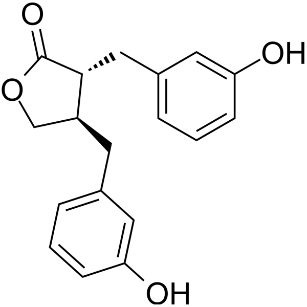
Enterolactone is a bioactive phenolic metabolite known as a mammalian lignan derived from dietary lignans. Enterolactone has estrogenic properties and anti-breast cancer activity. Enterolactone is a radiosensitizer for human breast cancer cell lines through impaired DNA repair and increased apoptosis.
商品は「研究用試薬」です。人や動物の医療用・臨床診断用・食品用の製品ではありません。
研究用途以外に使用した場合、当社は一切の責任を負いかねます。
- 純度 : 98.2%
- CAS 番号: 78473-71-9
- 分子式: C18H18O4
- 分子量:298.33
-
保管条件:Powder -20°C, 3 years ; In solvent -80°C, 6 months , -20°C, 1 month

To place orders, for customer services and technical support, please contact: MedChemExpress USA
Tel: 609-228-6898 E-mail: [email protected] [email protected]


-
 生物活性
生物活性
-
化学情報
-
溶剤 & 溶解度
- プロトコル
- 純度とドキュメンテーション
- 参考文献
-
 Help & FAQs


Endogenous Metabolite アイソフォーム固有の製品をすべて表示
More

生物活性
製品説明
Cellular Effect
|
Cell Line

|
Type | Value | Description | References |
|---|---|---|---|---|
| RAW264.7 | IC50 |
23.5 μM
Compound: Zerumboneoxide
|
Inhibition of LPS-induced nitric oxide production in mouse RAW264.7 cells measured after 20 hrs by Griess assay
Inhibition of LPS-induced nitric oxide production in mouse RAW264.7 cells measured after 20 hrs by Griess assay
|
[PMID: 30096653] |
体外実験
Enterolactone (25-75 μM; 48 hours) arrests the growth of MDA-MB-231 breast cancer cells in the ‘S’ phase[1]
Enterolactone (25-75 μM; 15 hours) triggers apoptosis in MDA-MB-231 breast cancer cells via caspase-3 activation[1].
Enterolactone inhibits TGF-β-induced migration of MDA-MB-231 breast cancer cells. Enterolactone inhibits TGF-β-induced invasion of MDA-MB-231 breast cancer cells through ECM. Enterolactone inhibits the TGF-β-induced EMT program in MDA-MB-231 breast cancer cells. Enterolactone reduces the formation of actin stress fibers by inhibiting the expression of CD44 and MAPK-p38. Enterolactone inhibits the ERK/NF-κB/Snail signaling pathway to revert TGF-β-induced EMT in MDA-MB-231 cells[1].
MedChemExpress (MCE) has not independently confirmed the accuracy of these methods. They are for reference only.
-
Cell Line:MDA-MB-231 cells
-
Concentration:25, 50, 75 μM
-
Incubation Time:48 hours
-
Result:There was a non-significant increase (~24%) in the S phase population following treatment with 25 μM EL, whereas there were significant increases (~34% and ~39%) following treatment with 50 and 75 μM EL, respectively.


化学情報
-
CAS 番号 78473-71-9
-
性状 Solid
-
分子量 298.33
-
分子式 C18H18O4
-
Color White to off-white
-
SMILES
O=C1OC[C@H](CC2=CC=CC(O)=C2)[C@H]1CC3=CC=CC(O)=C3
-
別名
エンテロラクトン
-
Structure Classification
-
Initial Source
-
輸送条件
Room temperature in continental US; may vary elsewhere.
-
保管条件
Powder -20°C 3 years In solvent -80°C 6 months -20°C 1 month


溶剤 & 溶解度
体外:
DMSO : 30 mg/mL (100.56 mM; Need ultrasonic and warming; Hygroscopic DMSO has a significant impact on the solubility of product, please use newly opened DMSO)
Please refer to the solubility information to select the appropriate solvent. Once prepared, please aliquot and store the solution to prevent product inactivation from repeated freeze-thaw cycles.
Storage method and period of stock solution: -80°C, 6 months; -20°C, 1 month. When stored at -80°C, please use it within 6 months. When stored at -20°C, please use it within 1 month.
Please refer to the solubility information to select the appropriate solvent. Once prepared, please aliquot and store the solution to prevent product inactivation from repeated freeze-thaw cycles.
Storage method and period of stock solution: -80°C, 6 months; -20°C, 1 month. When stored at -80°C, please use it within 6 months. When stored at -20°C, please use it within 1 month.
濃度 (開始) × 体積 (開始) = 濃度 (終了) × 体積 (終了)


純度とドキュメンテーション
-
データシート (281 KB)
-
SDS (393 KB)
- English - EN (393 KB)
- Français - FR (393 KB)
- Deutsch - DE (393 KB)
- Norwegian - NO (393 KB)
- Español - ES (393 KB)
- Swedish - SV (393 KB)
- Italian - IT (393 KB)
- Korean - KR (393 KB)
- Portuguese - PT (393 KB)
-
取扱説明書 (2659 KB)
参考文献
[1]. Bigdeli B, et al. Enterolactone: A novel radiosensitizer for human breast cancer cell lines through impaired DNA repair and increased apoptosis. Toxicol Appl Pharmacol. 2016;313:180-194. [Content Brief]
[2]. Mali AV, et al. Enterolactone modulates the ERK/NF-κB/Snail signaling pathway in triple-negative breast cancer cell line MDA-MB-231 to revert the TGF-β-induced epithelial-mesenchymal transition. Cancer Biol Med. 2018;15(2):137-156. [Content Brief]


Complete Stock Solution Preparation Table
Please refer to the solubility information to select the appropriate solvent. Once prepared, please aliquot and store the solution to prevent product inactivation from repeated freeze-thaw cycles.
Storage method and period of stock solution: -80°C, 6 months; -20°C, 1 month. When stored at -80°C, please use it within 6 months. When stored at -20°C, please use it within 1 month.
| Optional Solvent | Concentration Solvent Mass | 1 mg | 5 mg | 10 mg | 25 mg |
|---|---|---|---|---|---|
| DMSO | 1 mM | 3.3520 mL | 16.7600 mL | 33.5199 mL | 83.7998 mL |
| 5 mM | 0.6704 mL | 3.3520 mL | 6.7040 mL | 16.7600 mL | |
| 10 mM | 0.3352 mL | 1.6760 mL | 3.3520 mL | 8.3800 mL | |
| 15 mM | 0.2235 mL | 1.1173 mL | 2.2347 mL | 5.5867 mL | |
| 20 mM | 0.1676 mL | 0.8380 mL | 1.6760 mL | 4.1900 mL | |
| 25 mM | 0.1341 mL | 0.6704 mL | 1.3408 mL | 3.3520 mL | |
| 30 mM | 0.1117 mL | 0.5587 mL | 1.1173 mL | 2.7933 mL | |
| 40 mM | 0.0838 mL | 0.4190 mL | 0.8380 mL | 2.0950 mL | |
| 50 mM | 0.0670 mL | 0.3352 mL | 0.6704 mL | 1.6760 mL | |
| 60 mM | 0.0559 mL | 0.2793 mL | 0.5587 mL | 1.3967 mL | |
| 80 mM | 0.0419 mL | 0.2095 mL | 0.4190 mL | 1.0475 mL | |
| 100 mM | 0.0335 mL | 0.1676 mL | 0.3352 mL | 0.8380 mL |


Enterolactone 関連分類
HY-108692 関連分類
 Powered by Bioz