



チオアセトアミド
Based on 5 publication(s) in Google Scholar
Thioacetamide (TAA) is an indirect hepatotoxin and causes parenchymal cell necrosis. Thioacetamide requires metabolic activation by microsomal CYP2E1 to thioacetamide-S-oxide initially and then to thioacetamide-S-dioxide, which is a highly reactive metabolite, and its reactive metabolites covalently bind to proteins and lipids thereby causing oxidative stress and centrilobular necrosis. Thioacetamide can induce chronic liver fibrosis, encephalopathy and other events model.
商品は「研究用試薬」です。人や動物の医療用・臨床診断用・食品用の製品ではありません。
研究用途以外に使用した場合、当社は一切の責任を負いかねます。
- 純度 : 98.0%
- CAS 番号: 62-55-5
- 分子式: C2H5NS
- 分子量:75.13
-
保管条件:
4°C, stored under nitrogen
* In solvent : -80°C, 6 months; -20°C, 1 month (stored under nitrogen)


To place orders, for customer services and technical support, please contact: MedChemExpress USA
Tel: 609-228-6898 E-mail: [email protected] [email protected]


-
 生物活性
生物活性
-
化学情報
-
溶剤 & 溶解度
- プロトコル
- 純度とドキュメンテーション
- 参考文献
-
 Help & FAQs


-
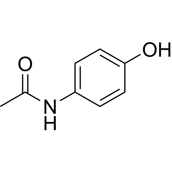
HY-66005
AcetaminophenCOX-2 Inhibitor -

HY-Y0320
Dimethyl sulfoxide, meets analytical specification of Ch.P.Aprotic Solvent -

HY-108749
Olive oilAnti-Inflammation Agent -
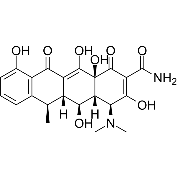
HY-N0565
DoxycyclineMMP Inhibitor -

HY-P2149
Concanavalin APlant Lectin -
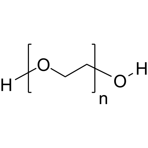
HY-Y0873
PEG300Neutral Polymer -
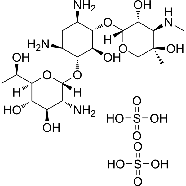
HY-17561
G-418 disulfateAminoglycoside Antibiotic -

HY-P0035
Insulin (human)Recombinant Human Insulin -
HY-12848
SAGSmoothened Agonist -

HY-P9907
TrastuzumabHER2 Blocking Antibody -

HY-E0264
Rodent Gnawing BlocksRodent Gnawing Block
MedChemExpress(MCE)の使用を引用している文献 Thioacetamide
More Customer Validation & Images

-
In Vivo Efficacy Study
-
Histological Imaging/Staining
-
IHC
-
WB
-
IHC


生物活性
製品説明
体外実験
Thioacetamide (TAA; 0-10000 μM; 24 h; WB-F344 cells) has cytotoxicity in a concentration-dependent manner[4].
Thioacetamide (TAA; 1000 and 10000 μM; 0-24 h; WB-F344 cells) has differentially-expressed genes in the early phases at low (1000 μM) and high (10000 μM) concentrations[4].
MedChemExpress (MCE) has not independently confirmed the accuracy of these methods. They are for reference only.
-
Cell Line:WB-F344 cells
-
Concentration:0-10000 μM
-
Incubation Time:24 hours
-
Result:Had 20% and 50% cell death at the 1000 and 10000 μM concentrations, respectively.
体内実験
-
Animal Model:Male ICR mice[2]
-
Dosage:100 mg/kg
-
Administration:Intraperitoneal injection; three times weekly for eight weeks
-
Result:Induced chronic liver fibrosis in male ICR mice and resulted in lower body weight, serum cholesterol and triglycerides as well as increased liver size, ALT, AST and LDH values.
-
Animal Model:Male C57BL/6 mice (20-25g, aged 8-12 weeks)[3]
-
Dosage:200, 600, and 1,200 mg/kg
-
Administration:Intraperitoneal injection; once
-
Result:Altered the neuropsychiatric state, motor behavior and reflex and sensory functions.
Increased in the glutamate release in the cerebral cortex of Hepatic encephalopathy (HE) mice.
Induced Disease Models
Please do not refer to only one article to determine the experimental conditions. It is recommended to determine the optimal experimental conditions (animal strain, age, dosage, frequency and cycle, detection time and indicators, etc.) through preliminary experiments before the formal experiment.
Thioacetamide can be used in animal modeling to create hepatic tumor and liver fibrosis models. As the dose of Thioacetamide increases, the half-life increases linearly, and the bioactivity of Thioacetamide in rats is characterized by zero-order or saturation kinetics[5][6].
Administration: 200 mg/kg • intraperitoneal injection • twice a week for 16 weeks
Tissue changes: Liver sections showed multiple cirrhotic nodules, abnormal arrangement of hepatic cords, central veins and portal veins separated by thick fibrous tissue, leukocyte infiltration, vascular congestion, obvious bile, and tubular hyperplasia. Strong blue-stained liver fibrosis was observed around cirrhotic or tumor nodules[5].
Administration: 150 mg/kg • intraperitoneal injection • twice per week for 12 weeks
MedChemExpress (MCE) has not independently confirmed the accuracy of these methods. They are for reference only.
臨床実験
| NCT Number | Sponsor | Condition | Start Date |
Phase

|
|---|---|---|---|---|
| NCT01329991 | Plexxikon| | 2011-05 | PHASE1 |


化学情報
-
CAS 番号 62-55-5
-
性状 Solid
-
分子量 75.13
-
分子式 C2H5NS
-
Color Off-white to light yellow
-
SMILES
CC(N)=S
-
別名
Thioacetamide; TAA; Acetothioamide; Thiacetamide
-
輸送条件
Room temperature in continental US; may vary elsewhere.
-
保管条件
4°C, stored under nitrogen
* In solvent : -80°C, 6 months; -20°C, 1 month (stored under nitrogen)


Publications (5)
-
Journal Impact Factor
-
Most Recent
-
Acta Pharm Sin B
P2Y14R activation facilitates liver regeneration via CREB/DNMT3b/Dact-2/ β-Catenin signals in acute liver failure. [Abstract]2025 Feb;15(2):919-933. PMID: 40177539 -
Commun Biol
Pantothenic acid ameliorates hepatic fibrosis by targeting IGFBP6 to regulate the TGF-β/SMADs pathway. [Abstract]2025 Jul 29;8(1):1127. PMID: 40730679
Thioacetamide purchased from MedChemExpress. Usage Cited in: Commun Biol. 2025 Jul 29;8(1):1127. [Abstract]
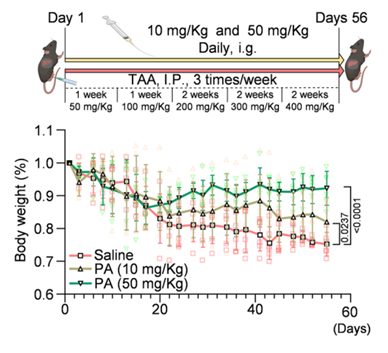
Weight loss in mice with hepatic fibrosis. Mice received intraperitoneal injections with gradually increasing concentrations of TAA (Thioacetamide) and were simultaneously treated with PA by gavage (n = 6).
Thioacetamide purchased from MedChemExpress. Usage Cited in: Commun Biol. 2025 Jul 29;8(1):1127. [Abstract]
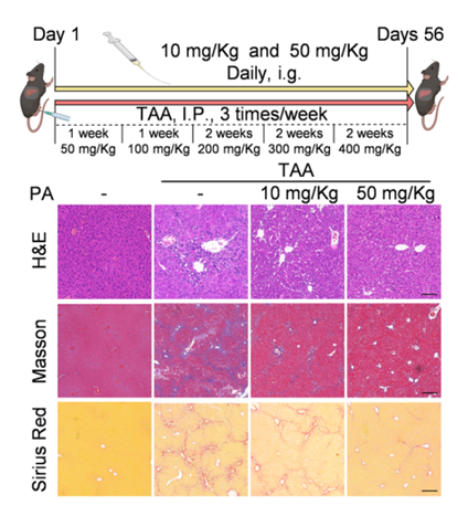
H&E staining, Masson’s trichrome staining, and Sirius red staining of hepatic sections (scale bar: H&E staining, 100 μm; Masson’s trichrome staining and Sirius red staining, 200 μm). Mice received intraperitoneal injections with gradually increasing concentrations of TAA (Thioacetamide) and were simultaneously treated with PA by gavage (n = 6).
Thioacetamide purchased from MedChemExpress. Usage Cited in: Commun Biol. 2025 Jul 29;8(1):1127. [Abstract]
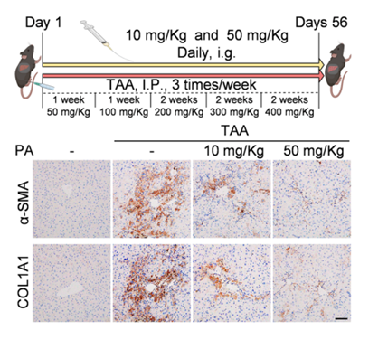
IHC was performed to detect the expression of α-SMA and COL1A1 in hepatic sections (scale bar: 50 μm) (n = 6). Mice received intraperitoneal injections with gradually increasing concentrations of TAA (Thioacetamide) and were simultaneously treated with PA by gavage (n = 6).
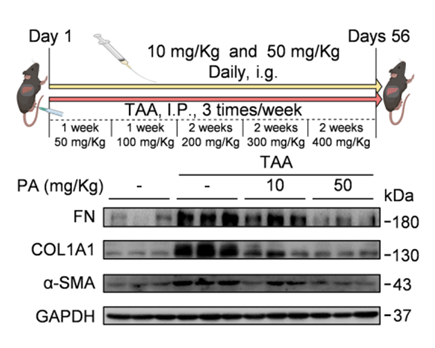
Thioacetamide purchased from MedChemExpress. Usage Cited in: Commun Biol. 2025 Jul 29;8(1):1127. [Abstract]
Western blotting of FN, COL1A1, and α-SMA protein expression in hepatic tissue homogenates. Mice received intraperitoneal injections with gradually increasing concentrations of TAA (Thioacetamide) and were simultaneously treated with PA by gavage (n = 6).
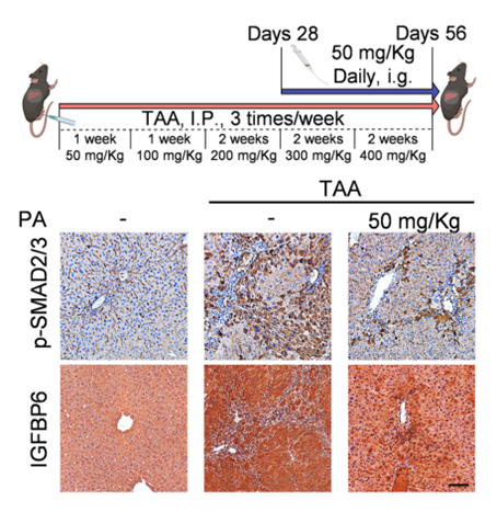
Thioacetamide purchased from MedChemExpress. Usage Cited in: Commun Biol. 2025 Jul 29;8(1):1127. [Abstract]
Mice received intraperitoneal TAA (Thioacetamide) injections at gradually increasing concentrations for 4 weeks, followed by oral gavage of PA for fibrosis treatment (n = 6). IHC (n = 6, scale bar: 50 μm).
-
Int Immunopharmacol
IRG1/Itaconate inhibits hepatic stellate cells ferroptosis and attenuates TAA-induced liver fibrosis by regulating SLC39A14 expression. [Abstract]2025 Jan 27:146:113945. PMID: 39724735 -
J Vasc Interv Radiol
Effect of Transarterial Embolization Combined with Chemotherapy on Regulatory T Cells and Vascular Endothelial Growth Factor in the Tumor Immune Microenvironment of Intrahepatic Cholangiocarcinoma. [Abstract]2026 Jan 13;37(4):107994. PMID: 41539595 -


溶剤 & 溶解度
体外:
DMSO : 100 mg/mL (1331.03 mM; Need ultrasonic; Hygroscopic DMSO has a significant impact on the solubility of product, please use newly opened DMSO)
H2O : 50 mg/mL (665.51 mM; Need ultrasonic)
Please refer to the solubility information to select the appropriate solvent. Once prepared, please aliquot and store the solution to prevent product inactivation from repeated freeze-thaw cycles.
Storage method and period of stock solution: -80°C, 6 months; -20°C, 1 month (stored under nitrogen). When stored at -80°C, please use it within 6 months. When stored at -20°C, please use it within 1 month.
* Note: If you choose water as the stock solution, please dilute it to the working solution, then filter and sterilize it with a 0.22 μm filter before use.
Please refer to the solubility information to select the appropriate solvent. Once prepared, please aliquot and store the solution to prevent product inactivation from repeated freeze-thaw cycles.
Storage method and period of stock solution: -80°C, 6 months; -20°C, 1 month (stored under nitrogen). When stored at -80°C, please use it within 6 months. When stored at -20°C, please use it within 1 month.
* Note: If you choose water as the stock solution, please dilute it to the working solution, then filter and sterilize it with a 0.22 μm filter before use.
濃度 (開始) × 体積 (開始) = 濃度 (終了) × 体積 (終了)
体内:
Select the appropriate dissolution method based on your experimental animal and administration route.
- For the following dissolution methods, please ensure to first prepare a clear stock solution using an In Vitro approach and then sequentially add co-solvents:
- To ensure reliable experimental results, the clarified stock solution can be appropriately stored based on storage conditions. As for the working solution for In Vivo experiments, it is recommended to prepare freshly and use it on the same day.
- The percentages shown for the solvents indicate their volumetric ratio in the final prepared solution. If precipitation or phase separation occurs during preparation, heat and/or sonication can be used to aid dissolution.
Add each solvent one by one: 10% DMSO 40% PEG300 5% Tween-80 45% Saline
Solubility: ≥ 2.5 mg/mL (33.28 mM); Clear solution
This protocol yields a clear solution of ≥ 2.5 mg/mL (saturation unknown).
Taking 1 mL working solution as an example, add 100 μL DMSO stock solution (25.0 mg/mL) to 400 μL PEG300, and mix evenly; then add 50 μL Tween-80 and mix evenly; then add 450 μL Saline to adjust the volume to 1 mL.
Preparation of Saline: Dissolve 0.9 g sodium chloride in ddH₂O and dilute to 100 mL to obtain a clear Saline solution.
Add each solvent one by one: 10% DMSO 90% (20% SBE-β-CD in Saline)
Solubility: ≥ 2.5 mg/mL (33.28 mM); Clear solution
This protocol yields a clear solution of ≥ 2.5 mg/mL (saturation unknown).
Taking 1 mL working solution as an example, add 100 μL DMSO stock solution (25.0 mg/mL) to 900 μL 20% SBE-β-CD in Saline, and mix evenly.
Preparation of 20% SBE-β-CD in Saline (4°C, storage for one week): 2 g SBE-β-CD powder is dissolved in 10 mL Saline, completely dissolve until clear.
In Vivo Dissolution Calculator
Please enter the basic information of animal experiments:
-
-
-
-
Recommended: Prepare an additional quantity of animals to account for potential losses during experiments.
Working solution concentration: 0.22 mg/mL
This product has good water solubility, please refer to the measured solubility data in water/PBS/Saline for details.


純度とドキュメンテーション
-
データシート (285 KB)
-
SDS (617 KB)
- English - EN (617 KB)
- Français - FR (617 KB)
- Deutsch - DE (617 KB)
- Norwegian - NO (617 KB)
- Español - ES (617 KB)
- Swedish - SV (617 KB)
- Italian - IT (617 KB)
- Korean - KR (617 KB)
- Portuguese - PT (617 KB)
-
取扱説明書 (2659 KB)
参考文献
[1]. Wallace MC, et, al. Standard operating procedures in experimental liver research: thioacetamide model in mice and rats. Lab Anim. 2015 Apr;49(1 Suppl):21-9. [Content Brief]
[2]. Chen IS, et, al. Hepatoprotection of silymarin against thioacetamide-induced chronic liver fibrosis. J Sci Food Agric. 2012 May;92(7):1441-7. [Content Brief]
[3]. Miranda AS, et, al. A thioacetamide-induced hepatic encephalopathy model in C57BL/6 mice: a behavioral and neurochemical study. Arq Neuropsiquiatr. 2010 Aug;68(4):597-602. [Content Brief]
[4]. Yeom HJ, et, al. Expression analysis of early response-related genes in rat liver epithelial cells exposed to thioacetamide in vitro. J Vet Med Sci. 2009 Jun;71(6):719-27. [Content Brief]
[5]. Ahmed G Abd Elhameed, et al. Saxagliptin defers thioacetamide-induced hepatocarcinogenesis in rats: A novel suppressive impact on Wnt/Hedgehog/Notch1 signaling. Environ Toxicol Pharmacol [Content Brief]
[6]. Mohamed E Shaker, et al. Nilotinib counteracts thioacetamide-induced hepatic oxidative stress and attenuates liver fibrosis progression. Fundam Clin Pharmacol. 2011 Apr;25(2):248-57. [Content Brief]


Complete Stock Solution Preparation Table
Please refer to the solubility information to select the appropriate solvent. Once prepared, please aliquot and store the solution to prevent product inactivation from repeated freeze-thaw cycles.
Storage method and period of stock solution: -80°C, 6 months; -20°C, 1 month (stored under nitrogen). When stored at -80°C, please use it within 6 months. When stored at -20°C, please use it within 1 month.
| Optional Solvent | Concentration Solvent Mass | 1 mg | 5 mg | 10 mg | 25 mg |
|---|---|---|---|---|---|
| H2O / DMSO | 1 mM | 13.3103 mL | 66.5513 mL | 133.1026 mL | 332.7566 mL |
| 5 mM | 2.6621 mL | 13.3103 mL | 26.6205 mL | 66.5513 mL | |
| 10 mM | 1.3310 mL | 6.6551 mL | 13.3103 mL | 33.2757 mL | |
| 15 mM | 0.8874 mL | 4.4368 mL | 8.8735 mL | 22.1838 mL | |
| 20 mM | 0.6655 mL | 3.3276 mL | 6.6551 mL | 16.6378 mL | |
| 25 mM | 0.5324 mL | 2.6621 mL | 5.3241 mL | 13.3103 mL | |
| 30 mM | 0.4437 mL | 2.2184 mL | 4.4368 mL | 11.0919 mL | |
| 40 mM | 0.3328 mL | 1.6638 mL | 3.3276 mL | 8.3189 mL | |
| 50 mM | 0.2662 mL | 1.3310 mL | 2.6621 mL | 6.6551 mL | |
| 60 mM | 0.2218 mL | 1.1092 mL | 2.2184 mL | 5.5459 mL | |
| 80 mM | 0.1664 mL | 0.8319 mL | 1.6638 mL | 4.1595 mL | |
| 100 mM | 0.1331 mL | 0.6655 mL | 1.3310 mL | 3.3276 mL |
* Note: If you choose water as the stock solution, please dilute it to the working solution, then filter and sterilize it with a 0.22 μm filter before use.


Thioacetamide 関連分類
HY-Y0698 関連分類
 Powered by Bioz