



AMG 511
Based on 5 publication(s) in Google Scholar
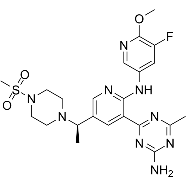
AMG 511 is a potent and orally available pan inhibitor of class I PI3Ks, with Kis of 4 nM, 6 nM, 2 nM and 1 nM for PI3Kα, β, δ and γ, respectively. AMG 511 significantly suppresses PI3K signaling that is indicated by p-Akt (Ser473) decrease. AMG 511 exhibits anti-tumor activity in mouse glioblastoma xenograft model.
연구목적의 판매만을 진행합니다. 환자를 대상으로 한 판매는 하지 않습니다.
- Purity : 99.76%
- CAS No.: 1253573-53-3
- 화학식: C22H28FN9O3S
- 분자량:517.58
-
보관:Powder -20°C, 3 years , 4°C, 2 years ; In solvent -80°C, 6 months , -20°C, 1 month

To place orders, for customer services and technical support, please contact: MedChemExpress USA
Tel: 609-228-6898 E-mail: [email protected] [email protected]


-
 Biological Activity
-
Chemical Information
-
용액&용해도
- Protocol
- 순도&문서
- References
-
 Help & FAQs


-
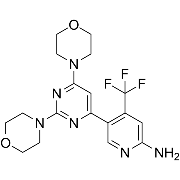
HY-70063
BuparlisibPI3K Inhibitor -
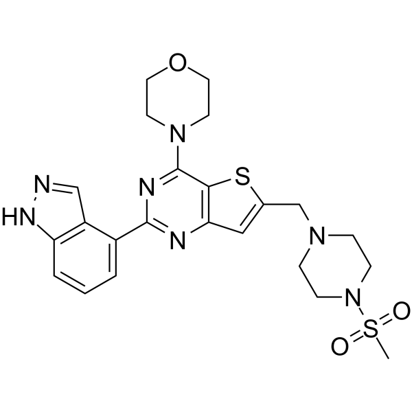
HY-50094
PictilisibPI3Kα/δ Inhibitor -
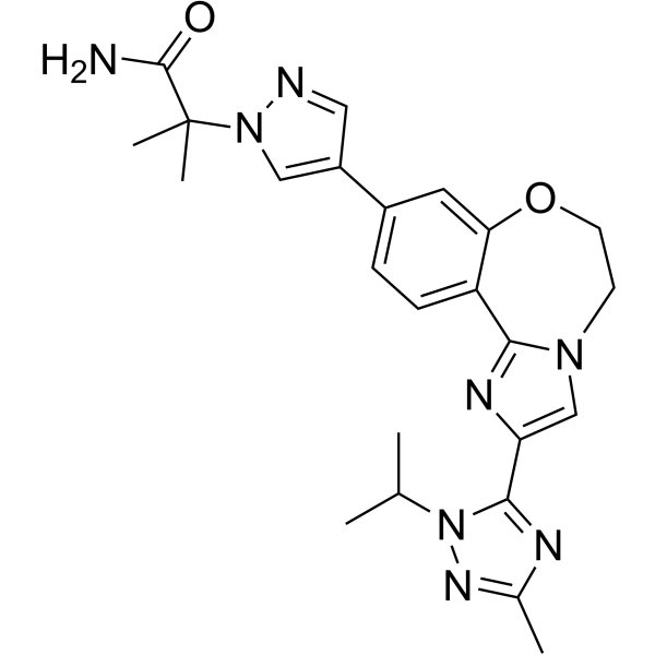
HY-13898
TaselisibPIK3CA Inhibitor -
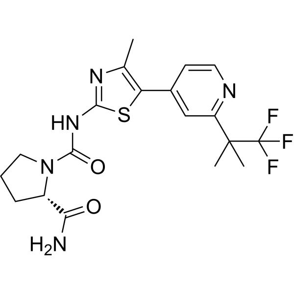
HY-15244
AlpelisibPI3Kα Inhibitor -

HY-50094R
Pictilisib (Standard)PI3Kα/δ Inhibitor -
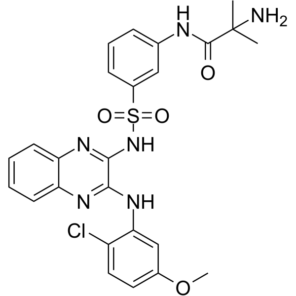
HY-16526
PilaralisibPI3K Inhibitor -
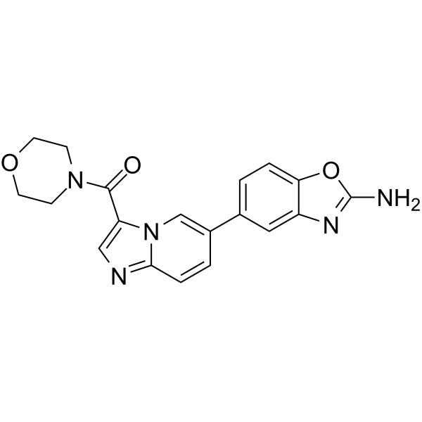
HY-12285
Serabelisibp110α Inhibitor -
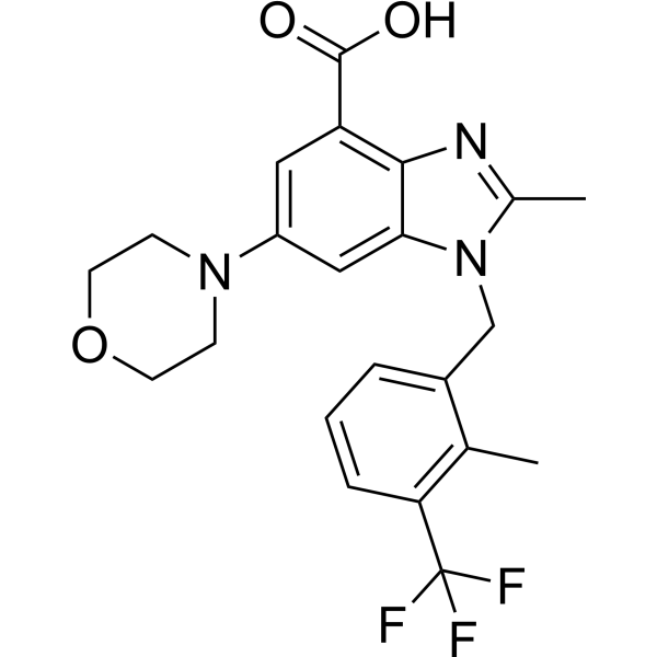
HY-15245
GSK2636771PI3Kβ Inhibitor -
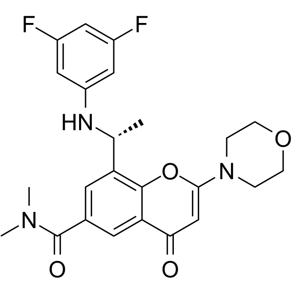
HY-12330
AZD8186PI3K Inhibitor -
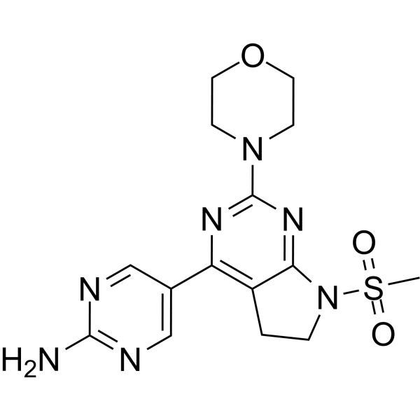
HY-15466
IzorlisibClass I PI3K Inhibitor -

HY-70063R
Buparlisib (Standard)PI3K Inhibitor -

HY-15244R
Alpelisib (Standard)Reference Standard
Publications Citing Use of MedChemExpress (MCE) AMG 511
More

Biological Activity
제품 설명
IC50 & Target
|
PI3Kα 4 nM (Ki) |
PI3Kβ 6 nM (Ki) |
PI3Kδ 2 nM (Ki) |
PI3Kγ 1 nM (Ki) |
Cellular Effect
|
Cell Line

|
Type | Value | Description | References |
|---|---|---|---|---|
| U-87MG ATCC | ED50 |
0.6 mg/kg
Compound: 31
|
Antitumor activity against human U87MG cells xenografted in po dosed CD1 nude mouse administered QD for 12 days
Antitumor activity against human U87MG cells xenografted in po dosed CD1 nude mouse administered QD for 12 days
|
[PMID: 22897589] |
| U-87MG ATCC | IC50 |
0.004 μM
Compound: 1, AMG 511
|
Inhibition of PI3K-mediated Akt S473 phosphorylation in human U87MG cells after 2 hrs by cell-based assay
Inhibition of PI3K-mediated Akt S473 phosphorylation in human U87MG cells after 2 hrs by cell-based assay
|
[PMID: 25466188] |
| U-87MG ATCC | IC50 |
4 nM
Compound: 31
|
Inhibition of PI3K-mediated AKT Ser473 phosphorylation in human U87MG cells after 2 hrs
Inhibition of PI3K-mediated AKT Ser473 phosphorylation in human U87MG cells after 2 hrs
|
[PMID: 22897589] |
In Vitro
In Vivo
AMG 511 shows excellent in vivo efficacy and pharmacokinetic profile[1].
MedChemExpress (MCE) has not independently confirmed the accuracy of these methods. They are for reference only.
-
Animal Model:Female CD1 NU/NU mice, with U87 MG glioblastoma xenograft model[1]
-
Dosage:1 mg/kg, 3 mg/kg, 10 mg/kg
-
Administration:Oral administration, daily, for 12 days
-
Result:Inhibited tumor growth.
-
Animal Model:Male Sprague-Dawley rats[1]
-
Dosage:1 mg/kg
-
Administration:Oral administration (Pharmacokinetic Analysis)
-
Result:Had a superior pharmacokinetic profile with low clearance (0.4 L/h/kg, 12% of liver blood flow), good oral bioavailability (F = 60%), and a commensurate high oral exposure (AUC = 5.0 μM·h).


Chemical Information
-
CAS No. 1253573-53-3
-
Appearance Solid
-
분자량 517.58
-
화학식 C22H28FN9O3S
-
Color Light yellow to yellow
-
SMILES
NC1=NC(C2=CC([C@H](N3CCN(S(=O)(C)=O)CC3)C)=CN=C2NC4=CC(F)=C(OC)N=C4)=NC(C)=N1
-
선적
Room temperature in continental US; may vary elsewhere.
-
보관
Powder -20°C 3 years 4°C 2 years In solvent -80°C 6 months -20°C 1 month


Publications (5)
-
Journal Impact Factor
-
Most Recent
-
Cancer Discov
Discovery of BBO-11818, a Potent and Selective Noncovalent Inhibitor of (ON) and (OFF) KRAS with Activity against Multiple Oncogenic Mutants. [Abstract]2026 Mar 6:OF1-OF20. PMID: 41790032 -
Cancer Lett
PI3K inhibitor impairs tumor progression and enhances sensitivity to anlotinib in anlotinib-resistant osteosarcoma. [Abstract]2022 Jun 28;536:215660. PMID: 35318116 -
Phytomedicine
Gastrodin promotes Alkbh5 nuclear localization and Gclm m6A demethylation to alleviate ferroptosis in ischemic stroke. [Abstract]2025 Nov:147:157177. PMID: 40839993 -
Leukemia
BET inhibitor-based combinations targeting novel dependencies in MECOM-rearranged (r) AML. [Abstract]2025 Dec 19. PMID: 41419608 -
Exp Ther Med
Hydroxysafflor Yellow A inhibits the viability and migration of vascular smooth muscle cells induced by serum from rats with chronic renal failure via inactivation of the PI3K/Akt signaling pathway. [Abstract]2021 Aug;22(2):850. PMID: 34149896


용액&용해도
In Vitro:
DMSO : 33.33 mg/mL (64.40 mM; Need ultrasonic; Hygroscopic DMSO has a significant impact on the solubility of product, please use newly opened DMSO)
Please refer to the solubility information to select the appropriate solvent. Once prepared, please aliquot and store the solution to prevent product inactivation from repeated freeze-thaw cycles.
Storage method and period of stock solution: -80°C, 6 months; -20°C, 1 month. When stored at -80°C, please use it within 6 months. When stored at -20°C, please use it within 1 month.
Please refer to the solubility information to select the appropriate solvent. Once prepared, please aliquot and store the solution to prevent product inactivation from repeated freeze-thaw cycles.
Storage method and period of stock solution: -80°C, 6 months; -20°C, 1 month. When stored at -80°C, please use it within 6 months. When stored at -20°C, please use it within 1 month.
Concentration (start) × Volume (start) = Concentration (final) × Volume (final)
In Vivo:
Select the appropriate dissolution method based on your experimental animal and administration route.
- For the following dissolution methods, please ensure to first prepare a clear stock solution using an In Vitro approach and then sequentially add co-solvents:
- To ensure reliable experimental results, the clarified stock solution can be appropriately stored based on storage conditions. As for the working solution for In Vivo experiments, it is recommended to prepare freshly and use it on the same day.
- The percentages shown for the solvents indicate their volumetric ratio in the final prepared solution. If precipitation or phase separation occurs during preparation, heat and/or sonication can be used to aid dissolution.
Add each solvent one by one: 10% DMSO 90% (20% SBE-β-CD in Saline)
Solubility: 2 mg/mL (3.86 mM); Suspended solution; Need ultrasonic
This protocol yields a suspended solution of 2 mg/mL. Suspended solution can be used for oral and intraperitoneal injection.
Taking 1 mL working solution as an example, add 100 μL DMSO stock solution (20.0 mg/mL) to 900 μL 20% SBE-β-CD in Saline, and mix evenly.
Preparation of 20% SBE-β-CD in Saline (4°C, storage for one week): 2 g SBE-β-CD powder is dissolved in 10 mL Saline, completely dissolve until clear.
In Vivo Dissolution Calculator
Please enter the basic information of animal experiments:
-
-
-
-
Recommended: Prepare an additional quantity of animals to account for potential losses during experiments.
Please enter your animal formula composition:
-
%DMSO +
Recommended: Keep the proportion of DMSO in working solution below 2% if your animal is weak.
-
%+
-
+%Tween-80 + +
-
%Saline +
The co-solvents required include: DMSO, . All of co-solvents are available by MedChemExpress (MCE). , Tween 80. All of co-solvents are available by MedChemExpress (MCE).
Working solution concentration: 0.22 mg/mL
Method for preparing stock solution: mg drug dissolved in μL DMSO. Stock solution concentration: mg/mL.
1. Take μL DMSO stock solution;
2. Add μL .
μL , mix evenly;
3. Then add μL Tween 80, mix evenly;
4. Then add μL
Please ensure that the stock solution in the first step is dissolved to a clear state, and add co-solvents in sequence. You can use ultrasonic heating (ultrasonic cleaner, recommended frequency 20-40 kHz), vortexing, etc. to assist dissolution.


순도&문서
-
Data Sheet (279 KB)
-
SDS (393 KB)
- English - EN (393 KB)
- Français - FR (393 KB)
- Deutsch - DE (393 KB)
- Norwegian - NO (393 KB)
- Español - ES (393 KB)
- Swedish - SV (393 KB)
- Italian - IT (393 KB)
- Korean - KR (393 KB)
- Portuguese - PT (393 KB)
-
Handling Instructions (2659 KB)
References


Complete Stock Solution Preparation Table
Please refer to the solubility information to select the appropriate solvent. Once prepared, please aliquot and store the solution to prevent product inactivation from repeated freeze-thaw cycles.
Storage method and period of stock solution: -80°C, 6 months; -20°C, 1 month. When stored at -80°C, please use it within 6 months. When stored at -20°C, please use it within 1 month.
| Optional Solvent | Concentration Solvent Mass | 1 mg | 5 mg | 10 mg | 25 mg |
|---|---|---|---|---|---|
| DMSO | 1 mM | 1.9321 mL | 9.6603 mL | 19.3207 mL | 48.3017 mL |
| 5 mM | 0.3864 mL | 1.9321 mL | 3.8641 mL | 9.6603 mL | |
| 10 mM | 0.1932 mL | 0.9660 mL | 1.9321 mL | 4.8302 mL | |
| 15 mM | 0.1288 mL | 0.6440 mL | 1.2880 mL | 3.2201 mL | |
| 20 mM | 0.0966 mL | 0.4830 mL | 0.9660 mL | 2.4151 mL | |
| 25 mM | 0.0773 mL | 0.3864 mL | 0.7728 mL | 1.9321 mL | |
| 30 mM | 0.0644 mL | 0.3220 mL | 0.6440 mL | 1.6101 mL | |
| 40 mM | 0.0483 mL | 0.2415 mL | 0.4830 mL | 1.2075 mL | |
| 50 mM | 0.0386 mL | 0.1932 mL | 0.3864 mL | 0.9660 mL | |
| 60 mM | 0.0322 mL | 0.1610 mL | 0.3220 mL | 0.8050 mL |


AMG 511 Related Classifications
HY-13440 Related Classifications
 Powered by Bioz