



Bixin
Based on 2 publication(s) in Google Scholar
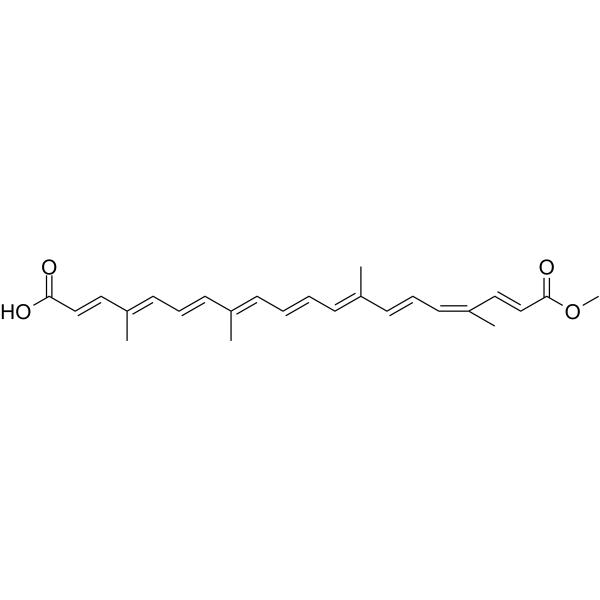
Bixin is an orally active carotenoid found in the seeds of Bixa orellana. Bixin induces apoptosis in cancer cells. Bixin possessing anti-inflammatory, anti-tumor and anti-oxidant activities. Bixin treatment ameliorated cardiac dysfunction through inhibiting fibrosis, inflammation and reactive oxygen species (ROS) generation.
연구목적의 판매만을 진행합니다. 환자를 대상으로 한 판매는 하지 않습니다.
- Purity : 98.61%
- CAS No.: 6983-79-5
- 화학식: C25H30O4
- 분자량:394.50
-
보관:
4°C, protect from light
* In solvent : -80°C, 6 months; -20°C, 1 month (protect from light)

To place orders, for customer services and technical support, please contact: MedChemExpress USA
Tel: 609-228-6898 E-mail: [email protected] [email protected]


-
 Biological Activity
-
Chemical Information
-
용액&용해도
- Protocol
- 순도&문서
- References
-
 Help & FAQs


-
HY-B2163
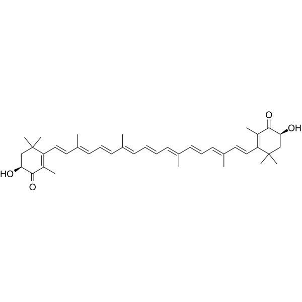
AstaxanthinAntioxidant -
HY-N0005
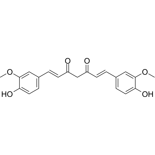
Curcuminp300 Histone Acetylatransferase Inhibitor -
HY-N6947
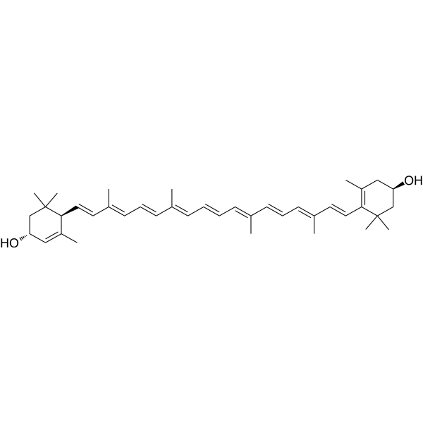
LuteinAnti-inflammatory Agent -
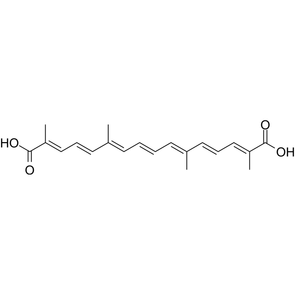
HY-N2072
CrocetinAnticancer/Neuroprotective Agent -
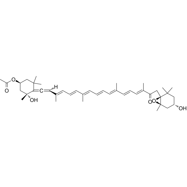
HY-N2302
FucoxanthinMarine Carotenoid -

HY-N6884R
Bixin (Standard)Reactive Oxygen Species Inhibitor -
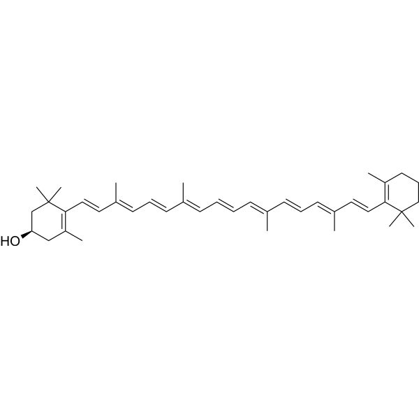
HY-108059
β-CryptoxanthinCarotenoid -

HY-W743654
Capsorubin -

HY-W743654R
Capsorubin (Standard) -
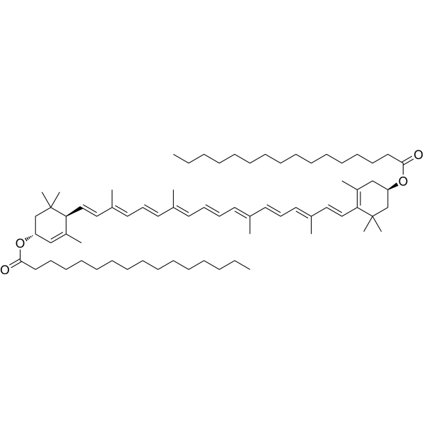
HY-N8731
Lutein dipalmitateLutein Ester -

HY-N11697
α-CryptoxanthinCarotenoid -

HY-W750723
RubixanthinCarotenoid
Publications Citing Use of MedChemExpress (MCE) Bixin
More Customer Validation & Images

-
IHC

-
WB

-
IF

-
Histological Imaging/Staining

-
In Vivo Efficacy Study


Biological Activity
제품 설명
IC50 & Target
ROS generation[1]
In Vitro
Bixin (20-80 μM, 24 h) improves Lipopolysaccharides (LPS) (HY-D1056)-induced fibrosis in H9C2 cells without cytotoxicity[1].
Bixin (20-80 μM, 24 h) reduces inflammation response via TLR4/NF-kB inactivation in H9C2 cells[1].
Bixin (20-80 μM, 24 h) down-regulates ROS production in LPS-induced H9C2 cells via activating Nrf2[1].
Bixin (0-500 μM, 48 h) has anti-proliferative activity on human lung cancer (A549), cervical cancer (HeLa), and breast cancer (MCF-7)[2].
MedChemExpress (MCE) has not independently confirmed the accuracy of these methods. They are for reference only.
-
Cell Line:H9C2 (Human cardiac muscle cell)
-
Concentration:2.5, 5, 10, 20, 80 μM
-
Incubation Time:0-72 h
-
Result:Showed no significant cytotoxicity to cardiac muscle cells H9C2.
-
Cell Line:H9C2 (Human cardiac muscle cell)
-
Concentration:20, 40 , 80 μM
-
Incubation Time:24 h
-
Result:Reduced LPS induction up-regulated fibrosis-related signals expression in a dose-dependent manner. Reduced nuclear factor of activated T cells isoforms c3 (NFATc3), GATA4 and A-type natriuretic peptides (ANP) protein expression levels. Reduced fTLR4, MyD88, p-IKKα, p-IκBα and p-NF-κB expression enhanced by LPS. Reduced fibrosis levels via collagen deposition down-regulation.
-
Cell Line:A549, HeLa, and MCF-7
-
Concentration:0-500 μM
-
Incubation Time:48 h
-
Result:Shows profound results on three types of cells, especially HeLa and MCF-7, with the crude extract having greater activity than the isolate. IC50 of crude extract were 185.54, 17.90, and 2.06 μM, while IC50 of isolated were 259.38, 43.87, and 14.38 μM against A549, HeLa, and MCF-7 cells, respectively.
-
Cell Line:H9C2 (Human cardiac muscle cell)
-
Concentration:20, 40 , 80 μM
-
Incubation Time:24 h
-
Result:Reversed SOD1, Nrf2, HO-1, CAT and GCLM mRNA levels in H9C2 cells restrained due to LPS treatment in a dose-dependent manner.
In Vivo
Bixin (50-200 mg/kg; p.o.; once a day for 14 weeks) restrains systemic metabolism disorder in high fat diet-induced mice[1].
Bixin (200 mg/kg, p.o.; once a day for 14 weeks) ameliorates high fat diet-induced heart failure in mice[1].
Bixin (50-200 mg/kg; p.o.; once a day for 14 weeks) reduces reactive oxygen species (ROS) generation in high fat diet-induced mice due to Nrf2 activation[1].
MedChemExpress (MCE) has not independently confirmed the accuracy of these methods. They are for reference only.
-
Animal Model:Male, 6-8 weeks, C57BL/6 mice weighed 20-25 g [1]
-
Dosage:200 mg/kg
-
Administration:Oral gavage (p.o.); once a day for 14 weeks
-
Result:Reduced mice heart size induced by high fat feeding from appearance, accompanied with decreased ratio of heart weight to body weight.
Reduced the diameter of myocytes.
Down-regulated fibrosis area in HFD-fed mice.
Reversed left ventricular systolic pressure (LVSP) stimulated by high fat diet.
Displayed suppressive role in controlling the percentage of ejection fraction (EF), fractional shortening (FS), and anterior wall thickness (AWT).
-
Animal Model:Male, 6-8 weeks, C57BL/6 mice weighed 20-25 g [1]
-
Dosage:50, 100, 200 mg/kg
-
Administration:Oral gavage (p.o.); once a day for 14 weeks
-
Result:Suppressed the insulin resistance induced by high fat diet (HFD) in a dose-dependent manner.
Reduced body weight caused by high fat diet in a dose-dependent manner.
Showed inhibitory role in low density lipoprotein cholesterol (LDLC), triglyceride (TG) and cholesterol (TC) levels.
Increased high density lipoprotein cholesterol (HDLC) due to HFD in a dose-dependent manner.
Reduced serum endotoxin in dose-dependent manner.
Impeded pro-inflammatory such as cytokines serum IL-1b, TNF-a and IL-18 releasing.
Reduced TLR4 and MyD88 expression from protein levels.
Could reverse HFD-induced heart injury through reducing ROS generation dependent on Nrf2 activation
- Fibrosis/Collagen Morphometry
- Collagen: Sirius Red Staining
- Detection of Apoptosis
- TUNEL staining for apoptotic DNA fragmentation
- Annexin V plus membrane-impermeant dye apoptosis staining
- ROS/oxidative-stress fluorescent staining
- Connective Tissue: Masson's Trichrome/Collagen Trichrome Staining
- Apoptosis Solutions
- Research Protocol for Inflammation-related Diseases
Clinical Trial
| NCT Number | Sponsor | Condition | Start Date |
Phase

|
|---|---|---|---|---|
| NCT01329991 | Plexxikon| | 2011-05 | PHASE1 |


Chemical Information
-
CAS No. 6983-79-5
-
Appearance Solid
-
분자량 394.50
-
화학식 C25H30O4
-
Color Brown to reddish brown
-
SMILES
OC(/C=C/C(C)=C/C=C/C(C)=C/C=C/C=C(C)/C=C/C=C(C)\C=C\C(OC)=O)=O
-
Structure Classification
-
Initial Source
-
선적
Room temperature in continental US; may vary elsewhere.
-
보관
4°C, protect from light
* In solvent : -80°C, 6 months; -20°C, 1 month (protect from light)


Publications (2)
-
Journal Impact Factor
-
Most Recent
-
Biomed Pharmacother
Bixin prevents tubulointerstitial fibrosis in hyperuricemic nephropathy via promoting PPARγ-NLRP3 interaction. [Abstract]2025 Dec:193:118745. PMID: 41232352
Bixin purchased from MedChemExpress. Usage Cited in: Biomed Pharmacother. 2025 Dec:193:118745. [Abstract]
Representative IHC images of PPARγ in kidneys treated with Bixin (20, 40 mg/kg, i.p.).
Bixin purchased from MedChemExpress. Usage Cited in: Biomed Pharmacother. 2025 Dec:193:118745. [Abstract]
Representative Western blots of PPARγ and quantification of the protein level treated with Bixin (10 μM).
Bixin purchased from MedChemExpress. Usage Cited in: Biomed Pharmacother. 2025 Dec:193:118745. [Abstract]
Representative IF images of NLRP3 (red)/ASC (green)/DAPI (blue) and NLRP3 (green)/csap1 (red)/DAPI (blue) treated with Bixin (10 μM).
Bixin purchased from MedChemExpress. Usage Cited in: Biomed Pharmacother. 2025 Dec:193:118745. [Abstract]
Representative images of HE staining in kidneys treated with Bixin (20, 40 mg/kg, i.p.).
Bixin purchased from MedChemExpress. Usage Cited in: Biomed Pharmacother. 2025 Dec:193:118745. [Abstract]
Dynamic changes of body weight treated with Bixin (20, 40 mg/kg, i.p.).
-


용액&용해도
In Vitro:
DMSO : 7.14 mg/mL (18.10 mM; Need ultrasonic; Hygroscopic DMSO has a significant impact on the solubility of product, please use newly opened DMSO)
Please refer to the solubility information to select the appropriate solvent. Once prepared, please aliquot and store the solution to prevent product inactivation from repeated freeze-thaw cycles.
Storage method and period of stock solution: -80°C, 6 months; -20°C, 1 month (protect from light). When stored at -80°C, please use it within 6 months. When stored at -20°C, please use it within 1 month.
Please refer to the solubility information to select the appropriate solvent. Once prepared, please aliquot and store the solution to prevent product inactivation from repeated freeze-thaw cycles.
Storage method and period of stock solution: -80°C, 6 months; -20°C, 1 month (protect from light). When stored at -80°C, please use it within 6 months. When stored at -20°C, please use it within 1 month.
Concentration (start) × Volume (start) = Concentration (final) × Volume (final)
In Vivo:
Select the appropriate dissolution method based on your experimental animal and administration route.
- For the following dissolution methods, please ensure to first prepare a clear stock solution using an In Vitro approach and then sequentially add co-solvents:
- To ensure reliable experimental results, the clarified stock solution can be appropriately stored based on storage conditions. As for the working solution for In Vivo experiments, it is recommended to prepare freshly and use it on the same day.
- The percentages shown for the solvents indicate their volumetric ratio in the final prepared solution. If precipitation or phase separation occurs during preparation, heat and/or sonication can be used to aid dissolution.
Add each solvent one by one: 10% DMSO 40% PEG300 5% Tween-80 45% Saline
Solubility: 0.71 mg/mL (1.80 mM); Suspended solution; Need ultrasonic
This protocol yields a suspended solution of 0.71 mg/mL. Suspended solution can be used for oral and intraperitoneal injection.
Taking 1 mL working solution as an example, add 100 μL DMSO stock solution (7.1 mg/mL) to 400 μL PEG300, and mix evenly; then add 50 μL Tween-80 and mix evenly; then add 450 μL Saline to adjust the volume to 1 mL.
Preparation of Saline: Dissolve 0.9 g sodium chloride in ddH₂O and dilute to 100 mL to obtain a clear Saline solution.
Add each solvent one by one: 10% DMSO 90% (20% SBE-β-CD in Saline)
Solubility: 0.71 mg/mL (1.80 mM); Suspended solution; Need ultrasonic
This protocol yields a suspended solution of 0.71 mg/mL. Suspended solution can be used for oral and intraperitoneal injection.
Taking 1 mL working solution as an example, add 100 μL DMSO stock solution (7.1 mg/mL) to 900 μL 20% SBE-β-CD in Saline, and mix evenly.
Preparation of 20% SBE-β-CD in Saline (4°C, storage for one week): 2 g SBE-β-CD powder is dissolved in 10 mL Saline, completely dissolve until clear.
In Vivo Dissolution Calculator
Please enter the basic information of animal experiments:
-
-
-
-
Recommended: Prepare an additional quantity of animals to account for potential losses during experiments.
Please enter your animal formula composition:
-
%DMSO +
Recommended: Keep the proportion of DMSO in working solution below 2% if your animal is weak.
-
%+
-
+%Tween-80 + +
-
%Saline +
The co-solvents required include: DMSO, . All of co-solvents are available by MedChemExpress (MCE). , Tween 80. All of co-solvents are available by MedChemExpress (MCE).
Working solution concentration: 0.22 mg/mL
Method for preparing stock solution: mg drug dissolved in μL DMSO. Stock solution concentration: mg/mL. * In solvent : -80°C, 6 months; -20°C, 1 month (protect from light)
1. Take μL DMSO stock solution;
2. Add μL .
μL , mix evenly;
3. Then add μL Tween 80, mix evenly;
4. Then add μL
Please ensure that the stock solution in the first step is dissolved to a clear state, and add co-solvents in sequence. You can use ultrasonic heating (ultrasonic cleaner, recommended frequency 20-40 kHz), vortexing, etc. to assist dissolution.


순도&문서
-
Data Sheet (277 KB)
-
SDS (393 KB)
- English - EN (393 KB)
- Français - FR (393 KB)
- Deutsch - DE (393 KB)
- Norwegian - NO (393 KB)
- Español - ES (393 KB)
- Swedish - SV (393 KB)
- Italian - IT (393 KB)
- Korean - KR (393 KB)
- Portuguese - PT (393 KB)
-
Handling Instructions (2659 KB)
References
[1]. Xu Z, et al. Bixin ameliorates high fat diet-induced cardiac injury in mice through inflammation and oxidative stress suppression. Biomed Pharmacother. 2017 May;89:991-1004. [Content Brief]
[2]. Kusmita L, et al. Bixa orellana L. carotenoids: antiproliferative activity on human lung cancer, breast cancer, and cervical cancer cells in vitro. Nat Prod Res. 2022 Dec;36(24):6421-6427. [Content Brief]


Complete Stock Solution Preparation Table
Please refer to the solubility information to select the appropriate solvent. Once prepared, please aliquot and store the solution to prevent product inactivation from repeated freeze-thaw cycles.
Storage method and period of stock solution: -80°C, 6 months; -20°C, 1 month (protect from light). When stored at -80°C, please use it within 6 months. When stored at -20°C, please use it within 1 month.
| Optional Solvent | Concentration Solvent Mass | 1 mg | 5 mg | 10 mg | 25 mg |
|---|---|---|---|---|---|
| DMSO | 1 mM | 2.5349 mL | 12.6743 mL | 25.3485 mL | 63.3714 mL |
| 5 mM | 0.5070 mL | 2.5349 mL | 5.0697 mL | 12.6743 mL | |
| 10 mM | 0.2535 mL | 1.2674 mL | 2.5349 mL | 6.3371 mL | |
| 15 mM | 0.1690 mL | 0.8450 mL | 1.6899 mL | 4.2248 mL |


Bixin Related Classifications
HY-N6884 Related Classifications
 Powered by Bioz