



COM503
COM503 is a high-affinity anti-IL18BP human monoclonal antibody with a Kd value of 0.291 pM. COM503 binds to IL18BP, blocks the IL18BP:IL18 interaction, displaces IL18 from preformed complexes, and enhances the activation and function of T cells and NK cells. COM503 induces anti-tumor effects by relieving the activity inhibition of endogenous IL18 in the tumor microenvironment. COM503 can be used for cancer research. The recommended isotype control is Human IgG4 (S228P) kappa, Isotype Control (HY-P99003).
연구목적의 판매만을 진행합니다. 환자를 대상으로 한 판매는 하지 않습니다.
-
보관:
Please store the product under the recommended conditions in the Certificate of Analysis.


To place orders, for customer services and technical support, please contact: MedChemExpress USA
Tel: 609-228-6898 E-mail: [email protected] [email protected]


-
 Biological Activity
-
Chemical Information
- Protocol
- 순도&문서
- References
-
 Help & FAQs




Biological Activity
제품 설명
Isotype
Human IgG4 kappa
Recommend Isotype Controls
Species Reactivity
Human
IC50 & Target
[1]|
IL18BP 0.291 pM (Kd) |
IL-18 |
In Vitro
COM503 binds to purified human IL18BP with high affinity (Kd = 0.291 pM), which is higher than the affinity between endogenous human IL18 and IL18BP (Kd = 0.441 pM)[1].
COM503 (1.25-10 μg/mL; 1 h (complex incubation at 37°C); RT or 37°C (assay incubation steps)) displaces IL18 from preformed recombinant human IL18:IL18BP complexes in a dose-dependent manner in vitro, with an EC50 of 14 nM[1].
COM503 (24 h) restores the activation of human melanoma CD8+ tumor-infiltrating lymphocytes (TILs) co-cultured with MEL624 cells and mediated by IL18, and increases the secretion levels of IFN-γ and TNF-α as well as the expression of CD137 in CD8+ TILs[1].
COM503 (24 h) restores IL18-mediated activation of isolated human NK cells blocked by preformed IL18:IL18BP complexes, and increases IFN-γ secretion levels[1].
COM503 (10 μg/mL; 24 h) restores IL18-mediated activation of CMV-reactive CD8+ T cells co-cultured with PD-L1-overexpressing MEL624 cells[1].
COM503 (24 h) restores IL18-mediated activation of human peripheral blood mononuclear cells (PBMCs) by blocking endogenously secreted IL18BP, and increases IFN-γ secretion levels with a mean EC50 of 1.6 nM[1].
COM503 (20 μg/mL; 24 h) enhances IFN-γ secretion by human melanoma CD8+ tumor-infiltrating lymphocytes (TILs) co-cultured with MEL624 cells through blocking endogenously secreted IL18BP at physiologically relevant IL18 concentrations[1].
MedChemExpress (MCE) has not independently confirmed the accuracy of these methods. They are for reference only.
Gene ID
Accession
O95998
Target
IL-18BP
Conjugated
Unconjugated
Reconsititution
The product can be reconstituted/diluted with sterile PBS or saline.
Format
-
Product Image
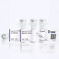
신청
ELISA, FACS, Functional assay


Chemical Information
-
Formulation
Please refer to the lot-specific COA for specific buffer information.
-
보관
Please store the product under the recommended conditions in the Certificate of Analysis.


순도&문서
References

Calculators
Concentration (start) × Volume (start) = Concentration (final) × Volume (final)


COM503 Related Classifications
HY-P992335 Related Classifications
 Powered by Bioz