



NDNA3
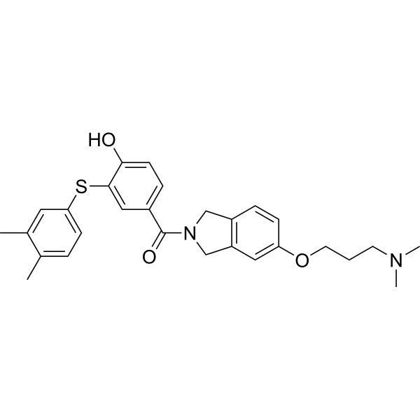
NDNA3 (compound 14) is a selective inhibitor of Hsp90α (IC50: 0.51 μM). NDNA3 is a permanently charged analog with low membrane permeability and low toxicity to Ovcar-8 (IC50: 12.66 μM) and MCF-10A (IC50: 11.72 μM) cells. NDNA3 prevents disruption of hERG channel maturation without generating a heat shock response or causing degradation of Hsp90α-dependent client proteins.
연구목적의 판매만을 진행합니다. 환자를 대상으로 한 판매는 하지 않습니다.
- 화학식: C28H32N2O3S
- 분자량:476.63
-
보관:
Please store the product under the recommended conditions in the Certificate of Analysis.

To place orders, for customer services and technical support, please contact: MedChemExpress USA
Tel: 609-228-6898 E-mail: [email protected] [email protected]


-
 Biological Activity
-
Chemical Information
- Protocol
- 순도&문서
- References
-
 Help & FAQs




Biological Activity
제품 설명
IC50 & Target
IC50: 0.51 μM (Hsp90α)[1]
Cellular Effect
|
Cell Line

|
Type | Value | Description | References |
|---|---|---|---|---|
| MCF-10A | IC50 |
11.72 μM
Compound: 14; NDNA3
|
Antiproliferative activity against human MCF-10A cells assessed as inhibition of cell growth incubated for 72 hrs by MTS assay
Antiproliferative activity against human MCF-10A cells assessed as inhibition of cell growth incubated for 72 hrs by MTS assay
|
[PMID: 37736193] |
| OVCAR-8 | IC50 |
12.66 μM
Compound: 14; NDNA3
|
Antiproliferative activity against human OVCAR-8 cells assessed as inhibition of cell growth incubated for 72 hrs by MTS assay
Antiproliferative activity against human OVCAR-8 cells assessed as inhibition of cell growth incubated for 72 hrs by MTS assay
|
[PMID: 37736193] |


Chemical Information
-
분자량 476.63
-
화학식 C28H32N2O3S
-
SMILES
CN(C)CCCOC1=CC2=C(CN(C(C3=CC=C(O)C(SC4=CC=C(C)C(C)=C4)=C3)=O)C2)C=C1
-
선적
Room temperature in continental US; may vary elsewhere.
-
보관
Please store the product under the recommended conditions in the Certificate of Analysis.


순도&문서
References

Calculators
Concentration (start) × Volume (start) = Concentration (final) × Volume (final)


NDNA3 Related Classifications
HY-149432 Related Classifications
 Powered by Bioz