



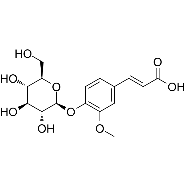
Lavandoside

Based on 1 Customer Validation
Lavandoside is an ABTS?+ free radical scavenger and a moderate inhibitor of xanthine oxidase (XO), with an IC50 of 71.6 μM for inhibiting NO production in LPS-induced macrophages. Lavandoside exerts its antioxidant and potential anti-inflammatory effects by directly scavenging free radicals and inhibiting XO activity, a mechanism related to the hydroxyl groups in its molecular structure. Lavandoside can be isolated from lavender and can be used in the development of natural antioxidants and in research on oxidative stress-related diseases and inflammation-related diseases.
연구목적의 판매만을 진행합니다. 환자를 대상으로 한 판매는 하지 않습니다.
- Purity : 99.98%
- CAS No.: 117405-51-3
- 화학식: C16H20O9
- 분자량:356.32
-
보관:
4°C, protect from light
* In solvent : -80°C, 6 months; -20°C, 1 month (protect from light)

To place orders, for customer services and technical support, please contact: MedChemExpress USA
Tel: 609-228-6898 E-mail: [email protected] [email protected]


-
 Biological Activity
-
Chemical Information
-
용액&용해도
- Protocol
- 순도&문서
- References
-
 Help & FAQs




Biological Activity
제품 설명
Cellular Effect
|
Cell Line

|
Type | Value | Description | References |
|---|---|---|---|---|
| RAW264.7 | IC50 |
71.6 μM
Compound: 13
|
Antiinflammatory activity in mouse RAW264.7 cells assessed as decrease in LPS-induced NO production after 24 hrs by Griess assay
Antiinflammatory activity in mouse RAW264.7 cells assessed as decrease in LPS-induced NO production after 24 hrs by Griess assay
|
[PMID: 25666824] |
In Vitro
Lavandoside (81.66 μg TE/mL) showed a free radical scavenging activity of 341 mAU in the ABTS free radical scavenging assay, an ORAC value of 37.44 μMol TE/mg in the ORAC assay, and a TEAC value of 12.14 μMol TE/mg in the TEAC assay, demonstrating antioxidant activity[1].
Lavandoside (4 μM, 20 μM, 100 μM; 24 h) showed no inhibitory activity on NO release in LPS+IFN-γ activated dendritic cells (D2SC/1) and was toxic to these cells[2].
Lavandoside (333.3 μM) showed moderate inhibitory activity in the xanthine oxidase (XO) inhibition assay[2].
MedChemExpress (MCE) has not independently confirmed the accuracy of these methods. They are for reference only.


Chemical Information
-
CAS No. 117405-51-3
-
Appearance Solid
-
분자량 356.32
-
화학식 C16H20O9
-
Color White to off-white
-
SMILES
O=C(O)/C=C/C1=CC(OC)=C(C=C1)O[C@H]2[C@H](O)[C@@H](O)[C@H](O)[C@@H](CO)O2
-
Structure Classification
-
Initial Source
-
선적
Room temperature in continental US; may vary elsewhere.
-
보관
4°C, protect from light
* In solvent : -80°C, 6 months; -20°C, 1 month (protect from light)


용액&용해도
In Vitro:
DMSO : 50 mg/mL (140.32 mM; Need ultrasonic; Hygroscopic DMSO has a significant impact on the solubility of product, please use newly opened DMSO)
Please refer to the solubility information to select the appropriate solvent. Once prepared, please aliquot and store the solution to prevent product inactivation from repeated freeze-thaw cycles.
Storage method and period of stock solution: -80°C, 6 months; -20°C, 1 month (protect from light). When stored at -80°C, please use it within 6 months. When stored at -20°C, please use it within 1 month.
Please refer to the solubility information to select the appropriate solvent. Once prepared, please aliquot and store the solution to prevent product inactivation from repeated freeze-thaw cycles.
Storage method and period of stock solution: -80°C, 6 months; -20°C, 1 month (protect from light). When stored at -80°C, please use it within 6 months. When stored at -20°C, please use it within 1 month.
Concentration (start) × Volume (start) = Concentration (final) × Volume (final)


순도&문서
-
Data Sheet (273 KB)
-
SDS (393 KB)
- English - EN (393 KB)
- Français - FR (393 KB)
- Deutsch - DE (393 KB)
- Norwegian - NO (393 KB)
- Español - ES (393 KB)
- Swedish - SV (393 KB)
- Italian - IT (393 KB)
- Korean - KR (393 KB)
- Portuguese - PT (393 KB)
-
Handling Instructions (2659 KB)
References
[1]. Chemam Y, et al. On-Line Screening, Isolation and Identification of Antioxidant Compounds of Helianthemum ruficomum. Molecules. 2017 Feb 8;22(2):239. [Content Brief]
[2]. Fu YP, et al. Polysaccharides and Bioactive Phenolics from Aconitum septentrionale Roots. Chem Biodivers. 2023 Aug;20(8):e202300161. [Content Brief]


Complete Stock Solution Preparation Table
Please refer to the solubility information to select the appropriate solvent. Once prepared, please aliquot and store the solution to prevent product inactivation from repeated freeze-thaw cycles.
Storage method and period of stock solution: -80°C, 6 months; -20°C, 1 month (protect from light). When stored at -80°C, please use it within 6 months. When stored at -20°C, please use it within 1 month.
| Optional Solvent | Concentration Solvent Mass | 1 mg | 5 mg | 10 mg | 25 mg |
|---|---|---|---|---|---|
| DMSO | 1 mM | 2.8065 mL | 14.0323 mL | 28.0647 mL | 70.1617 mL |
| 5 mM | 0.5613 mL | 2.8065 mL | 5.6129 mL | 14.0323 mL | |
| 10 mM | 0.2806 mL | 1.4032 mL | 2.8065 mL | 7.0162 mL | |
| 15 mM | 0.1871 mL | 0.9355 mL | 1.8710 mL | 4.6774 mL | |
| 20 mM | 0.1403 mL | 0.7016 mL | 1.4032 mL | 3.5081 mL | |
| 25 mM | 0.1123 mL | 0.5613 mL | 1.1226 mL | 2.8065 mL | |
| 30 mM | 0.0935 mL | 0.4677 mL | 0.9355 mL | 2.3387 mL | |
| 40 mM | 0.0702 mL | 0.3508 mL | 0.7016 mL | 1.7540 mL | |
| 50 mM | 0.0561 mL | 0.2806 mL | 0.5613 mL | 1.4032 mL | |
| 60 mM | 0.0468 mL | 0.2339 mL | 0.4677 mL | 1.1694 mL | |
| 80 mM | 0.0351 mL | 0.1754 mL | 0.3508 mL | 0.8770 mL | |
| 100 mM | 0.0281 mL | 0.1403 mL | 0.2806 mL | 0.7016 mL |


Lavandoside Related Classifications
HY-W047187 Related Classifications
 Powered by Bioz