



MOTS-c (mouse)
Based on 2 publication(s) in Google Scholar
MOTS-c (mouse) is a mitochondrial-derived polypeptide that can be used to regulates pancreatic cell function. MOTS-c (mouse) reduces insulin secretion and expression in INS-1E cells, and enhances glucagon secretion and expression in αTC-1 cells. MOTS-c (mouse) decreases the apoptosis in INS-1E and αTC-1. MOTS-c (mouse) counteracts diet-induced obesity and insulin resistance through AMPK activation.
For research use only. We do not sell to patients.
- Purity : 97.33%
- CAS No.: 2209854-03-3
- Formula: C69H99N13O17S2
- Molecular Weight:1446.73
-
Storage:
Sealed storage, away from moisture and light.
Powder -80°C, 2 years , -20°C, 1 year* In solvent : -80°C, 6 months; -20°C, 1 month (sealed storage, away from moisture and light)


To place orders, for customer services and technical support, please contact: MedChemExpress USA
Tel: 609-228-6898 E-mail: [email protected] [email protected]


-
 Biological Activity
-
Chemical Information
-
Solvent & Solubility
- Protocols
- Purity & Documentation
- References
-
 Help & FAQs


Publications Citing Use of MedChemExpress (MCE) MOTS-c (mouse)
MoreAll AMPK Isoforms
More

Biological Activity
Description


Chemical Information
-
CAS No. 2209854-03-3
-
Appearance Solid
-
Molecular Weight 1446.73
-
Formula C69H99N13O17S2
-
Color White to off-white
-
Sequence
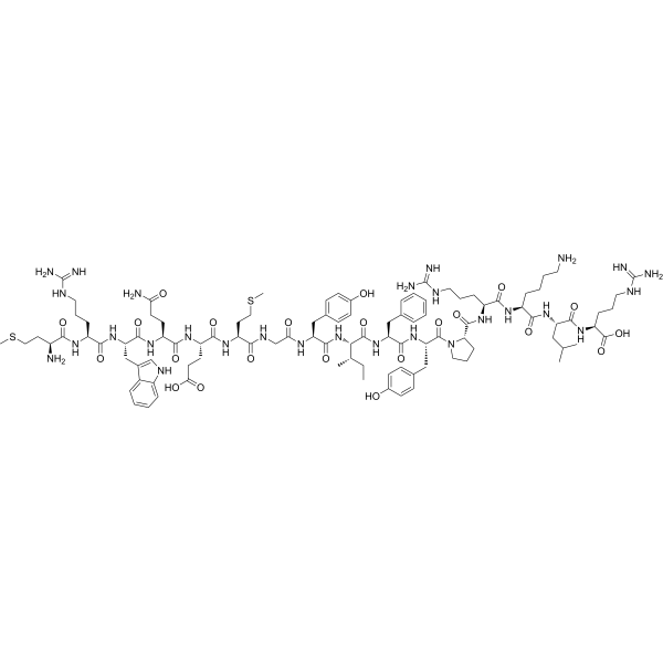
Met-Lys-Trp-Glu-Glu-Met-Gly-Tyr-Ile-Phe-Leu
-
Sequence Shortening
MKWEEMGYIFL
-
Shipping
Room temperature in continental US; may vary elsewhere.
-
Storage
Sealed storage, away from moisture and light
Powder -80°C 2 years -20°C 1 year * In solvent : -80°C, 6 months; -20°C, 1 month (sealed storage, away from moisture and light)


Publications (2)
-
Journal Impact Factor
-
Most Recent
-
Free Radic Biol Med
MOTS-c attenuates mitochondrial dysfunction induces pyroptosis and cartilage degradation in osteoarthritis via an Nrf2-Dependent Mechanism. [Abstract]2025 Oct 1:241:717-731. PMID: 41043625 -
Int J Neurosci
A mitochondrial-derived peptide MOTS-c contributes to the protective effect against brain injury associated with LPS-induced sepsis by strengthening the blood-brain barrier's ultrastructure. [Abstract]2025 Aug 5:1-14. PMID: 40753494


Solvent & Solubility
In Vitro:
H2O : 33.33 mg/mL (23.04 mM; ultrasonic and adjust pH to 9 with NH3·H2O)
Please refer to the solubility information to select the appropriate solvent. Once prepared, please aliquot and store the solution to prevent product inactivation from repeated freeze-thaw cycles.
Storage method and period of stock solution: -80°C, 6 months; -20°C, 1 month (sealed storage, away from moisture and light). When stored at -80°C, please use it within 6 months. When stored at -20°C, please use it within 1 month.
* Note: If you choose water as the stock solution, please dilute it to the working solution, then filter and sterilize it with a 0.22 μm filter before use.
Please refer to the solubility information to select the appropriate solvent. Once prepared, please aliquot and store the solution to prevent product inactivation from repeated freeze-thaw cycles.
Storage method and period of stock solution: -80°C, 6 months; -20°C, 1 month (sealed storage, away from moisture and light). When stored at -80°C, please use it within 6 months. When stored at -20°C, please use it within 1 month.
* Note: If you choose water as the stock solution, please dilute it to the working solution, then filter and sterilize it with a 0.22 μm filter before use.
Concentration (start) × Volume (start) = Concentration (final) × Volume (final)


Purity & Documentation
-
Data Sheet (273 KB)
-
SDS (252 KB)
- English - EN (252 KB)
- Français - FR (252 KB)
- Deutsch - DE (252 KB)
- Norwegian - NO (252 KB)
- Español - ES (252 KB)
- Swedish - SV (252 KB)
- Italian - IT (252 KB)
- Korean - KR (252 KB)
- Portuguese - PT (252 KB)
-
Handling Instructions (2659 KB)
References


Complete Stock Solution Preparation Table
Please refer to the solubility information to select the appropriate solvent. Once prepared, please aliquot and store the solution to prevent product inactivation from repeated freeze-thaw cycles.
Storage method and period of stock solution: -80°C, 6 months; -20°C, 1 month (sealed storage, away from moisture and light). When stored at -80°C, please use it within 6 months. When stored at -20°C, please use it within 1 month.
| Optional Solvent | Concentration Solvent Mass | 1 mg | 5 mg | 10 mg | 25 mg |
|---|---|---|---|---|---|
| H2O | 1 mM | 0.6912 mL | 3.4561 mL | 6.9121 mL | 17.2803 mL |
| 5 mM | 0.1382 mL | 0.6912 mL | 1.3824 mL | 3.4561 mL | |
| 10 mM | 0.0691 mL | 0.3456 mL | 0.6912 mL | 1.7280 mL | |
| 15 mM | 0.0461 mL | 0.2304 mL | 0.4608 mL | 1.1520 mL | |
| 20 mM | 0.0346 mL | 0.1728 mL | 0.3456 mL | 0.8640 mL |
* Note: If you choose water as the stock solution, please dilute it to the working solution, then filter and sterilize it with a 0.22 μm filter before use.


MOTS-c (mouse) Related Classifications
HY-P10769 Related Classifications
 Powered by Bioz