



NS1-IN-1
Based on 1 publication(s) in Google Scholar
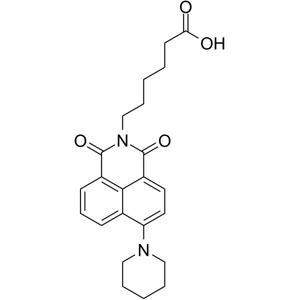
NS1-IN-1 (REDD1 inducer-1) is a naphthalimide compound and an NS1 inhibitor. NS1-IN-1 antagonizes influenza A virus NS1-mediated inhibition of host gene expression and induces REDD1 expression. NS1-IN-1 suppresses the mTORC1 signaling pathway via a TSC1-TSC2 dependent mechanism. NS1-IN-1 exhibits broad antiviral activity against influenza virus and vesicular stomatitis virus (VSV), with low toxicity.
For research use only. We do not sell to patients.
- CAS No.: 181373-35-3
- Formula: C23H26N2O4
- Molecular Weight:394.46
-
Storage:
Please store the product under the recommended conditions in the Certificate of Analysis.

To place orders, for customer services and technical support, please contact: MedChemExpress USA
Tel: 609-228-6898 E-mail: [email protected] [email protected]


-
 Biological Activity
-
Chemical Information
- Protocols
- Purity & Documentation
- References
-
 Help & FAQs


Publications Citing Use of MedChemExpress (MCE) NS1-IN-1
More

Biological Activity
Description
In Vitro
NS1-IN-1 (compound 3) (25-50 μM; 24-30 h) reduces influenza virus replication and viral protein expression (NP, M1, etc.) in MDCK cells with low cytotoxicity, consistent with cell viability assay showing CC50/IC50 ≈ 31[1].
NS1-IN-1 (30 μM; 4-9 h) inhibits influenza viral protein expression and induces REDD1 in A549 cells, while suppressing mTORC1 signaling via a TSC1-TSC2 dependent mechanism[1].
NS1-IN-1 (30 μM; up to 18 h) transcriptionally induces REDD1 mRNA without activating IFN-β or ISGs, confirming a non-interferon antiviral mechanism[1].
NS1-IN-1 (25-50 μM; 24 h) also inhibits VSV infection in MDCK cells, with antiviral activity dependent on the REDD1-TSC1-TSC2-mTORC1 axis[1].
MedChemExpress (MCE) has not independently confirmed the accuracy of these methods. They are for reference only. Further protocols information, click here.
-
Cell Line:MDCK, MEF, HBEC cells
-
Concentration:0.2-100 μM
-
Incubation Time:24-72 h
-
Result:Showed low cytotoxicity in host cells, with CC50/IC50 selectivity ratio of approximately 31.
-
Cell Line:MDCK, A549 cells
-
Concentration:30 μM
-
Incubation Time:4-9 h post-infection
-
Result:Downregulated influenza viral proteins (NS1, NP, M1, etc.) and induced REDD1 protein (6-8 fold).
Reduced S6K phosphorylation at Thr389 (mTORC1 inhibition) without directly affecting AKT in early stages.
-
Cell Line:A549 cell
-
Concentration:30 μM
-
Incubation Time:Various time points (up to 18 h pretreatment + infection)
-
Result:Transcriptionally induced REDD1 mRNA expression; induction was abolished by actinomycin D.
Did not induce IFN-β or interferon-stimulated genes.


Chemical Information
-
CAS No. 181373-35-3
-
Molecular Weight 394.46
-
Formula C23H26N2O4
-
SMILES
O=C(O)CCCCCN(C(C1=CC=C(N2CCCCC2)C3=CC=CC4=C13)=O)C4=O
-
Synonyms
REDD1 inducer-1
-
Shipping
Room temperature in continental US; may vary elsewhere.
-
Storage
Please store the product under the recommended conditions in the Certificate of Analysis.


Publications (1)
-
Journal Impact Factor
-
Most Recent
-
J Neuroinflammation
Microglial SIRT1 activation attenuates synapse loss in retinal inner plexiform layer via mTORC1 inhibition. [Abstract]2023 Sep 5;20(1):202. PMID: 37670386

Protocols
-
RT-PCR
Reverse transcription technology uses RNA as a template to synthesize DNA. RT-PCR is simple, specific and sensitive, and can be used to detect gene expression levels and expression differences in cells; detect RNA virus content; clone cDNA sequences of specific genes.
-
RNA extraction experimental
By lysing cells, releasing RNA, and removing impurities such as proteins and DNA, high-purity RNA products are finally obtained. The commonly used traditional method is the guanidine isothiocyanate/phenol/chloroform method (Trizol), which is suitable for a variety of animal materials including animal tissues, microorganisms, cultured cells, etc., and most plant materials.
-
Research Protocol for Infectious Diseases
Infectious-disease experiments test how pathogens interact with host barriers, innate immune receptors, inflammatory signaling, pathogen replication, and tissue injury; pattern-recognition receptors such as TLRs, RIG-I-like receptors, NOD-like receptors, and inflammasomes detect microbial molecules and activate NF-κB, interferon, and cytokine responses. The central hypothesis is that infection severity reflects the balance between pathogen burden and host response: protective inflammation restricts pathogen growth, whereas excessive or mislocalized inflammation contributes to tissue damage and disease phenotype. Unresolved questions include which host pathways are protective versus pathogenic, why some infection models fail to translate to human disease, and which combined readouts best predict clinically relevant infection outcomes.
-
Real Time qPCR (Q-PCR)
Real-time quantitative PCR (qPCR) quantifies an amplifiable nucleic-acid target by monitoring fluorescence during PCR cycling rather than measuring product only after amplification. The increase in fluorescence tracks accumulation of PCR product, and the quantification cycle (Cq; historically also Ct/CP) is related to the initial amount of target: samples containing more starting target generally reach the defined fluorescence threshold in fewer cycles.


Purity & Documentation
References

Calculators
Concentration (start) × Volume (start) = Concentration (final) × Volume (final)


NS1-IN-1 Related Classifications
HY-134922 Related Classifications
 Powered by Bioz