



Bexobrutideg
Based on 4 publication(s) in Google Scholar
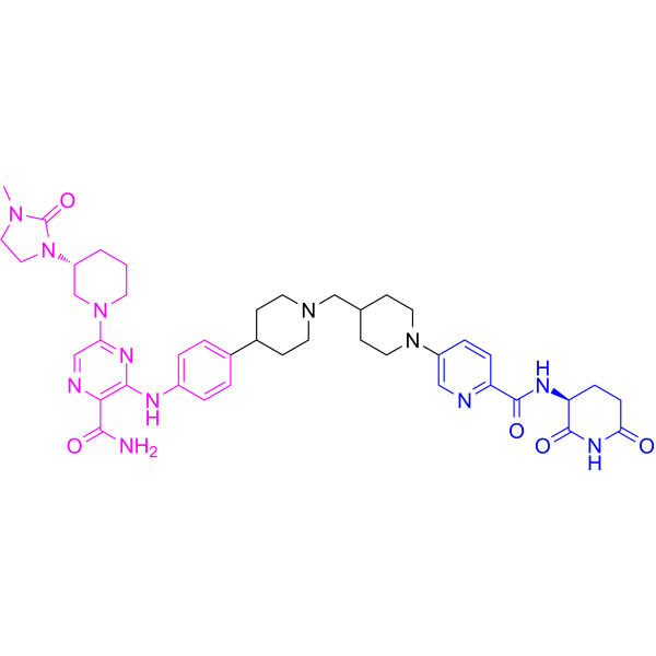
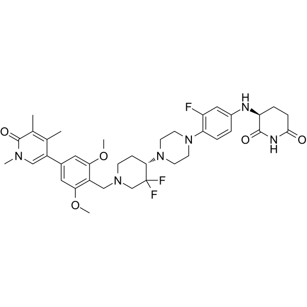
Bexobrutideg (NX-5948) is an orally active chimeric targeting molecule (CTM) that induces specific BTK protein degradation by the cereblon E3 ligase (CRBN) complex without degradation of other cereblon neo-substrates. Bexobrutideg mediates potent anti-inflammatory activity via BTK degradation with resultant inhibition of B cell activation. Bexobrutideg exhibits potent tumor growth inhibition in TMD8 xenograft models that contain either wild-type BTK or BTKi-resistant mutations. Bexobrutideg is efficacious in a mouse collageninduced arthritis (CIA) model. Bexobrutideg can cross the blood brain barrier (BBB). Bexobrutideg is a PROTAC composed of the ligand for target protein, a linker, and a cereblon E3 ligase (CRBN) complex (Red: BTK ligand (HY-170324); Blue: CRBN ligand (HY-171893); Black: linker).
(Pink: Btk ligand (HY-170324); Blue: Cereblon ligand (HY-171893); Black: linker).
商品は「研究用試薬」です。人や動物の医療用・臨床診断用・食品用の製品ではありません。
研究用途以外に使用した場合、当社は一切の責任を負いかねます。
- 純度 : 98.25%
- CAS 番号: 2649400-34-8
- 分子式: C42H54N12O5
- 分子量:806.96
-
保管条件:Powder -20°C, 3 years , 4°C, 2 years ; In solvent -80°C, 6 months , -20°C, 1 month

To place orders, for customer services and technical support, please contact: MedChemExpress USA
Tel: 609-228-6898 E-mail: [email protected] [email protected]


-
 生物活性
生物活性
-
化学情報
-
溶剤 & 溶解度
- プロトコル
- 純度とドキュメンテーション
- 参考文献
-
 Help & FAQs


-
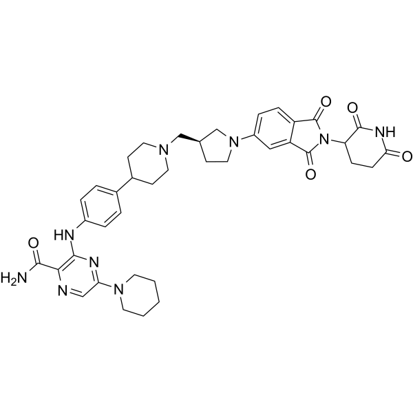
HY-153220
ZelebrudomideBTK PROTAC Degrader -
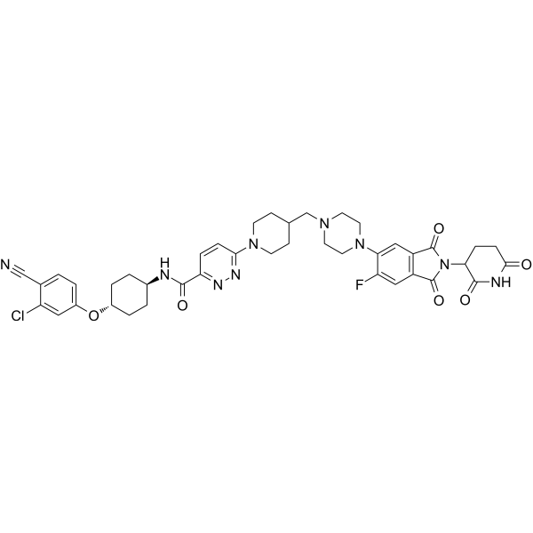
HY-138641
BavdegalutamideAR PROTAC Degrader -
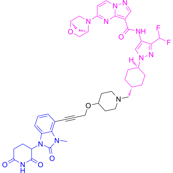
HY-145483
KT-474PROTAC IRAK4 Degrader -
HY-138642
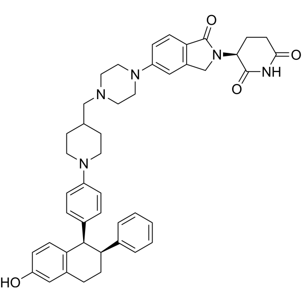
VepdegestrantPROTAC ER Degrader -
HY-160141
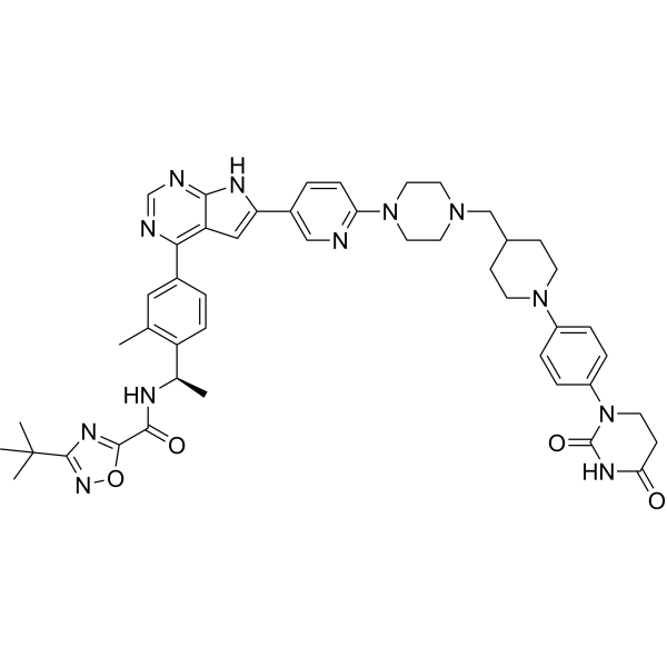
CatadegbrutinibBTK Degrader -
HY-153342
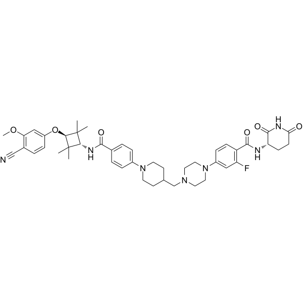
LuxdegalutamidePROTAC -
HY-130604
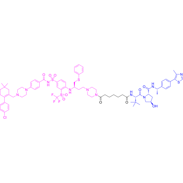
DT2216PROTAC BCL-XL Degrader -
HY-158101
BMS-986365PROTAC AR Degrader -
HY-153367
FHD-609BRD9 PROTAC Degrader -
HY-158105
ARV-393BCL6 PROTAC Degrader -
HY-153368
ZomiradomideIRAK4 PROTAC Degrader -
HY-145925B
CFT8634BRD9 PROTAC Degrader
MedChemExpress(MCE)の使用を引用している文献 Bexobrutideg
MorePROTACs アイソフォーム固有の製品をすべて表示
More

生物活性
製品説明
|
Cereblon |
体外実験
Bexobrutideg (NX-5948) (BTK-IN-24; 0.0001-1000 nM; 4 h) is a potent degrader of BTK in primary human B cells (DC50=0.34 nM) and inhibits BCR signaling[1]. NX-5948 induces the degradation of BTK (DC50< 1 nM) in lymphoma cell lines and PBMCs[3].
Bexobrutideg (NX-5948) (10 nM; 0.25, 0.5, 1, 2, 4, 6, 18, 24 h) catalyzes rapid BTK degradation within 1 hour and is complete within 2 hours in Ramos cells[1].
MedChemExpress (MCE) has not independently confirmed the accuracy of these methods. They are for reference only.
体内実験
Bexobrutideg (NX-5948) (3, 10, 30 mg/kg; po) causes dose- and time-dependent reduction in BTK levels in circulating murine and non-human primate, cynomolgus monkey B cells[1].
MedChemExpress (MCE) has not independently confirmed the accuracy of these methods. They are for reference only.
-
Animal Model:Mouse collagen-induced arthritis (CIA) model[1]
-
Dosage:10, 30 mg/kg
-
Administration:PO; daily; Day 18 to 36
-
Result:Showed efficacious and well-tolerated in a mouse CIA model.
- Collagen-Induced Arthritis
- Cotton Pellet Granuloma
- Carrageenan-Induced Paw Edema
- Patient-Derived Xenograft (PDX)
- Patient-Derived Orthotopic Xenograft (PDOX)
- Splenic/Portal-Vein Liver Metastasis Xenograft
- Research Protocol for Inflammation-related Diseases
- Subcutaneous Cell-Line-Derived Xenograft
- Orthotopic Cell-Line Xenograft
- Intraperitoneal/Peritoneal Dissemination Xenograft
臨床実験
| NCT Number | Sponsor | Condition | Start Date |
Phase

|
|---|---|---|---|---|
| NCT01329991 | Plexxikon| | 2011-05 | PHASE1 |


化学情報
-
CAS 番号 2649400-34-8
-
性状 Solid
-
分子量 806.96
-
分子式 C42H54N12O5
-
Color Off-white to yellow
-
SMILES
NC(C1=C(N=C(N2C[C@H](N3C(N(CC3)C)=O)CCC2)C=N1)NC4=CC=C(C5CCN(CC5)CC6CCN(C7=CN=C(C(N[C@@H]8C(NC(CC8)=O)=O)=O)C=C7)CC6)C=C4)=O
-
別名
NX-5948; BTK-IN-24
-
輸送条件
Room temperature in continental US; may vary elsewhere.
-
保管条件
Powder -20°C 3 years 4°C 2 years In solvent -80°C 6 months -20°C 1 month


Publications (4)
-
Journal Impact Factor
-
Most Recent
-
Blood Adv
PLCG2 Exon-Skipped Variants: Insights into Their Potential Role in Chronic Lymphocytic Leukemia. [Abstract]2026 Jun 5:bloodadvances.2025019077. PMID: 42263669 -
-
-
bioRxiv
2025 May 10:2025.05.07.652612. PMID: 40654846


溶剤 & 溶解度
体外:
DMSO : 100 mg/mL (123.92 mM; Need ultrasonic; Hygroscopic DMSO has a significant impact on the solubility of product, please use newly opened DMSO)
Please refer to the solubility information to select the appropriate solvent. Once prepared, please aliquot and store the solution to prevent product inactivation from repeated freeze-thaw cycles.
Storage method and period of stock solution: -80°C, 6 months; -20°C, 1 month. When stored at -80°C, please use it within 6 months. When stored at -20°C, please use it within 1 month.
Please refer to the solubility information to select the appropriate solvent. Once prepared, please aliquot and store the solution to prevent product inactivation from repeated freeze-thaw cycles.
Storage method and period of stock solution: -80°C, 6 months; -20°C, 1 month. When stored at -80°C, please use it within 6 months. When stored at -20°C, please use it within 1 month.
濃度 (開始) × 体積 (開始) = 濃度 (終了) × 体積 (終了)
体内:
Select the appropriate dissolution method based on your experimental animal and administration route.
- For the following dissolution methods, please ensure to first prepare a clear stock solution using an In Vitro approach and then sequentially add co-solvents:
- To ensure reliable experimental results, the clarified stock solution can be appropriately stored based on storage conditions. As for the working solution for In Vivo experiments, it is recommended to prepare freshly and use it on the same day.
- The percentages shown for the solvents indicate their volumetric ratio in the final prepared solution. If precipitation or phase separation occurs during preparation, heat and/or sonication can be used to aid dissolution.
Add each solvent one by one: 10% DMSO 40% PEG300 5% Tween-80 45% Saline
Solubility: ≥ 3.33 mg/mL (4.13 mM); Clear solution
This protocol yields a clear solution of ≥ 3.33 mg/mL (saturation unknown).
Taking 1 mL working solution as an example, add 100 μL DMSO stock solution (33.3 mg/mL) to 400 μL PEG300, and mix evenly; then add 50 μL Tween-80 and mix evenly; then add 450 μL Saline to adjust the volume to 1 mL.
Preparation of Saline: Dissolve 0.9 g sodium chloride in ddH₂O and dilute to 100 mL to obtain a clear Saline solution.
Add each solvent one by one: 10% DMSO 90% (20% SBE-β-CD in Saline)
Solubility: ≥ 3.33 mg/mL (4.13 mM); Clear solution
This protocol yields a clear solution of ≥ 3.33 mg/mL (saturation unknown).
Taking 1 mL working solution as an example, add 100 μL DMSO stock solution (33.3 mg/mL) to 900 μL 20% SBE-β-CD in Saline, and mix evenly.
Preparation of 20% SBE-β-CD in Saline (4°C, storage for one week): 2 g SBE-β-CD powder is dissolved in 10 mL Saline, completely dissolve until clear.
In Vivo Dissolution Calculator
Please enter the basic information of animal experiments:
-
-
-
-
Recommended: Prepare an additional quantity of animals to account for potential losses during experiments.
Please enter your animal formula composition:
-
%DMSO +
Recommended: Keep the proportion of DMSO in working solution below 2% if your animal is weak.
-
%+
-
+%Tween-80 + +
-
%Saline +
The co-solvents required include: DMSO, . All of co-solvents are available by MedChemExpress (MCE). , Tween 80. All of co-solvents are available by MedChemExpress (MCE).
Working solution concentration: 0.22 mg/mL
Method for preparing stock solution: mg drug dissolved in μL DMSO. Stock solution concentration: mg/mL.
1. Take μL DMSO stock solution;
2. Add μL .
μL , mix evenly;
3. Then add μL Tween 80, mix evenly;
4. Then add μL
Please ensure that the stock solution in the first step is dissolved to a clear state, and add co-solvents in sequence. You can use ultrasonic heating (ultrasonic cleaner, recommended frequency 20-40 kHz), vortexing, etc. to assist dissolution.


純度とドキュメンテーション
-
データシート (274 KB)
-
SDS (251 KB)
- English - EN (251 KB)
- Français - FR (251 KB)
- Deutsch - DE (251 KB)
- Norwegian - NO (251 KB)
- Español - ES (251 KB)
- Swedish - SV (251 KB)
- Italian - IT (251 KB)
- Korean - KR (251 KB)
- Portuguese - PT (251 KB)
-
取扱説明書 (2659 KB)
参考文献


Complete Stock Solution Preparation Table
Please refer to the solubility information to select the appropriate solvent. Once prepared, please aliquot and store the solution to prevent product inactivation from repeated freeze-thaw cycles.
Storage method and period of stock solution: -80°C, 6 months; -20°C, 1 month. When stored at -80°C, please use it within 6 months. When stored at -20°C, please use it within 1 month.
| Optional Solvent | Concentration Solvent Mass | 1 mg | 5 mg | 10 mg | 25 mg |
|---|---|---|---|---|---|
| DMSO | 1 mM | 1.2392 mL | 6.1961 mL | 12.3922 mL | 30.9805 mL |
| 5 mM | 0.2478 mL | 1.2392 mL | 2.4784 mL | 6.1961 mL | |
| 10 mM | 0.1239 mL | 0.6196 mL | 1.2392 mL | 3.0980 mL | |
| 15 mM | 0.0826 mL | 0.4131 mL | 0.8261 mL | 2.0654 mL | |
| 20 mM | 0.0620 mL | 0.3098 mL | 0.6196 mL | 1.5490 mL | |
| 25 mM | 0.0496 mL | 0.2478 mL | 0.4957 mL | 1.2392 mL | |
| 30 mM | 0.0413 mL | 0.2065 mL | 0.4131 mL | 1.0327 mL | |
| 40 mM | 0.0310 mL | 0.1549 mL | 0.3098 mL | 0.7745 mL | |
| 50 mM | 0.0248 mL | 0.1239 mL | 0.2478 mL | 0.6196 mL | |
| 60 mM | 0.0207 mL | 0.1033 mL | 0.2065 mL | 0.5163 mL | |
| 80 mM | 0.0155 mL | 0.0775 mL | 0.1549 mL | 0.3873 mL | |
| 100 mM | 0.0124 mL | 0.0620 mL | 0.1239 mL | 0.3098 mL |


Bexobrutideg 関連分類
HY-153321 関連分類
 Powered by Bioz