



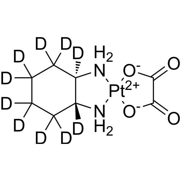
Oxaliplatin-d10
Oxaliplatin-d10 is a deuterium labeled Oxaliplatin (HY-17371). Oxaliplatin is a DNA synthesis inhibitor. Oxaliplatin causes DNA crosslinking damage, prevents DNA replication and transcription and induces apoptosis. Oxaliplatin can be used for cancer research.
Nos produits utilisent uniquement pour la recherche. Nous ne vendons pas aux patients.
- CAS No.: 1132819-16-9
- Formule: C8H4D10N2O4Pt
- Masse moléculaire:407.35
-
Stockage:
Please store the product under the recommended conditions in the Certificate of Analysis.

To place orders, for customer services and technical support, please contact: MedChemExpress USA
Tel: 609-228-6898 E-mail: [email protected] [email protected]


-
 Activité biologique
-
Chemical Information
- Protocole
- Pureté et documentation
- Références
-
 Help & FAQs


-
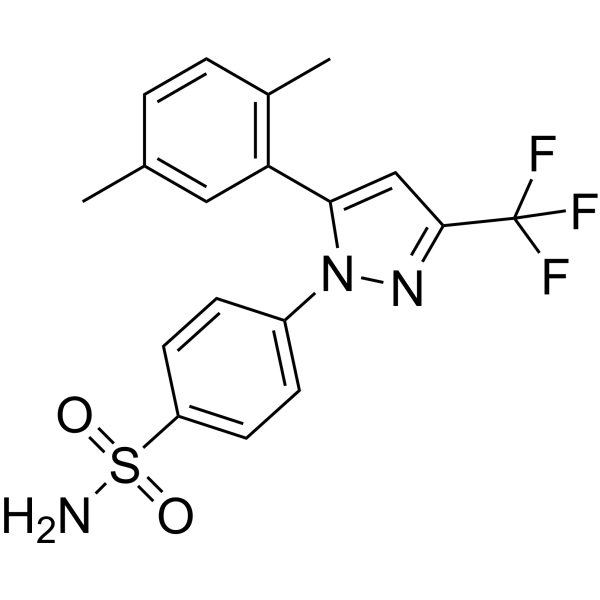
HY-W010995
2,5-DimethylcelecoxibWnt/β-catenin inhibitor -
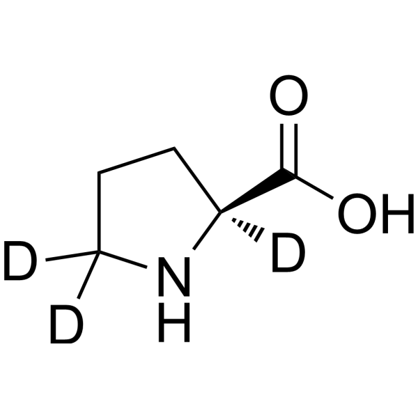
HY-Y0252S1
L-Proline-d3Stable Isotope -
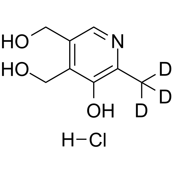
HY-N0682S
Pyridoxine-d3 hydrochlorideStable Isotope -
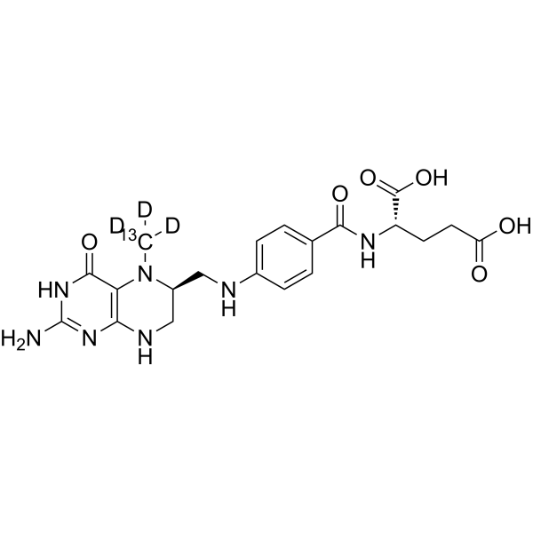
HY-14781S1
Levomefolic acid-13C,d3Stable Isotope -
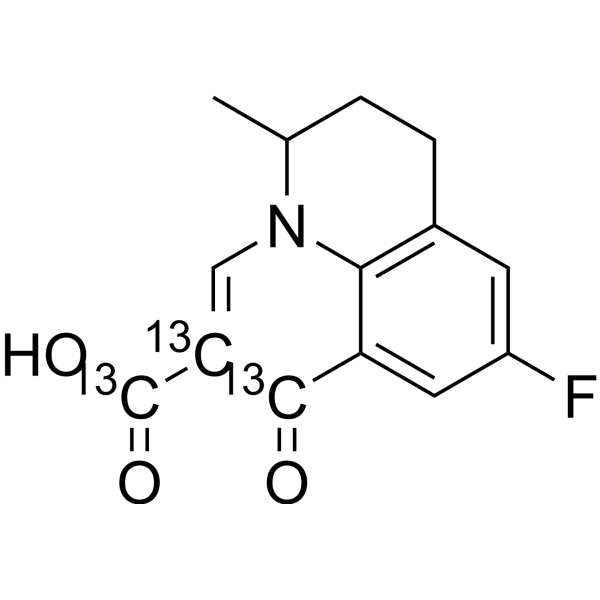
HY-B0526S
Flumequine-13C3Stable Isotope -
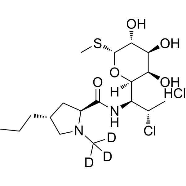
HY-B1455S
Clindamycin-d3 hydrochlorideStable Isotope -
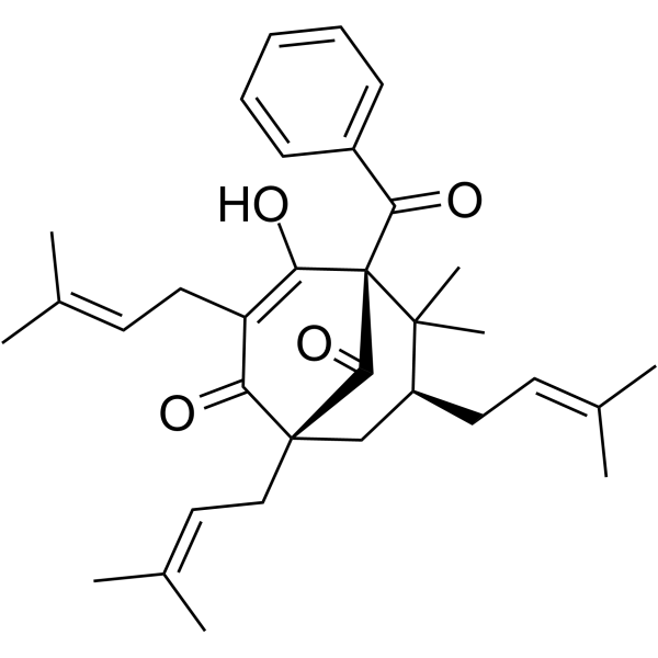
HY-121458
NemorosoneAntiproliferative Anticarcinogen
Voir tous les produits spécifiques à Isoform DNA/RNA Synthesis
More

Activité biologique
Description
In Vitro
Stable heavy isotopes of hydrogen, carbon, and other elements have been incorporated into drug molecules, largely as tracers for quantitation during the drug development process. Deuteration has gained attention because of its potential to affect the pharmacokinetic and metabolic profiles of drugs[1].
MedChemExpress (MCE) has not independently confirmed the accuracy of these methods. They are for reference only.
Application
1. This compound can be used as a tracer
2. This compound can be used as an internal standard for quantitative analysis by NMR, GC-MS, or LC-MS.


Chemical Information
-
CAS No. 1132819-16-9
-
Unlabeled CAS 61825-94-3
-
Masse moléculaire 407.35
-
Formule C8H4D10N2O4Pt
-
SMILES
O=C1[O-][Pt+2]2([O-]C1=O)[NH2][C@@]3([2H])[C@](C([2H])([2H])C([2H])([2H])C([2H])([2H])C3([2H])[2H])([2H])[NH2]2
-
Livraison
Room temperature in continental US; may vary elsewhere.
-
Stockage
Please store the product under the recommended conditions in the Certificate of Analysis.


Pureté et documentation
-
Fiche technique (280 KB)
-
SDS (252 KB)
- Français - FR (252 KB)
- Deutsch - DE (252 KB)
- Norwegian - NO (252 KB)
- Español - ES (252 KB)
- Swedish - SV (252 KB)
- Italian - IT (252 KB)
- Korean - KR (252 KB)
- Portuguese - PT (252 KB)
-
Instruction de manipulation (2659 KB)
Références
[1]. Russak EM, et al. Impact of Deuterium Substitution on the Pharmacokinetics of Pharmaceuticals. Ann Pharmacother. 2019 Feb;53(2):211-246. [Content Brief]
[2]. Garrett MJ, et, al. Capecitabine, Oxaliplatin, and Bevacizumab (BCapOx) Regimen for Metastatic Colorectal Cancer. Hosp Pharm. 2017 May;52(5):341-347. [Content Brief]
[3]. Yi Yao, et al. Comparative proteomic analysis of colon cancer cells in response to oxaliplatin treatment. Biochim Biophys Acta. 2009 Oct;1794(10):1433-40. [Content Brief]
[4]. Park GY, et al. Phenanthriplatin, a monofunctional DNA-binding platinum anticancer drug candidate with unusual potency and cellular activity profile. Proc Natl Acad Sci U S A. 2012 Jul 24;109(30):11987-92. [Content Brief]
[5]. Mohammed MQ, et al. Oxaliplatin is active in vitro against human melanoma cell lines: comparison with NSC 119875 and NSC 241240. Anticancer Drugs. 2000 Nov;11(10):859-63. [Content Brief]
[6]. Raymond E, et al. Oxaliplatin: a review of preclinical and clinical studies. Ann Oncol. 1998 Oct;9(10):1053-71. [Content Brief]
[7]. Wang Z, et al. Oxaliplatin induces apoptosis in hepatocellular carcinoma cells and inhibits tumor growth. Expert Opin Investig Drugs. 2009 Nov;18(11):1595-604 [Content Brief]
[8]. Pendyala L, et al. In vitro cytotoxicity, protein binding, red blood cell partitioning, and biotransformation of oxaliplatin. Cancer Res. 1993 Dec 15;53(24):5970-6. [Content Brief]
[9]. Mathé G, et al. Oxalato-platinum or 1-OHP, a third-generation platinum complex: an experimental and clinical appraisal and preliminary comparison with cis-platinum. Biomed Pharmacother. 1989;43(4):237-50. [Content Brief]
[10]. Schellingerhout D, et al. Impairment of retrograde neuronal transport in oxaliplatin-induced neuropathy demonstrated by molecular imaging. PLoS One. 2012;7(9):e45776. doi: 10.1371/journal.pone.0045776. Epub 2012 Sep 20. [Content Brief]

Calculators
Concentration (start) × Volume (start) = Concentration (final) × Volume (final)


Oxaliplatin-d10 Related Classifications
HY-17371S Related Classifications
 Powered by Bioz