



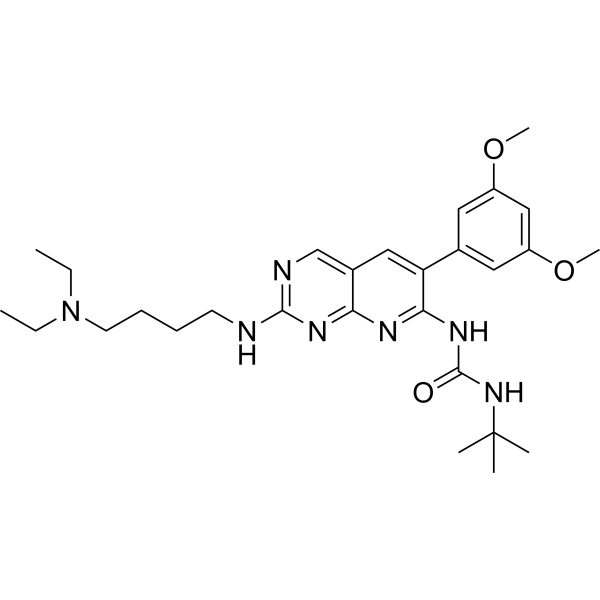
PD173074 GMP is PD173074 (HY-10321) produced by using GMP guidelines. GMP small molecules works appropriately as an auxiliary reagent for cell therapy manufacture. PD173074 is an orally active FGFR inhibitor that targets the transphosphorylation of FGFR1 and FGFR2 and blocks the FGF signaling pathway. By reducing the phosphorylation level of SMAD2 and altering the expression of Nodal/Activin target genes, PD173074 eliminates endothelial differentiation potential, thereby inhibiting the formation of capillary-like structures. PD173074 blocks the proliferation and colony formation of tumor cells and increases intratumoral cell apoptosis. PD173074 successfully reverses FGF-2-induced chemoresistance to enhance the effect of cisplatin (HY-17394) in small cell lung cancer models. PD173074 can be applied to research related to critical limb ischemia and small cell lung cancer.
For research use only. We do not sell to patients.
- CAS No.: 219580-11-7
- Formula: C28H41N7O3
- Molecular Weight:523.67
-
Storage:
Please store the product under the recommended conditions in the Certificate of Analysis.


To place orders, for customer services and technical support, please contact: MedChemExpress USA
Tel: 609-228-6898 E-mail: [email protected] [email protected]


-
 Biological Activity
-
Chemical Information
- Protocols
- Purity & Documentation
- References
-
 Help & FAQs


-
HY-10321
PD173074FGFR1 Inhibitor -
HY-13311
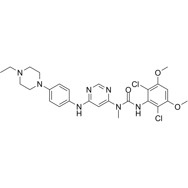
InfigratinibFGFR Inhibitor -

HY-10321R
PD173074 (Standard)FGFR1 Inhibitor -
HY-50904
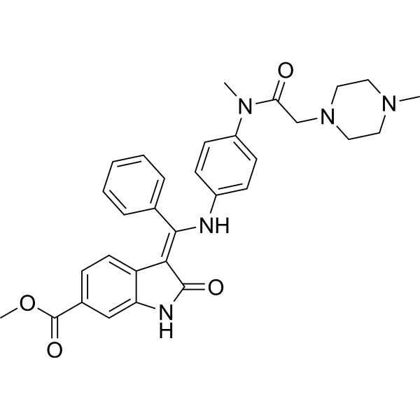
NintedanibPDGFR/VEGFR/FGFR Inhibitor -
HY-109099
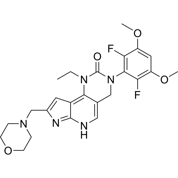
PemigatinibFGFR Inhibitor -
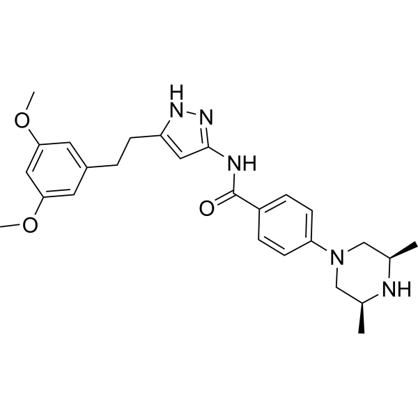
HY-13330
FexagratinibFGFR Inhibitor -
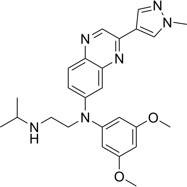
HY-18708
ErdafitinibFGFR Inhibitor -
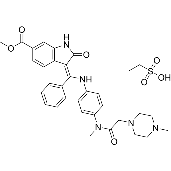
HY-11106
Nintedanib esylatePDGFR/VEGFR/FGFR Inhibitor -
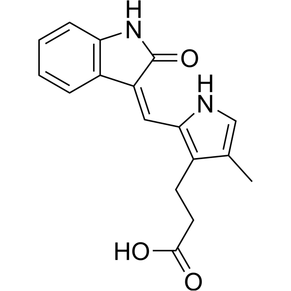
HY-10407
SU 5402Tyrosine Kinase Inhibitor -
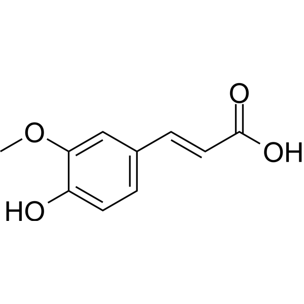
HY-N0060
Ferulic acidFGFR1 Inhibitor -
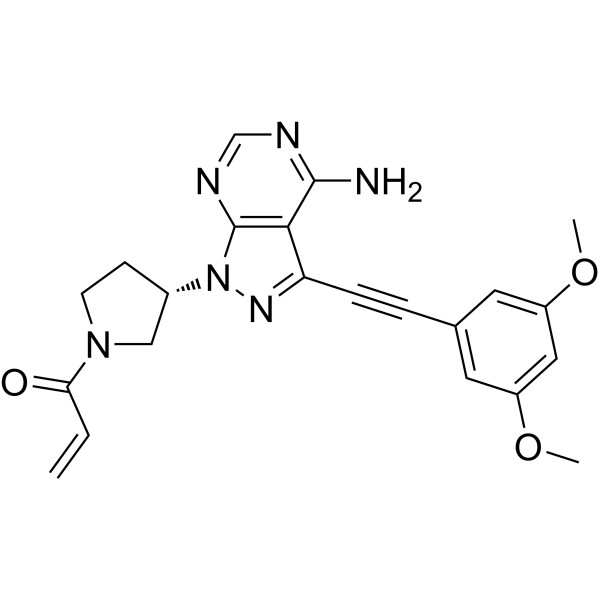
HY-100818
FutibatinibFGFR Inhibitor -
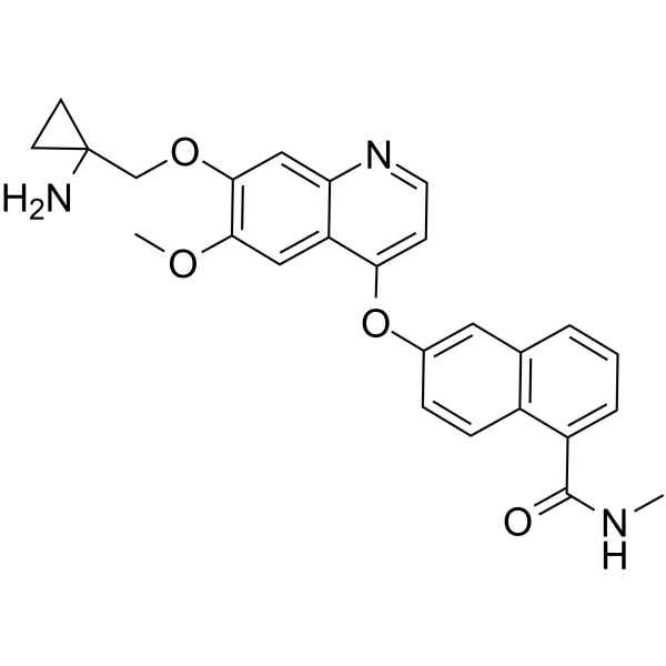
HY-15391
LucitanibVEGFR/FGFR Inhibitor


Biological Activity
Description
IC50 & Target
|
FGFR1 |
FGFR2 |
Cellular Effect
|
Cell Line

|
Type | Value | Description | References |
|---|---|---|---|---|
| NCI-H520 | IC50 |
281 nM
Compound: PD173074
|
Antiproliferative activity against human FGFR1-amplified NCI-H520 cells after 7 days by SRB assay
Antiproliferative activity against human FGFR1-amplified NCI-H520 cells after 7 days by SRB assay
|
[PMID: 28521156] |
| RT-112 | GI50 |
0.015 μM
Compound: PD173074
|
Antiproliferative activity against human RT112 cells after 72 hrs by MTT assay
Antiproliferative activity against human RT112 cells after 72 hrs by MTT assay
|
[PMID: 27599742] |
| SUM185PE | IC50 |
20 nM
Compound: 58; PD173074
|
Anticancer activity against human SUM185PE cells assessed as cell growth inhibition incubated for 6 days by CellTiter96 analysis
Anticancer activity against human SUM185PE cells assessed as cell growth inhibition incubated for 6 days by CellTiter96 analysis
|
[PMID: 33650861] |
| SW780 | IC50 |
84.3 nM
Compound: PD173074
|
Antiproliferative activity against human FGFR3-amplified SW780 cells after 5 days by SRB assay
Antiproliferative activity against human FGFR3-amplified SW780 cells after 5 days by SRB assay
|
[PMID: 28521156] |
In Vitro
PD173074 GMP (GMP) (100 nmol/L; 3-hour pre-incubation, followed by 10-day incubation with repeated 3-hour pre-incubations every 3 days) blocks FGF-induced endothelial differentiation of human adipose-derived stem cells, including reducing CD31 and eNOS mRNA expression, preventing capillary-like structure formation on Matrigel, and eliminating eNOS protein expression[1].
PD173074 GMP (GMP) (200 nM; 6 h) upregulates LEFTY1, LEFTY2, and FST and downregulates CRIPTO, NODAL, INHBA, INHBB, and NANOG in on-feeder human iPSCs, with no effect on NOMO1, NOMO2, NOMO3, NCLN, or OCT4[2].
PD173074 GMP (GMP) (200 nM; 6 h) downregulates LEFTY1, NODAL, and NANOG and upregulates FST in feeder-less human iPSCs, with no effect on CRIPTO, LEFTY2, or OCT4[2].
PD173074 GMP (GMP) (200 nM; 2-6 h) induces time-dependent downregulation of LEFTY1 (starting at 2 h), NODAL (at 6 h), LEFTY2 (at 6 h), and NANOG (starting at 4 h) in feeder-less human iPSCs, with no effect on CRIPTO or OCT4 over 6 h[2].
PD173074 GMP (GMP) (200 nM; 24 h) downregulates CRIPTO, LEFTY1, and NANOG in feeder-less human iPSCs[2].
PD173074 GMP (GMP) (200 nM; 6 h) reduces phosphorylation of SMAD2 and FGFR in feeder-less human iPSCs, with no effect on total SMAD2/3 or CRIPTO protein levels[2].
PD173074 GMP (GMP) (200 nM; 6 h) does not change the perinuclear localization of CRIPTO in feeder-less human iPSCs[2].
MedChemExpress (MCE) has not independently confirmed the accuracy of these methods. They are for reference only.
-
Cell Line:activin A-pretreated feeder-less human induced pluripotent stem cells (iPSCs)
-
Concentration:200 nM
-
Incubation Time:6 h (following 24 h pretreatment with 10 ng/mL activin A)
-
Result:Significantly increased the relative expression levels of LEFTY1 and LEFTY2.
Significantly decreased the relative expression level of NANOG.
Caused no significant change in the relative expression levels of CRIPTO, NODAL, and OCT4.
-
Cell Line:feeder-less human induced pluripotent stem cells (iPSCs)
-
Concentration:200 nM
-
Incubation Time:2 h; 4 h; 6 h
-
Result:Caused significant downregulation of LEFTY1 expression at 2 h, with further downregulation at 4 h and 6 h.
Significantly downregulated NODAL expression at 6 h.
Significantly downregulated NANOG expression starting at 4 h.
Caused no significant change in CRIPTO or OCT4 expression at any time point up to 6 h.
Downregulated LEFTY2 expression at 6 h.
-
Cell Line:feeder-less human induced pluripotent stem cells (iPSCs)
-
Concentration:200 nM
-
Incubation Time:6 h
-
Result:Significantly decreased the relative signal strength of phosphorylated SMAD2 (pSMAD2) and phosphorylated FGF receptor (pFGFR; ~60 kDa).
Caused no significant change in the relative signal strength of total SMAD2/3 or CRIPTO (25 kDa).
Did not alter the size or abundance of CRIPTO protein.
In Vivo
PD173074 GMP (50 mg/kg; p.o.; once daily; 14 d): In nude mouse models inoculated with H-69 human small cell lung cancer cells, it reduces intratumoral proliferation in the early stage of treatment, and this change can be detected by [18F] FLT-PET in vivo imaging between 7 and 14 days of treatment[3].
MedChemExpress (MCE) has not independently confirmed the accuracy of these methods. They are for reference only.
-
Animal Model:Nude mice, bearing H-510 human small cell lung cancer xenografts[3]
-
Dosage:50 mg/kg
-
Administration:p.o.; once daily; 28 d
-
Result:Impaired tumor growth to a similar extent as single-agent cisplatin, increased the median survival of tumor-bearing mice compared with control animals, and did not affect tumor vasculature or lymph node metastasis.
- Kinase activity and phosphorylation assays
- Western Blot
- RNA extraction experimental
- Human pluripotent stem cell endothelial-cell differentiation
- Detection of Apoptosis
- TUNEL staining for apoptotic DNA fragmentation
- Annexin V plus membrane-impermeant dye apoptosis staining
- Apoptosis Solutions
- Protocol for Kinase activity and phosphorylation assays
- Soft Agar Colony Formation Assay
- Colony Formation (Clonogenic) Assay
- Cell Viability Determination by MTT Colorimetric Assay


Chemical Information
-
CAS No. 219580-11-7
-
Molecular Weight 523.67
-
Formula C28H41N7O3
-
SMILES
O=C(NC1=NC2=NC(NCCCCN(CC)CC)=NC=C2C=C1C3=CC(OC)=CC(OC)=C3)NC(C)(C)C
-
Shipping
Room temperature in continental US; may vary elsewhere.
-
Storage
Please store the product under the recommended conditions in the Certificate of Analysis.


Purity & Documentation
References

Calculators
Concentration (start) × Volume (start) = Concentration (final) × Volume (final)


PD173074 (GMP) Related Classifications
HY-10321G Related Classifications
 Powered by Bioz