



(Rac)-GSK-3484862

Based on 1 Customer Validation
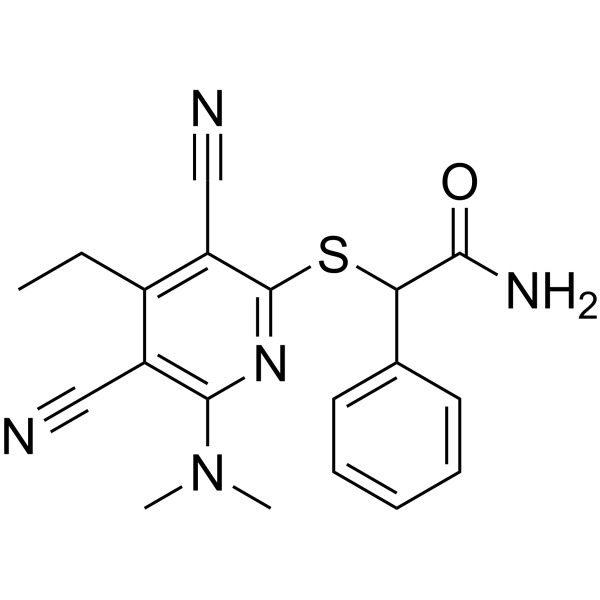
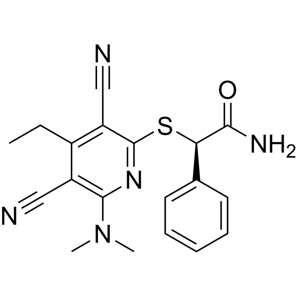
(Rac)-GSK-3484862 (GSK3482364) is a selective, orally active, reversible DNA methyltransferase 1 (DNMT1) inhibitor with an IC50 of 0.4 μM. (Rac)-GSK-3484862 inhibits the methyltransferase activity of DNMT1 independently of DNA incorporation, reduces global DNA methylation levels, and relieves the transcriptional repression of HBG1 and HBG2 genes. (Rac)-GSK-3484862 increases fetal hemoglobin levels and the proportion of red blood cells expressing fetal hemoglobin in erythroid progenitor cells and a transgenic mouse model of sickle cell disease. (Rac)-GSK-3484862 can be used in studies related to sickle cell disease.
For research use only. We do not sell to patients.
- Purity : 98.76%
- CAS No.: 2170136-02-2
- Formula: C19H19N5OS
- Molecular Weight:365.45
-
Storage:Powder -20°C, 3 years , 4°C, 2 years ; In solvent -80°C, 6 months , -20°C, 1 month

To place orders, for customer services and technical support, please contact: MedChemExpress USA
Tel: 609-228-6898 E-mail: [email protected] [email protected]


-
 Biological Activity
-
Chemical Information
-
Solvent & Solubility
- Protocols
- Purity & Documentation
- References
-
 Help & FAQs


All DNA Methyltransferase Isoforms
More

Biological Activity
Description
IC50 & Target
[1]|
DNMT1 0.4 μM (IC50) |
In Vitro
(Rac)-GSK-3484862 (0.01-10 μM; 5 days) induces dose-dependent genome-wide and HBG1/HBG2 promoter hypomethylation, upregulation of HBG1/HBG2 mRNA, and HbF protein expression in human bone marrow-derived EPCs, with an IC50 of 0.24 μM for genome-wide DNA hypomethylation[1].
(Rac)-GSK-3484862 (1.0 μM; 18 days) increases the number of HbF-positive cells to more than 4-fold the original level in human bone marrow CD34+ cells induced to differentiate into reticulocytes over 18 days, without affecting the maturation of erythroid cells[1].
(Rac)-GSK-3484862 reduces the methylation activity of DNMT1, reactivates the globin genes HBG1 and HBG2, upregulates the expression of HbF, and increases the proportion of HbF-containing red blood cells in an unspecified in vitro cell system[2].
MedChemExpress (MCE) has not independently confirmed the accuracy of these methods. They are for reference only. Further protocols information, click here.
In Vivo
(Rac)-GSK-3484862 reduces DNMT1 activity, reactivates globin genes, and increases HbF expression and HbF-containing erythrocytes in transgenic murine models of sickle cell disease[2].
MedChemExpress (MCE) has not independently confirmed the accuracy of these methods. They are for reference only.
-
Animal Model:B6;129-HBAtm1(HBA)Tow/HBBtm2(HBG1,HBB)Tow/J (male and female, 6-8 weeks old, sickle cell disease transgenic model)[1]
-
Dosage:3.7-100 mg/kg (12-day dosing); 3.7-100 mg/kg (2-week intermittent dosing); 3.7-100 mg/kg (4-week intermittent dosing)
-
Administration:p.o.; b.i.d. on weekdays; 12 days, 2 weeks, or 4 weeks
-
Result:Increased HbF levels to 3.3% (from vehicle 0.4%) at 10 mg/kg.
Increased F-cells to 22.7% (from vehicle 4.3%) at 10 mg/kg.
Increased HbF 10.3-fold relative to vehicle (to 5.3%) at 50 mg/kg.
Increased F-cells 8.4-fold relative to vehicle (to 35.5%) at 50 mg/kg.
Increased HbF dose-dependently up to 9-fold versus vehicle across 3.7-100 mg/kg doses, reaching a plateau at 33.3 mg/kg and 100 mg/kg.
Reduced -53 bp HBG1 promoter methylation by a maximum 25% at 33.3 mg/kg in bone marrow.
Increased HBG1 mRNA levels up to 29-fold versus vehicle across all dose groups in bone marrow.
Maintained unchanged HBB and HBA1 mRNA levels in bone marrow.
Showed no significant changes in peripheral blood cell counts (RBC, platelets, neutrophils, lymphocytes, monocytes) at any dose compared to vehicle.
Showed no abnormalities in cellularity or hematopoietic cell composition in sternal bone marrow histology at 100 mg/kg.
Produced robust HbF induction with no body weight changes during intermittent dosing (b.i.d. on 3 weekdays) for 2 or 4 weeks.
Showed a small non-significant increase in HbF with 4-week intermittent dosing compared to 2-week dosing.


Chemical Information
-
CAS No. 2170136-02-2
-
Appearance Solid
-
Molecular Weight 365.45
-
Formula C19H19N5OS
-
Color Brown to orange
-
SMILES
O=C(N)C(SC1=NC(N(C)C)=C(C#N)C(CC)=C1C#N)C2=CC=CC=C2
-
Synonyms
GSK3482364
-
Shipping
Room temperature in continental US; may vary elsewhere.
-
Storage
Powder -20°C 3 years 4°C 2 years In solvent -80°C 6 months -20°C 1 month


Solvent & Solubility
In Vitro:
DMSO : 50 mg/mL (136.82 mM; ultrasonic and warming and heat to 60°C; Hygroscopic DMSO has a significant impact on the solubility of product, please use newly opened DMSO)
Please refer to the solubility information to select the appropriate solvent. Once prepared, please aliquot and store the solution to prevent product inactivation from repeated freeze-thaw cycles.
Storage method and period of stock solution: -80°C, 6 months; -20°C, 1 month. When stored at -80°C, please use it within 6 months. When stored at -20°C, please use it within 1 month.
Please refer to the solubility information to select the appropriate solvent. Once prepared, please aliquot and store the solution to prevent product inactivation from repeated freeze-thaw cycles.
Storage method and period of stock solution: -80°C, 6 months; -20°C, 1 month. When stored at -80°C, please use it within 6 months. When stored at -20°C, please use it within 1 month.
Concentration (start) × Volume (start) = Concentration (final) × Volume (final)

Protocols
-
Cell Cytotoxicity Assay
Cytotoxicity assays are usually based on the assessment of cell membrane damage, which can also be indirectly detected by measuring cell viability. Detection methods include MTT assay, CKK-8 assay, LDH assay and ATP assay, etc.
-
Research Protocol for Epigenomic Data Analysis
Epigenomic data analysis identifies genome-wide regulatory features that influence gene expression, chromatin state, and phenotype without changing the underlying DNA sequence. In this strategy, the core regulatory layer includes chromatin accessibility, transcription-factor or histone-mark occupancy, DNA methylation, and chromatin-state patterns; these features are measured by sequencing-based assays and interpreted as regulatory elements, promoters, enhancers, repressive domains, methylated cytosines, or candidate phenotype-associated chromatin programs. The literature links epigenomic features to phenotype by showing that functional genomic elements can be mapped across human cell types and tissues, and that integrated epigenomic maps reveal cell-type-specific regulatory programs. ENCODE integrated transcription, chromatin accessibility, transcription-factor occupancy, and histone modification data to annotate functional elements in the human genome, while the Roadmap Epigenomics Co
-
How to Choose the Right Model Animal
Choosing the right model animal is a validity-driven decision in which the species, strain, sex, age, genetic background, disease-induction method, outcome measures, and welfare burden must match the scientific question rather than laboratory tradition or convenience. A model should be selected by judging face validity, construct validity, and predictive validity: whether it resembles the human phenotype, whether it reproduces relevant mechanisms, and whether results are likely to predict human biology or treatment response. Animal studies often fail to translate because of species differences, weak disease resemblance, poor experimental design, inadequate reporting, publication bias, and underuse of randomization, blinding, and sample-size justification. Unresolved questions include how to rank competing models objectively, how much human-disease complexity must be reproduced for a given objective, and when non-animal systems such as organoids, ex vivo tissue, or computational models


Purity & Documentation
-
Data Sheet (291 KB)
-
SDS (254 KB)
- English - EN (254 KB)
- Français - FR (254 KB)
- Deutsch - DE (254 KB)
- Norwegian - NO (254 KB)
- Español - ES (254 KB)
- Swedish - SV (254 KB)
- Italian - IT (254 KB)
- Korean - KR (254 KB)
- Portuguese - PT (254 KB)
-
Handling Instructions (2659 KB)
References
[1].
Gilmartin AG, et al. In vitro and in vivo induction of fetal hemoglobin with a reversible and selective DNMT1 inhibitor. Haematologica. 2021 Jul 1;106(7):1979-1987.
[Content Brief]
[2]. Ala C, et al. Discovery of potent DNMT1 inhibitors against sickle cell disease using structural-based virtual screening, MM-GBSA and molecular dynamics simulation-based approaches. Journal of biomolecular structure & dynamics. 2024;42(1):261-273. [Content Brief]


Complete Stock Solution Preparation Table
Please refer to the solubility information to select the appropriate solvent. Once prepared, please aliquot and store the solution to prevent product inactivation from repeated freeze-thaw cycles.
Storage method and period of stock solution: -80°C, 6 months; -20°C, 1 month. When stored at -80°C, please use it within 6 months. When stored at -20°C, please use it within 1 month.
| Optional Solvent | Concentration Solvent Mass | 1 mg | 5 mg | 10 mg | 25 mg |
|---|---|---|---|---|---|
| DMSO | 1 mM | 2.7364 mL | 13.6818 mL | 27.3635 mL | 68.4088 mL |
| 5 mM | 0.5473 mL | 2.7364 mL | 5.4727 mL | 13.6818 mL | |
| 10 mM | 0.2736 mL | 1.3682 mL | 2.7364 mL | 6.8409 mL | |
| 15 mM | 0.1824 mL | 0.9121 mL | 1.8242 mL | 4.5606 mL | |
| 20 mM | 0.1368 mL | 0.6841 mL | 1.3682 mL | 3.4204 mL | |
| 25 mM | 0.1095 mL | 0.5473 mL | 1.0945 mL | 2.7364 mL | |
| 30 mM | 0.0912 mL | 0.4561 mL | 0.9121 mL | 2.2803 mL | |
| 40 mM | 0.0684 mL | 0.3420 mL | 0.6841 mL | 1.7102 mL | |
| 50 mM | 0.0547 mL | 0.2736 mL | 0.5473 mL | 1.3682 mL | |
| 60 mM | 0.0456 mL | 0.2280 mL | 0.4561 mL | 1.1401 mL | |
| 80 mM | 0.0342 mL | 0.1710 mL | 0.3420 mL | 0.8551 mL | |
| 100 mM | 0.0274 mL | 0.1368 mL | 0.2736 mL | 0.6841 mL |


(Rac)-GSK-3484862 Related Classifications
HY-135146A Related Classifications
 Powered by Bioz