



IL-10R beta Protein, Mouse (HEK293, His)
Based on 1 Customer Validation
IL-10R beta protein is an IL10 cytokine surface receptor involved in IL10-mediated inflammation and immune regulation, as well as viral defense responses. IL-10R beta Protein, Mouse (HEK293, His) is expressed by HEK 293 cells and has a transmembrane region (W223-Y251) with a His tag at the C-terminus.
- Species: Mouse
- Source: HEK293
-
Storage:Stored at -20°C for 2 years from date of receipt. After reconstitution, it is stable at 4°C for 1 week or -20°C for longer (with carrier protein). It is recommended to freeze aliquots at -20°C or -80°C for extended storage.


To place orders, for customer services and technical support, please contact: MedChemExpress USA
Tel: 609-228-6898 E-mail: [email protected] [email protected]


-
 Biological Activity
-
Technical Parameters
-
Product Properties
- Documentation
- References
- Help & FAQs




Biological Activity
Description
IL-10R beta protein is an IL10 cytokine surface receptor involved in IL10-mediated inflammation and immune regulation, as well as viral defense responses. IL-10R beta Protein, Mouse (HEK293, His) is expressed by HEK 293 cells and has a transmembrane region (W223-Y251) with a His tag at the C-terminus[3].
Background
IL-10 receptor complex consists of a heterodimer of four transmembrane chains, two of which form IL-10R alpha and two of which form IL-10R beta. IL-10R beta, originally known as the orphan receptor CRF2-4, is an essential accessory subunit for the active interleukin 10 receptor complex[1].
IL-10R beta can bind to IL-10 via the JAK-STAT pathway, and activation of the IL-10 receptor complex leads to phosphorylation of the receptor-associated proteins TYK and JAK-1, and ultimately to activation of the transcription factors STAT3, STAT1, and STAT5, which regulate the expression of IL-10-responsive genes, including c-myc, bcl-2, and bcl-xL, initiating signal transduction[2].
IL-10R beta is expressed on most cell types and is also a shared cell surface receptor required for the activation of five class II cytokines (IL-10, IL-22, IL-26, IL-28 and IFNL1), which play a key role in host defense, immune regulation. Among them, the IFNLR1/IL10R beta dimer is the receptor for the cytokine ligands IFNL2 and IFNL3 and mediates their antiviral activity[3].
In Vitro
Increasing IL-1R beta levels but not IL-22Rα levels in HEK293T cells is sufficient to reduce the degree of STAT3 bias induced by IL-22 variant 22-B3. It is shown that different STAT1/3 activation thresholds downstream of IL-22 can be regulated by IL-1R beta affinity as well as IL-1R beta expression, resulting in tissue-selective agonism of individual IL-22 receptor ligands[4].
In Vivo
Transfer of CD4 T cells from IL-1R beta -/- mice into CCR9-/- mice can not induce cells producing ovalbumin (via oral administration)-specific IL-1, suggesting that intestine-homing CD4 T cells need to be exposed to IL-1 signaling in the intestine to become fully tolerant to IL-1 in the oral immunological tolerance (OT)[5].
IL-1R beta expression is closely correlated with IL-22 variant 22-B3 signaling intensity, and IL-1R beta expression is highest in the pancreas, followed by the colon, skin, and then the liver in C57BL/6J mice, suggesting that IL-1R beta expression levels are a major determinant of the type of STAT signaling induced by some IL-22 agonists[4].
Verified Bioactivity
Measured by its binding ability in a functional ELISA. When Mouse IL-10 R alpha is present at 1 µg/mL can bind Mouse IL-10 R beta in the presence of 1 µg/mL Mouse IL-10. The ED50 for this effect is 1.098 μg/mL.
MCE Validation Data
-
Bioactivity - ELISA
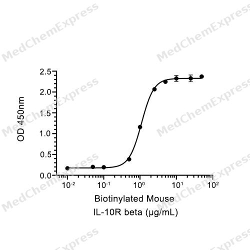
Bioactivity - ELISA


Technical Parameters
-
Species Mouse
-
Source HEK293
-
Tag C-6*His
-
Accession
Q8VHM7 (M22-S222)
-
Molecular Construction
-
N-term
-
IL-10Rβ (M22-S222)
Accession # Q8VHM7 -
His
-
C-term
-
-
Protein Length
Partial
-
Synonyms
IL10RB; CDW210B; Prev. D21S58; IL-10 Receptor Subunit Beta; Prev. D21S66; IL-10R Subunit Beta; Prev. CRFB4; IL-10R Subunit 2; IL-10R2; IL-10RB; CRF2-4; Cytokine Receptor Family II, Member 4; Interleukin-10 Receptor Subunit Beta; Cytokine Receptor Class-II
-
AA Sequence
MIPPPEKVRMNSVNFKNILQWEVPAFPKTNLTFTAQYESYRSFQDHCKRTASTQCDFSHLSKYGDYTVRVRAELADEHSEWVNVTFCPVEDTIIGPPEMQIESLAESLHLRFSAPQIENEPETWTLKNIYDSWAYRVQYWKNGTNEKFQVVSPYDSEVLRNLEPWTTYCIQVQGFLLDQNRTGEWSEPICERTGNDEITPS
-
Molecular Weight
Approximately 35-45 kDa, based on SDS-PAGE under reducing conditions, due to the glycosylation.
-
Purity
≥ 95%, as determined by reducing SDS-PAGE.


Product Properties
Lyophilized powder.
Lyophilized from a 0.22 μm filtered solution of PBS, pH 7.4.
<1 EU/μg, determined by LAL method.
It is not recommended to reconstitute to a concentration less than 100 μg/mL in ddH2O. For long term storage it is recommended to add a carrier protein (0.1% BSA, 5% HSA, 10% FBS or 5% Trehalose).
Stored at -20°C for 2 years from date of receipt. After reconstitution, it is stable at 4°C for 1 week or -20°C for longer (with carrier protein). It is recommended to freeze aliquots at -20°C or -80°C for extended storage.
Room temperature in continental US; may vary elsewhere.


Documentation
-
Data Sheet (264 KB)
-
SDS (254 KB)
- English - EN (254 KB)
- Français - FR (254 KB)
- Deutsch - DE (254 KB)
- Norwegian - NO (254 KB)
- Español - ES (254 KB)
- Swedish - SV (254 KB)
- Italian - IT (254 KB)
- Korean - KR (254 KB)
- Portuguese - PT (254 KB)
-
Handling Instructions (2659 KB)


References
[1]. Sung-Il Yoon, et al. Structure and mechanism of receptor sharing by the IL-10R2 common chain. Structure. 2010 May 12;18(5):638-48. [Content Brief]
[2]. J Shi, et al. IL10 inhibits starvation-induced autophagy in hypertrophic scar fibroblasts via cross talk between the IL10-IL10R-STAT3 and IL10-AKT-mTOR pathways. Cell Death Dis. 2016 Mar 10;7(3):e2133. [Content Brief]
[3]. Paul Sheppard, et al. IL-28, IL-29 and their class II cytokine receptor IL-28R. Nat Immunol. 2003 Jan;4(1):63-8. [Content Brief]
[4]. Robert A Saxton, et al. The tissue protective functions of interleukin-22 can be decoupled from pro-inflammatory actions through structure-based design. Immunity. 2021 Apr 13;54(4):660-672.e9. [Content Brief]
[5]. Barbara Cassani, et al. Gut-tropic T cells that express integrin α4β7 and CCR9 are required for induction of oral immune tolerance in mice. Gastroenterology. 2011 Dec;141(6):2109-18. [Content Brief]

Calculators
Concentration (start) × Volume (start) = Concentration (final) × Volume (final)


 Powered by Bioz

