



RECQL5-IN-1

Based on 1 Customer Validation
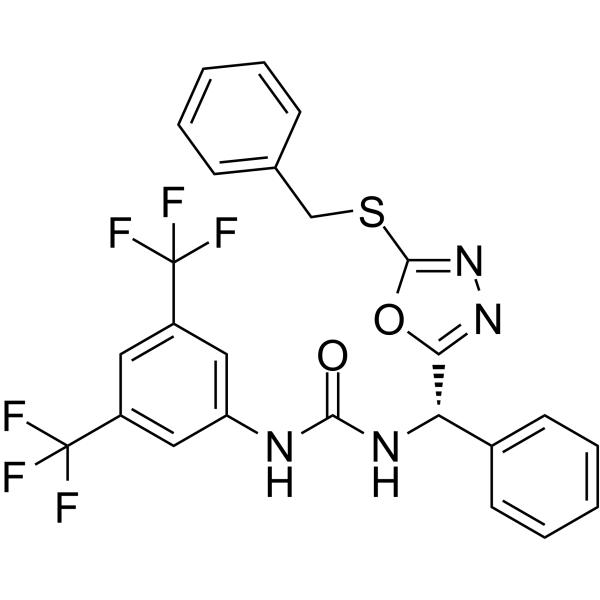
RECQL5-IN-1 (Compound 4a) acts as an orally effective RECQL5 inhibitor (targeting both enzymatic and nonenzymatic domain). RECQL5-IN-1 is a potent inhibitor of RECQL5 helicase activity (IC50=46.3 nM), stabilizes the interaction between RECQL5-RAD51 proteins, causes RAD51 aggregation and homologous recombination repair (HRR) inhibition, thereby exhibiting selective cytotoxicity in RECQL5-expressing cancer cells.
For research use only. We do not sell to patients.
- Purity : 98.40%
- CAS No.: 2718170-45-5
- Formula: C25H18F6N4O2S
- Molecular Weight:552.49
-
Storage:Powder -20°C, 3 years , 4°C, 2 years ; In solvent -80°C, 6 months , -20°C, 1 month

To place orders, for customer services and technical support, please contact: MedChemExpress USA
Tel: 609-228-6898 E-mail: [email protected] [email protected]


-
 Biological Activity
-
Chemical Information
-
Solvent & Solubility
- Protocols
- Purity & Documentation
- References
-
 Help & FAQs



All DNA/RNA Synthesis Isoforms
More

Biological Activity
Description
IC50 & Target
|
Helicase |
Cellular Effect
|
Cell Line

|
Type | Value | Description | References |
|---|---|---|---|---|
| MCF7 | IC50 |
19.6 μM
Compound: 4a
|
Cytotoxicity against RECQL5 knock out human MCF7 cells assessed as inhibition of colony formation measured after 8 to 10 days by clonogenic survival assay
Cytotoxicity against RECQL5 knock out human MCF7 cells assessed as inhibition of colony formation measured after 8 to 10 days by clonogenic survival assay
|
[PMID: 33529023] |
| MCF7 | IC50 |
4.8 μM
Compound: 4a
|
Cytotoxicity against human MCF7 cells expressing wild type RECQL5 assessed as inhibition of colony formation measured after 8 to 10 days by clonogenic survival assay
Cytotoxicity against human MCF7 cells expressing wild type RECQL5 assessed as inhibition of colony formation measured after 8 to 10 days by clonogenic survival assay
|
[PMID: 33529023] |
In Vitro
RECQL5-IN-1 (0-50 μM, 24-72 h) causes the accumulation of double-strand breaks (DSBs), inhibits MCF-7 and MCF10A with IC20s of 8.4 μM and 33.4 μM. RECQL5-IN-1 inhibits the colony formation of RECQL5-wildtype cell[1].
MedChemExpress (MCE) has not independently confirmed the accuracy of these methods. They are for reference only. Further protocols information, click here.
-
Cell Line:MCF-7 and MCF10A
-
Concentration:0-50 μM
-
Incubation Time:72 h
-
Result:Inhibited the cell viability.
In Vivo
MedChemExpress (MCE) has not independently confirmed the accuracy of these methods. They are for reference only.
-
Animal Model:MCF-7 xenograft mouse models[1]
-
Dosage:50 mg/kg
-
Administration:po, every two days for 30 days
-
Result:Inhibited tumor growth.


Chemical Information
-
CAS No. 2718170-45-5
-
Appearance Solid
-
Molecular Weight 552.49
-
Formula C25H18F6N4O2S
-
Color White to off-white
-
SMILES
FC(F)(F)C1=CC(C(F)(F)F)=CC(NC(N[C@@H](C2=CC=CC=C2)C3=NN=C(SCC4=CC=CC=C4)O3)=O)=C1
-
Shipping
Room temperature in continental US; may vary elsewhere.
-
Storage
Powder -20°C 3 years 4°C 2 years In solvent -80°C 6 months -20°C 1 month


Solvent & Solubility
In Vitro:
DMSO : 250 mg/mL (452.50 mM; Need ultrasonic; Hygroscopic DMSO has a significant impact on the solubility of product, please use newly opened DMSO)
Please refer to the solubility information to select the appropriate solvent. Once prepared, please aliquot and store the solution to prevent product inactivation from repeated freeze-thaw cycles.
Storage method and period of stock solution: -80°C, 6 months; -20°C, 1 month. When stored at -80°C, please use it within 6 months. When stored at -20°C, please use it within 1 month.
Please refer to the solubility information to select the appropriate solvent. Once prepared, please aliquot and store the solution to prevent product inactivation from repeated freeze-thaw cycles.
Storage method and period of stock solution: -80°C, 6 months; -20°C, 1 month. When stored at -80°C, please use it within 6 months. When stored at -20°C, please use it within 1 month.
Concentration (start) × Volume (start) = Concentration (final) × Volume (final)

Protocols
-
CRISPR-Cas9 HDR knock-in/precise editing
CRISPR-Cas9 HDR knock-in uses a guide RNA to direct Cas9 to a genomic target adjacent to a PAM, where Cas9 creates a double-strand break; if a donor DNA template with homology to the cut region is present, cellular HDR can copy the donor sequence into the genome, producing a precise substitution, tag, reporter, or insertion rather than an indel. The readout is the fraction of alleles or cells carrying the intended donor-derived edit, measured by junction PCR, restriction-fragment analysis, Sanger sequencing, amplicon deep sequencing, flow cytometry for reporter knock-in, or clone genotyping; NHEJ indels and partial or non-HDR insertions are measured in parallel because they compete with or confound precise HDR outcomes.
-
Cell Cytotoxicity Assay
Cytotoxicity assays are usually based on the assessment of cell membrane damage, which can also be indirectly detected by measuring cell viability. Detection methods include MTT assay, CKK-8 assay, LDH assay and ATP assay, etc.
-
Mammalian live/dead viability and cytotoxicity staining
Live/dead viability and cytotoxicity staining assays are based on the simultaneous detection of intracellular esterase activity in metabolically active (viable) cells and membrane integrity loss in non-viable cells. In commonly used dual-staining approaches, membrane-permeant fluorogenic substrates are converted by intracellular esterases into fluorescent products in live cells, while impermeant DNA-binding dyes selectively enter cells with compromised plasma membranes and label nucleic acids in dead or dying cells, enabling discrimination between viable and non-viable populations by fluorescence microscopy or flow cytometry.
-
Cell Viability Determination by MTT Colorimetric Assay
The following protocol uses the MTT colorimetric assay as a classic literature-established method for assessing cell viability/metabolic activity in cultured mammalian cells. MTT[3-(4,5-dimethylthiazol-2-yl)-2,5-diphenyltetrazolium bromide] is reduced by metabolically active cells to a colored formazan product; the amount of formazan is quantified spectrophotometrically and provides an indirect measure of metabolically active viable cells. Importantly, MTT reduction reflects cellular oxidoreductase/metabolic activity rather than an absolute direct count of living cells, so changes in cellular metabolism can alter the signal independently of cell number.


Purity & Documentation
-
Data Sheet (278 KB)
-
SDS (254 KB)
- English - EN (254 KB)
- Français - FR (254 KB)
- Deutsch - DE (254 KB)
- Norwegian - NO (254 KB)
- Español - ES (254 KB)
- Swedish - SV (254 KB)
- Italian - IT (254 KB)
- Korean - KR (254 KB)
- Portuguese - PT (254 KB)
-
Handling Instructions (2659 KB)
References


Complete Stock Solution Preparation Table
Please refer to the solubility information to select the appropriate solvent. Once prepared, please aliquot and store the solution to prevent product inactivation from repeated freeze-thaw cycles.
Storage method and period of stock solution: -80°C, 6 months; -20°C, 1 month. When stored at -80°C, please use it within 6 months. When stored at -20°C, please use it within 1 month.
| Optional Solvent | Concentration Solvent Mass | 1 mg | 5 mg | 10 mg | 25 mg |
|---|---|---|---|---|---|
| DMSO | 1 mM | 1.8100 mL | 9.0499 mL | 18.0999 mL | 45.2497 mL |
| 5 mM | 0.3620 mL | 1.8100 mL | 3.6200 mL | 9.0499 mL | |
| 10 mM | 0.1810 mL | 0.9050 mL | 1.8100 mL | 4.5250 mL | |
| 15 mM | 0.1207 mL | 0.6033 mL | 1.2067 mL | 3.0166 mL | |
| 20 mM | 0.0905 mL | 0.4525 mL | 0.9050 mL | 2.2625 mL | |
| 25 mM | 0.0724 mL | 0.3620 mL | 0.7240 mL | 1.8100 mL | |
| 30 mM | 0.0603 mL | 0.3017 mL | 0.6033 mL | 1.5083 mL | |
| 40 mM | 0.0452 mL | 0.2262 mL | 0.4525 mL | 1.1312 mL | |
| 50 mM | 0.0362 mL | 0.1810 mL | 0.3620 mL | 0.9050 mL | |
| 60 mM | 0.0302 mL | 0.1508 mL | 0.3017 mL | 0.7542 mL | |
| 80 mM | 0.0226 mL | 0.1131 mL | 0.2262 mL | 0.5656 mL | |
| 100 mM | 0.0181 mL | 0.0905 mL | 0.1810 mL | 0.4525 mL |


RECQL5-IN-1 Related Classifications
HY-145685 Related Classifications
 Powered by Bioz