



Rosuvastatin-d3
Based on 1 publication(s) in Google Scholar
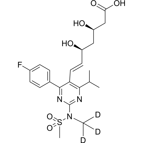
Rosuvastatin-d3 is the deuterated-labeled Rosuvastatin (HY-17504A). Rosuvastatin (ZD 4522) is a competitive HMG-CoA reductase inhibitor with an IC50 of 11 nM. Rosuvastatin potently blocks hERG current with an IC50 of 195 nM, delayed cardiac repolarization, and thereby prolonged action potential durations (APDs) and corrected QT interval (QTc) intervals. Rosuvastatin reduces the expression of the mature hERG and the interaction of heat shock protein 70 (Hsp70) with the hERG protein. Rosuvastatin effectively lowers low-density lipoprotein (LDL) cholesterol, triglycerides, and C-reactive protein levels.
For research use only. We do not sell to patients.
- CAS No.: 1133429-16-9
- Formula: C22H25D3FN3O6S
- Molecular Weight:484.56
-
Storage:
Please store the product under the recommended conditions in the Certificate of Analysis.

To place orders, for customer services and technical support, please contact: MedChemExpress USA
Tel: 609-228-6898 E-mail: [email protected] [email protected]


-
 Biological Activity
-
Chemical Information
- Protocols
- Purity & Documentation
- References
-
 Help & FAQs


Publications Citing Use of MedChemExpress (MCE) Rosuvastatin-d3
More

Biological Activity
Description
IC50 & Target
In Vitro
Stable heavy isotopes of hydrogen, carbon, and other elements have been incorporated into drug molecules, largely as tracers for quantitation during the drug development process. Deuteration has gained attention because of its potential to affect the pharmacokinetic and metabolic profiles of drugs.
MedChemExpress (MCE) has not independently confirmed the accuracy of these methods. They are for reference only.
Application
1. This compound can be used as a tracer
2. This compound can be used as an internal standard for quantitative analysis by NMR, GC-MS, or LC-MS.


Chemical Information
-
CAS No. 1133429-16-9
-
Unlabeled CAS 287714-41-4
-
Molecular Weight 484.56
-
Formula C22H25D3FN3O6S
-
SMILES
FC(C=C1)=CC=C1C2=NC(N(C([2H])([2H])[2H])S(=O)(C)=O)=NC(C(C)C)=C2/C=C/[C@@H](O)C[C@@H](O)CC(O)=O
-
Synonyms
ZD 4522 d3
-
Shipping
Room temperature in continental US; may vary elsewhere.
-
Storage
Please store the product under the recommended conditions in the Certificate of Analysis.


Publications (1)
-
Journal Impact Factor
-
Most Recent


Purity & Documentation
References
[1]. Watanabe, M., et al., Synthesis and biological activity of methanesulfonamide pyrimidine- and N-methanesulfonyl pyrrole-substituted 3,5-dihydroxy-6-heptenoates, a novel series of HMG-CoA reductase inhibitors. Bioorg Med Chem, 1997. 5(2): p. 437-44. [Content Brief]
[2]. Plante I, et al. Rosuvastatin blocks hERG current and prolongs cardiac repolarization. J Pharm Sci. 2012 Feb;101(2):868-78. [Content Brief]
[3]. Feng PF, et al. Intracellular Mechanism of Rosuvastatin-Induced Decrease in Mature hERG Protein Expression on Membrane. Mol Pharm. 2019 Apr 1;16(4):1477-1488. [Content Brief]
[4]. Carswell C.I., et al. Rosuvastatin. Drugs, 2002. 62(14): p. 2075-85; discussion 2086-7. [Content Brief]

Calculators
Concentration (start) × Volume (start) = Concentration (final) × Volume (final)


Rosuvastatin-d3 Related Classifications
HY-17504AS Related Classifications
 Powered by Bioz