42 Results for "

live cell staining

" in MedChemExpress (MCE) Product Catalog:
Products (42)

42 Results for "live cell staining" in MCE Product Catalog:

Cat. No.: HY-DY1061
CAS No.: 158757-84-7
BODIPY 558/568 C12 (solution) is a BODIPY dye. BODIPY dye is a small molecule dye with strong ultraviolet absorption ability, its fluorescence peak is relatively sharp, and the quantum yield is high. They are relatively insensitive to the polarity and pH of the environment and are relatively stable under different physiological conditions. Due to its structural asymmetry, BODIPY derives a variety of structural products. BODIPY lipid droplet dyes can well pass through the cell membrane into the cell, and localize the neutral lipids in the cell to specifically stain the lipid droplets, which can be used for labeling of live cells and fixed cells . Maximum excitation/emission wavelength: 558/568 nm .
Solvent and concentration: DMSO: 2 mM
loading...
    loading...
Cat. No.: HY-D1075
CAS No.: 36536-22-8
Synonyms: HIDC
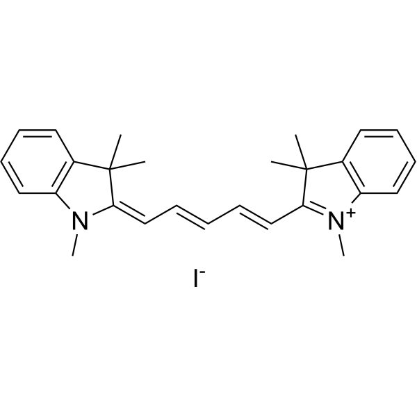
1,1',3,3,3',3'-Hexamethylindodicarbocyanine iodide is a carbocyanine dye that stains mitochondria of live cells.
loading...
    loading...
Cat. No.: HY-107864
CAS No.: 15905-32-5
Purity:  99.07%
Synonyms: Erythrosin extra bluish free acid; Tetraiodofluorescein
Research Areas:  

Infection

Erythrosine B free acid​ (Erythrosin extra bluish free acid; Tetraiodofluorescein) is a visible red dye and insecticide with colorimetric and fluorescent properties. Erythrosine B free acid serves as a membrane-impermeant live-cell dye for Gram-positive and Gram-negative bacteria, primarily staining dead cells with damaged plasma membranes. Erythrosine B free acid exhibits photosensitive larvicidal toxicity against Culex pipiens quinquefasciatus larvae. Erythrosine B free acid is used in live/dead bacterial assays in colorimetric and fluorescent measurements for low-, medium-, and high-throughput experiments, as well as in research related to mosquito-borne diseases and food coloring .
loading...
    loading...
Cat. No.: HY-D1385
CAS No.: 2410614-07-0
Target:  

Fluorescent Dye

Research Areas:  

Others

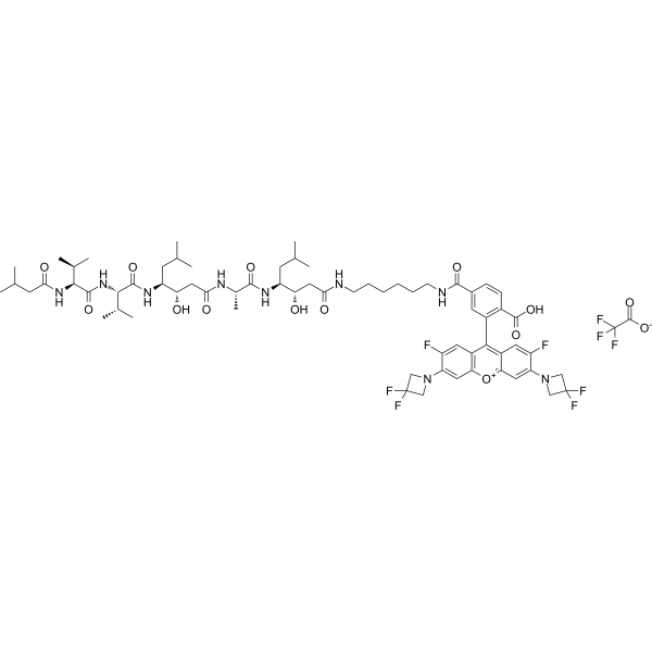
JF526–Pepstatin A TFA is a fluorescent dye that can be used for lysosomal staining in live cells. The excitation maximum is 530 nm and the emission maximum is 549 nm .
loading...
    loading...
Cat. No.: HY-D0259R
CAS No.: 16423-68-0
Synonyms: Erythrosin extra bluish (Standard); Tetraiodofluorescein disodium (Standard)
Research Areas:  

Infection

Erythrosine B (Standard) (Erythrosin extra bluish (Standard); Tetraiodofluorescein disodium (Standard)) is the analytical standard of Erythrosine B (HY-D0259). This product is intended for research and analytical applications. Erythrosine B (Erythrosin extra bluish; Tetraiodofluorescein) is a visible red dye and insecticide with colorimetric and fluorescent properties. Erythrosine B serves as a membrane-impermeant live-cell dye for Gram-positive and Gram-negative bacteria, primarily staining dead cells with damaged plasma membranes. Erythrosine B exhibits photosensitive larvicidal toxicity against Culex pipiens quinquefasciatus larvae. Erythrosine B is used in live/dead bacterial assays in colorimetric and fluorescent measurements for low-, medium-, and high-throughput experiments, as well as in research related to mosquito-borne diseases and food coloring .
loading...
    loading...
Cat. No.: HY-DY2020
Target:  

Fluorescent Dye

Research Areas:  

Others

Neutral Red Staining Solution (for Live Cells) is a vital stain used for vital staining of peripheral blood cells, and it can also be used for staining other living cells or tissues. Neutral Red Staining Solution (for Live Cells) accumulates in cytoplasmic vacuoles, lysosomes of living cells, as well as plant/fungal vacuoles. It binds to anionic sites, suberin, lignin, cell walls, DNA and phenolic substances, and can intercalate into DNA. Neutral Red Staining Solution (for Live Cells) stains the cytoplasm of specific phytoplankton and estuarine zooplankton, while dead cells are not stained, enabling viability determination of such organisms .
loading...
    loading...
Cat. No.: HY-DY2022
Target:  

Fluorescent Dye

Research Areas:  

Others

Eosin-Nigrosin Sperm Viability Staining Solution is a differential viability staining solution used to distinguish live/dead sperm and other cells, consisting of Eosin Y (HY-D0505) and Nigrosin (HY-W250145). Eosin-Nigrosin Sperm Viability Staining Solution cannot penetrate the membrane of live cells; it stains dead cells dark pink and uses nigrosin to form a contrasting dark background. Eosin-Nigrosin Sperm Viability Staining Solution can distinguish live sperm, dead sperm and sperm with neck leakage, and can stain dead sperm or sperm affected by excessive microbial growth .
loading...
    loading...
Cat. No.: HY-K1076

Annexin V-mCherry Apoptosis Detection Kit provides a rapid and convenient method to detect cell apoptosis and necrosis. After staining, live cells show little or no fluorescence, apoptosis cells and necrosis cells show red fluorescence.

loading...
    loading...
Cat. No.: HY-D3172
CAS No.: 1180112-32-6
Synonyms: Glucagon Yellow
BD-105 (Glucagon Yellow) is a glucagon-binding fluorescent probe with a KD value of 13.3 μM. BD-105 exhibits changes in fluorescence intensity upon interaction with glucagon, and colocalizes with glucagon in cells and tissues. BD-105 is a selective cell stain that labels glucagon-secreting cells without staining insulin-secreting cells or non-endocrine control cells. BD-105 serves as an imaging reagent for glucagon in live cells and tissues (Ex/Em = 540 nm/580 nm) .
loading...
    loading...
Cat. No.: HY-D3410
CAS No.: 2991899-29-5
CDr17 is a GLUT1 substrate and selective fluorescent dye staining M1 microphages. CDr17 utilizes the Gating-Oriented Live-cell Distinction (GOLD) mechanism to enter M1 macrophages (Ex/Em = 646/662 nm) .
loading...
    loading...
Cat. No.: HY-D3459
SYTO-9 is a cell-permeable fluorescent dye that can be used for nucleic acid staining. The fluorescence intensity of SYTO-9 increases significantly upon binding to RNA and DNA . SYTO-9 can stain RNA and DNA in live and dead eukaryotic cells, as well as in Gram-positive and Gram-negative bacteria. SYTO-9 is applicable for fluorescence microscopy, flow cytometry, fluorophotometry, and bacterial counting in aquatic systems (RNA: Ex/Em = 486/501 nm; DNA: Ex/Em = 485/498 nm) .
loading...
    loading...
Cat. No.: HY-107864A
CAS No.: 12227-78-0
Synonyms: Erythrosin extra bluish aluminum; Tetraiodofluorescein aluminum
Research Areas:  

Infection

Erythrosine B aluminum​ (Erythrosin extra bluish aluminum; Tetraiodofluorescein aluminum) is a visible red dye and insecticide with colorimetric and fluorescent properties. Erythrosine B aluminum serves as a membrane-impermeant live-cell dye for Gram-positive and Gram-negative bacteria, primarily staining dead cells with damaged plasma membranes. Erythrosine B aluminum exhibits photosensitive larvicidal toxicity against Culex pipiens quinquefasciatus larvae. Erythrosine B aluminum is used in live/dead bacterial assays in colorimetric and fluorescent measurements for low-, medium-, and high-throughput experiments, as well as in research related to mosquito-borne diseases and food coloring .
loading...
    loading...
Cat. No.: HY-172754
ICAAc is a solvatochromic fluorophore with reduced basicity. ICAAc demonstrates significant solvatochromic behavior across solvents of varying polarity, with a large dipole moment difference and low quantum yield in water, making it a tunable solvatochromic fluorophore. ICAAc enables pH sensing via UV-vis/fluorescence detection and in microenvironments including sodium lauryl sulfate micelle Stern layers. ICAAc acts as a supravital cell stain for epifluorescence imaging of live cancer cells .
loading...
    loading...
Cat. No.: HY-D3525
Target:  

Fluorescent Dye

Research Areas:  

Others

BDL-E5 is a fluorescent probe for detecting live induced pluripotent stem cells at the early reprogramming stage. Its detection mechanism involves specific binding to authentic reprogramming cells, which exhibit increased expression of pluripotency and epithelial genes and decreased expression of mesenchymal genes compared to non-reprogramming cells; it appears to co-localize more significantly with the Golgi complex than other organelle markers in reprogramming cells, and it does not require washing after staining to reduce background signals; fluorescence is generated upon binding to these early reprogramming cells, with positive cells appearing 7 days before iPS colonies are visible and stain positive for the conventional pluripotent marker TRA-1-60, and sorting BDL-E5-positive cells enriches populations that generate higher numbers and quality of iPS colonies. Its detection wavelength is Ex/Em = 578/599 nm .
loading...
    loading...
Cat. No.: HY-107864R
CAS No.: 15905-32-5
Synonyms: Erythrosin extra bluish free acid (Standard); Tetraiodofluorescein (Standard)
Research Areas:  

Infection

Erythrosine B free acid (Standard) (Erythrosin extra bluish free acid (Standard); Tetraiodofluorescein (Standard)) is the analytical standard of Erythrosine B free acid (HY-107864). This product is intended for research and analytical applications. Erythrosine B free acid​ (Erythrosin extra bluish free acid; Tetraiodofluorescein) is a visible red dye and insecticide with colorimetric and fluorescent properties. Erythrosine B free acid serves as a membrane-impermeant live-cell dye for Gram-positive and Gram-negative bacteria, primarily staining dead cells with damaged plasma membranes. Erythrosine B free acid exhibits photosensitive larvicidal toxicity against Culex pipiens quinquefasciatus larvae. Erythrosine B free acid is used in live/dead bacterial assays in colorimetric and fluorescent measurements for low-, medium-, and high-throughput experiments, as well as in research related to mosquito-borne diseases and food coloring .
loading...
    loading...
Cat. No.: HY-D3430
PTPA-QM is a low-cytotoxicity Aβ protein probe that can be used for live-cell imaging and tissue section staining (Lys. Ex/Em = 385/615 nm). PTPA-QM can intercalate into the β-sheet layered structure of β-amyloid fibrils, form intermolecular interactions with amino acid residues, restrict intramolecular rotation and trigger fluorescence activation for imaging purposes. PTPA-QM is applicable to Alzheimer's disease-related research. Maximum excitation/emission wavelength: 448/605 nm .
loading...
    loading...
Cat. No.: HY-D3163
Target:  

Fluorescent Dye

Research Areas:  

Others

Endo-pH is a cell-permeable, pH-responsive fluorescent probe based on BODIPY, with a pKa of 5.08. When pH decreases from 7.32 to 4.97, Endo-pH undergoes protonation under acidic conditions, resulting in a 79-fold increase in fluorescence intensity at 786 nm. The detection wavelengths of its protonated form are Ex/Em=688/786 nm, while the absorption wavelength of its non-protonated form is 745 nm. Endo-pH can be used for staining late endosomes in live cells and determining the fusion time of late endosomes and lysosomes .
loading...
    loading...
Cat. No.: HY-KD1025

The kit uses the Halo-tag principle to specifically label living cell structures.

loading...
    loading...
Cat. No.: HY-DY1090
CAS No.: 216434-81-0
BODIPY 493/503 methyl bromide (solution) is a BODIPY dye. BODIPY dye is a small molecule dye with strong ultraviolet absorption ability, its fluorescence peak is relatively sharp, and the quantum yield is high. They are relatively insensitive to the polarity and pH of the environment and are relatively stable under different physiological conditions. Due to its structural asymmetry, BODIPY derives a variety of structural products. BODIPY lipid droplet dyes can well pass through the cell membrane into the cell, and localize the neutral lipids in the cell to specifically stain the lipid droplets, which can be used for labeling of live cells and fixed cells . Maximum excitation/emission wavelength: 493/503 nm .
Solvent and concentration: DMSO: 5 mM
loading...
    loading...
Cat. No.: HY-D3197
CDg16 is a selective fluorescent dye targeting SLC18B1 (λabs/λem=458/544 nm) that is actively transported into lysosomal vesicles of activated macrophages independent of the endocytic pathway. CDg16 enables highly specific vesicle localization in live cells. CDg16 exhibits no cytotoxicity and accurately distinguishes activated M1 and M2 subsets from different origins. CDg16 shows low background staining in non-activated cells and normal organs, making it suitable for time-lapse imaging. In preclinical animal models of inflammatory sites, atherosclerotic plaques and liver inflammation, CDg16 allows visualization of activated macrophages. CDg16 can be used to study inflammation-related diseases and atherosclerosis .
loading...
    loading...

