13 Results for "

anionic dye

" in MedChemExpress (MCE) Product Catalog:
Products (13)

13 Results for "anionic dye" in MCE Product Catalog:

1
1 Cited Publications
Art. -Nr.: HY-D0232
CAS. Nr.: 6104-59-2
Synonyms: Brilliant Blue R
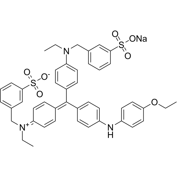
Brilliant blue R250 (Brilliant Blue R), an anionic dye, is the most popular stain to detect proteins resolved in SDS-PAGE gels .
loading...
    loading...
Art. -Nr.: HY-D0031
CAS. Nr.: 196504-57-1
Synonyms: APTS
Target:  

Fluorescent Dye

Forschungsgebiete:  

Others

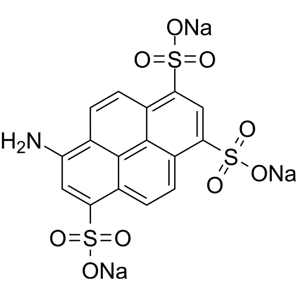
8-Aminopyrene-1,3,6-trisulfonic acid trisodium (APTS) is an anionic fluorescent dye (excitation/emission: 425/503 nm in pH 7.4 PBS). The fluorescence intensity of 8-Aminopyrene-1,3,6-trisulfonic acid trisodium remains stable at pH 4-10. 8-Aminopyrene-1,3,6-trisulfonic acid trisodium retains its fluorescence properties when immobilized in a hydrogel by copolymerization. 8-Aminopyrene-1,3,6-trisulfonic acid trisodium can be used for glucose sensing .
loading...
    loading...
Art. -Nr.: HY-W110929
CAS. Nr.: 129-17-9
Synonyms: Xylene blue; C.I. Acid blue 1
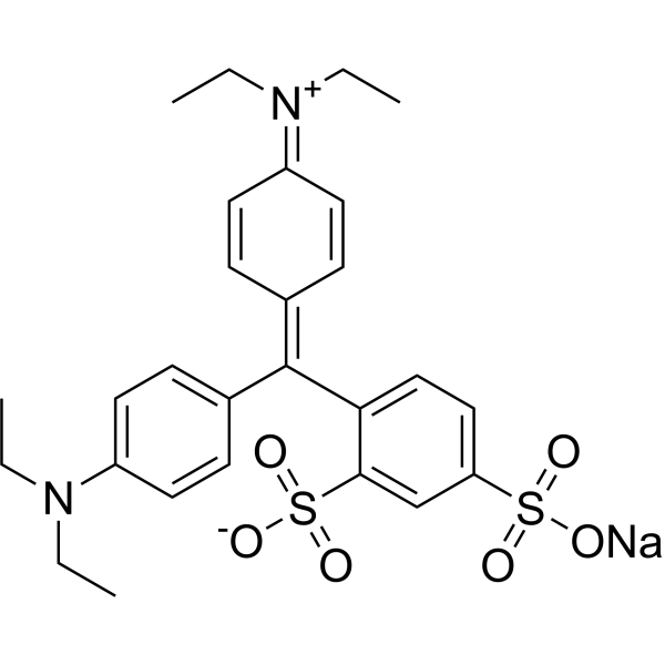
Acid blue 1 (Xylene blue) is an anionic triarylmethane dye and substrate for photocatalytic degradation .
loading...
    loading...
Art. -Nr.: HY-W110910
CAS. Nr.: 1787-61-7
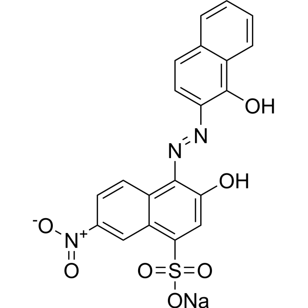
Eriochrome black T, Indicator is a complexing agent for metal ions (e.g., Ca 2+, Mg 2+) and is used as an indicator in complexometric titrations. Eriochrome black T, Indicator forms colored complexes with metal ions through covalent coordination bonds, and indicates the endpoint of the titration by color change. Eriochrome black T, Indicator can be used as an anionic azo dye in photocatalytic degradation studies to evaluate the performance of photocatalysts. The reaction solution of Eriochrome black T, Indicator combined with Mg 2+ is initially purple. During loop-mediated isothermal amplification (LAMP), the color changes from purple to sky blue due to the consumption of Mg 2+ by the formation of magnesium pyrophosphate, indicating a positive reaction. The optimal concentration of Eriochrome black T, Indicator in LAMP is 60 μM, and the detection limit for Mycobacterium tuberculosis is 1 pg DNA/reaction .
loading...
    loading...
Art. -Nr.: HY-D1091
CAS. Nr.: 47623-98-3
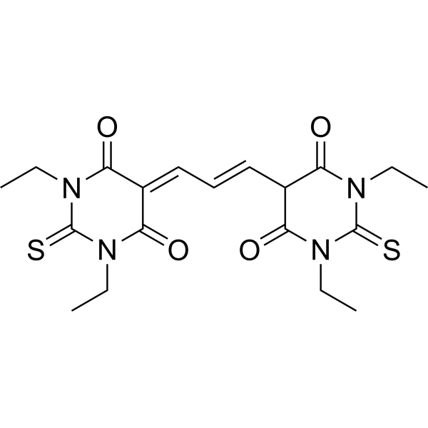
DiSBAC2(3) is an anionic membrane-potential-sensitive dye. DiSBAC2(3) is a plasma membrane potential indicator .
loading...
    loading...
Art. -Nr.: HY-D0929
CAS. Nr.: 523-44-4
Synonyms: α-Naphthol Orange
Target:  

Fluorescent Dye

Forschungsgebiete:  

Others

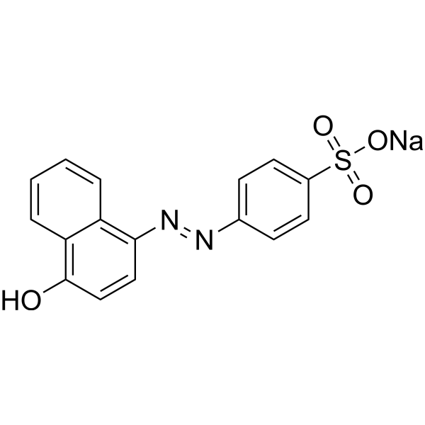
Orange I (α-Naphthol Orange) is an anionic azo dye that can form a complex with Methylene blue (HY-14536) and cause fluorescence quenching. The equilibrium constant of the complex of Orange I and Methylene blue is 79900 mol -1·dm 3. Orange I can change the absorption spectrum of itself and Methylene blue .
loading...
    loading...
Art. -Nr.: HY-Y0425
CAS. Nr.: 623-27-8
Terephthalaldehyde is a crosslinking agent. Terephthalaldehyde forms a crosslinked structure inside the gelatin matrix by forming Schiff base imines with the amino groups of gelatin, thereby constructing a three-dimensional network. Terephthalaldehyde improves the hydrophobicity of the gelatin matrix, delays water vapor penetration and enhances the liquid water resistance of gelatin films. Terephthalaldehyde can be used as a crosslinking agent to prepare crosslinked chitosan hydrogel (CAAT) via ultrasound-induced synthesis. Terephthalaldehyde helps CAAT hydrogels selectively adsorb anionic dyes from aqueous media, including multi-component systems containing cationic dyes. Terephthalaldehyde serves as a starting material for the synthesis of bis-heterocyclic compounds (including bis-thiazole and bis-triazolopyrimidine compounds) .
loading...
    loading...
Art. -Nr.: HY-W344938
CAS. Nr.: 8004-92-0
Synonyms: Quinoline yellow
C.I. Acid yellow 3 is an anionic quinolinone dye that is commonly used as a food additive, but also in the cosmetic and pharmaceutical industries .
loading...
    loading...
Art. -Nr.: HY-D0307AR
CAS. Nr.: 915-67-3
Synonyms: Acid Red 27 (Standard); Azorubin S (Standard); FD & C Red dye No. 2 (Standard)
Amaranth (Standard) is the analytical standard of Amaranth. This product is intended for research and analytical applications. Amaranth is a dark red to purple azo dye used as a food dye and cosmetics. Amaranth is an anionic dye that can be applied to natural and synthetic fibers, leather, paper, and phenol-formaldehyde resins.
loading...
    loading...
Art. -Nr.: HY-D0009
CAS. Nr.: 62625-32-5
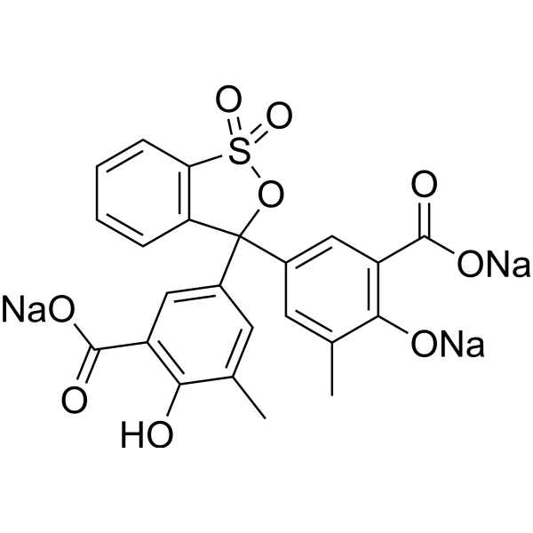
Bromocresol green sodium is an anionic dye. Bromocresol green sodium can be used for pH indication and DNA agarose gel electrophoresis. Bromocresol green sodium is also used in mammalian albumin measurement. Bromocresol green sodium deprotonates and produces the monoanionic form of yellow colour at lower pH (acidic condition), and produces dianionic blue colour at the basic condition .
loading...
    loading...
Art. -Nr.: HY-B1533A
CAS. Nr.: 3861-73-2
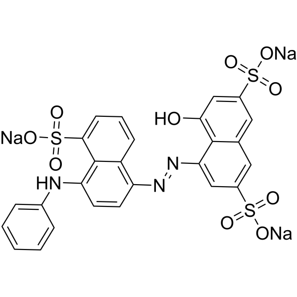
Anazolene trisodium is an anionic dye, a textile azo dye with a maximum absorption wavelength of λmax = 571 nm .
loading...
    loading...
Art. -Nr.: HY-W344938R
CAS. Nr.: 8004-92-0
Synonyms: Quinoline yellow (Standard)
C.I. Acid yellow 3 (Standard) is the analytical standard of C.I. Acid yellow 3. This product is intended for research and analytical applications. C.I. Acid yellow 3 is an anionic quinolinone dye that is commonly used as a food additive, but also in the cosmetic and pharmaceutical industries .
loading...
    loading...
Art. -Nr.: HY-K0612

MCE Weigert Elastin Staining Kit combines Weigert Resorcinol Fuchsin Staining Solution with Van Gieson (VG) Staining Solution. Weigert Resorcinol Fuchsin Solution is mainly used for staining elastic fibers, Van Gieson (VG) Solution is used for collagen fiber staining. The principle of VG staining is based on differences in the size of anionic dye molecules and the permeability of tissues. Picric acid (PA) has the smallest molecular weight and preferentially enters dense structures. Ponceau Red or acid fuchsin has a relatively larger molecular weight and primarily binds to collagen fibers, while light green has the largest molecular weight and stains other tissue components. After VG staining, collagen fibers appear red, while muscle fibers and cytoplasm appear yellow, enabling a clear distinction between tissue components.

loading...
    loading...
  • 1

