



Tat-beclin 1 TFA
Based on 7 publication(s) in Google Scholar
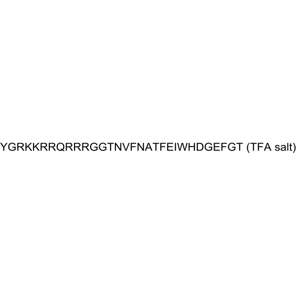
Tat-beclin 1 TFA, a peptide derived from a region of the autophagy protein (beclin 1), is a potent inducer of autophagy and interacts with negative regulator of autophagy, GAPR-1 (GLIPR2). Tat-beclin 1 TFA decreases the accumulation of polyglutamine expansion protein aggregates and the replication of several pathogens (including HIV-1) in vitro, and reduces mortality in mice infected with chikungunya (CHIKV) or West Nile virus (WNV).
For research use only. We do not sell to patients.
- Formula: C166H252F3N57O47
- Molecular Weight:3855.12
-
Storage:
Please store the product under the recommended conditions in the Certificate of Analysis.

To place orders, for customer services and technical support, please contact: MedChemExpress USA
Tel: 609-228-6898 E-mail: [email protected] [email protected]


-
 Biological Activity
-
Chemical Information
-
Solvent & Solubility
- Protocols
- Purity & Documentation
- References
-
 Help & FAQs


Publications Citing Use of MedChemExpress (MCE) Tat-beclin 1 TFA
More- Autophagy. 2025 Oct;21(10):2168-2191. [Abstract]
- Clin Transl Med. 2024 Nov;14(11):e70082. [Abstract]
- Aging Cell. 2026 Aug;25(8):e70644.
- Int Immunopharmacol. 2025 Dec 3:166:115600. [Abstract]
- Sci Rep. 2025 Oct 22;15(1):36952. [Abstract]
- Bone. 2025 Mar:192:117348. [Abstract]
- Biomed Pharmacother. 2022 Oct:154:113606. [Abstract]


Biological Activity
Description
IC50 & Target
[1]|
HIV-1 |
In Vitro
Tat-beclin 1 TFA (10, 30, 50 μM; 24 hours) induces autophagy and results in a dose-dependent decrease in amounts of p62, a selective autophagy substrate, and a dose-dependent conversion of the non-lipidated form of LC3, LC3-I, to the lipidated, autophagosome-associated form of LC3, LC3-II, in multiple cell lines and primary murine embryonic fibroblasts (MEFs)[1].
Tat-beclin 1 TFA (10 μM; 2-4 hours post-infection) decreases the intracellular survival of L. monocytogenes in primary murine bone-marrow-derived macrophages (BMDMs)[1].
MedChemExpress (MCE) has not independently confirmed the accuracy of these methods. They are for reference only. Further protocols information, click here.
In Vivo
MedChemExpress (MCE) has not independently confirmed the accuracy of these methods. They are for reference only.


Chemical Information
-
Molecular Weight 3855.12
-
Formula C166H252F3N57O47
-
Shipping
Room temperature in continental US; may vary elsewhere.
-
Storage
Please store the product under the recommended conditions in the Certificate of Analysis.


Publications (7)
-
Journal Impact Factor
-
Most Recent
-
Autophagy
METTL3-dependent m6A modification of SNAP29 induces "autophagy-mitochondrial crisis" in the ischemic microenvironment after soft tissue transplantation. [Abstract]2025 Oct;21(10):2168-2191. PMID: 40340690 -
Clin Transl Med
Targeting endoplasmic reticulum stress-induced lymphatic dysfunction for mitigating bisphosphonate-related osteonecrosis. [Abstract]2024 Nov;14(11):e70082. PMID: 39521624 -
-
Int Immunopharmacol
2025 Dec 3:166:115600. PMID: 41045801 -
Sci Rep
Lysosomal homeostasis regulates myocardial ischemia-reperfusion injury through autophagy pathway. [Abstract]2025 Oct 22;15(1):36952. PMID: 41125732 -
Bone
2025 Mar:192:117348. PMID: 39617111 -
Biomed Pharmacother
Novel cardioprotective mechanism for Empagliflozin in nondiabetic myocardial infarction with acute hyperglycemia. [Abstract]2022 Oct:154:113606. PMID: 36030589


Solvent & Solubility
In Vitro
H2O
Peptide Solubility and Storage Guidelines:
1. Calculate the length of the peptide.
2. Calculate the overall charge of the entire peptide according to the following table:
| Contents | Assign value | |
|---|---|---|
| Acidic amino acid | Asp (D), Glu (E), and the C-terminal -COOH. | -1 |
| Basic amino acid | Arg (R), Lys (K), His (H), and the N-terminal -NH2 | +1 |
| Neutral amino acid | Gly (G), Ala (A), Leu (L), Ile (I), Val (V), Cys (C), Met (M), Thr (T), Ser (S), Phe (F), Tyr (Y), Trp (W), Pro (P), Asn (N), Gln (Q) | 0 |
3. Recommended solution:
| Overall charge of peptide | Details |
|---|---|
| Negative (<0) |
1. Try to dissolve the peptide in water first. 2. If water fails, add NH4OH (<50 μL). 3. If the peptide still does not dissolve, add DMSO (50-100 μL) to solubilize the peptide. |
| Positive (>0) |
1. Try to dissolve the peptide in water first. 2. If water fails, try dissolving the peptide in a 10%-30% acetic acid solution. 3. If the peptide still does not dissolve, try dissolving the peptide in a small amount of DMSO. |
| Zero (=0) |
1. Try to dissolve the peptide in organic solvent (acetonitrile, methanol, etc.) first. 2. For very hydrophobic peptides, try dissolving the peptide in a small amount of DMSO, and then dilute the solution with water to the desired concentration. |

Protocols
-
Autophagy
Autophagy is a process in which eukaryotic cells use lysosomes to degrade their own cytoplasmic proteins and damaged organelles under the regulation of autophagy related gene (Atg). Microtubule-associated proteins light chain 3 (LC3) is recognized as autophagy marker, which transfers from cytoplasmic LC3 (LC3-I) to membrane type (LC3-II). LC3-II/I ratio could be detected by Western Blot and fluorescence microscopy.
-
Lysosome and acidic-vesicle live-cell staining
Lysosome and acidic-vesicle live-cell staining detects acidic intracellular compartments by using membrane-permeant acidotropic probes that accumulate in low-pH vesicles, including lysosomes, late endosomes, autolysosomes, and acidic phagosomes. LysoTracker staining is commonly used as an intensity-based readout of acidic lysosomal compartment abundance or enlargement, while acridine orange produces green fluorescence in less concentrated compartments and red fluorescence after concentration-dependent accumulation in acidic vesicular organelles. Loss or reduction of acridine-orange red signal can be used as a readout of lysosomal membrane permeabilization or reduced acidic-vesicle integrity. This protocol is designed for live cultured cells and can be adapted for fluorescence microscopy, high-content imaging, plate-reader readout, or flow cytometry when the selected literature supports the readout. Because these dyes report acidotropic accumulation rather than lysosome identity alone,
-
Macroautophagy Solutions
Macroautophagy is a conserved lysosome-dependent degradation pathway in which cytoplasmic material is sequestered into double-membrane autophagosomes and delivered to lysosomes for degradation and recycling. The pathway supports cellular homeostasis during nutrient limitation, organelle stress, protein-aggregate accumulation, infection, differentiation, and tissue remodeling by coupling cargo sequestration, autophagosome maturation, lysosomal fusion, and degradation of cargo-derived macromolecules. The core molecular sequence includes initiation by nutrient- and stress-regulated autophagy machinery, autophagosome nucleation, LC3/ATG8-family conjugation to autophagosomal membranes, cargo selection through receptors such as SQSTM1/p62, autophagosome-lysosome fusion, and lysosomal degradation. LC3 was identified as a mammalian homolog of yeast Atg8 that localizes to autophagosomal membranes after processing, and p62/SQSTM1 was shown to connect ubiquitinated cargo with autophagic degradati


Purity & Documentation
References

Calculators
Concentration (start) × Volume (start) = Concentration (final) × Volume (final)


Tat-beclin 1 TFA Related Classifications
HY-P2260A Related Classifications
 Powered by Bioz