



Tubulozole
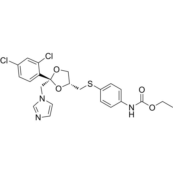
Tubulozole (R 46846) is an orally active inhibitor of tubulin polymerization with an ID50 of 0.34 μM. Tubulozole induces activation of the Chk1 kinase and phosphorylation of ERK1/2, which is required for microtubule formation. Tubulozole arrests Mitosis at the metaphase stage. Tubulozole causes cells to accumulate in the radiosensitive mitotic phase of the cell cycle. Tubulozole exerts anticancer activity against colon cancer. Tubulozole produces a radiosensitizing effect in mouse tumor models and enhances the tumor growth delay effect when combined with γ-ray irradiation. Tubulozole is used in studies related to colon cancer, fascioliasis, MO4 fibrosarcoma, and Lewis lung carcinoma.
For research use only. We do not sell to patients.
- CAS No.: 84697-22-3
- Formula: C23H23Cl2N3O4S
- Molecular Weight:508.42
-
Storage:
Please store the product under the recommended conditions in the Certificate of Analysis.

To place orders, for customer services and technical support, please contact: MedChemExpress USA
Tel: 609-228-6898 E-mail: [email protected] [email protected]


-
 Biological Activity
-
Chemical Information
- Protocols
- Purity & Documentation
- References
-
 Help & FAQs




Biological Activity
Description
IC50 & Target
[1]|
Chk1 |
ERK1 |
ERK2 |
In Vitro
Tubulozole (10 μM; 6 h) selectively induces robust Chk1 kinase activation in colon adenocarcinoma COLO 205 cells, while producing minimal Chk1 activation in normal human colon epithelial CRL cells[1].
Tubulozole (10 μM; 3-6 h) induces rapid, early phosphorylation of Chk1 (Ser-345) and Bad (Ser-155) in colon adenocarcinoma COLO 205 cells, followed by enhanced 14-3-3β binding to both phosphorylated Cdc25C and phosphorylated Bad[1].
Tubulozole (10 μM) drives the translocation of phosphorylated Cdc25C (Ser-216) from the nucleus to the cytoplasm in colon adenocarcinoma COLO 205 cells[1].
Tubulozole inhibits purified tubulin polymerization in vitro with an ID50 of 0.34 μM[4].
MedChemExpress (MCE) has not independently confirmed the accuracy of these methods. They are for reference only. Further protocols information, click here.
-
Cell Line:Human colon adenocarcinoma COLO 205 cells
-
Concentration:10 μM
-
Incubation Time:3 h, 6 h
-
Result:Induces phosphorylation of Chk1 at Ser-345 as early as 3 h post-exposure, with no change in total Chk1 protein expression.
Induces phosphorylation of Bad at Ser-155 as early as 3 h post-exposure.
Significantly increases the binding of phosphorylated Cdc25C (Ser-216) and phosphorylated Bad (Ser-155) to 14-3-3β protein at 6 h of treatment.
In Vivo
MedChemExpress (MCE) has not independently confirmed the accuracy of these methods. They are for reference only.
-
Animal Model:CDF1 (DBA/2× C3H) hybrid mice (male, 18-22 g body weight)[4]
-
Dosage:160 mg/kg
-
Administration:oral gavage; single dose
-
Result:Produce initial subcutaneous MO4 fibrosarcoma tumor regression equivalent to the initial tumor regression achieved with 80 mg/kg erbulozole administered at its optimal 2-hour pretreatment interval before 10 Gy gamma irradiation.
Confirm the 6-hour pretreatment window as the established optimal time point for Tubulozole (hydrochloride) to exert maximum tumor radiation enhancement effects in this model, a time interval that is non-optimal for erbulozole which shows negative excess growth delay at 4 and 6 hour pretreatment intervals.


Chemical Information
-
CAS No. 84697-22-3
-
Molecular Weight 508.42
-
Formula C23H23Cl2N3O4S
-
SMILES
O=C(NC1=CC=C(C=C1)SC[C@@H]2O[C@@](C3=CC=C(C=C3Cl)Cl)(OC2)CN4C=CN=C4)OCC
-
Synonyms
R 46846
-
Shipping
Room temperature in continental US; may vary elsewhere.
-
Storage
Please store the product under the recommended conditions in the Certificate of Analysis.

Protocols
-
Kinase activity and phosphorylation assays
Kinase activity assays measure the ability of kinases to transfer phosphate groups from ATP to specific substrates, while phosphorylation assays detect the presence and levels of phosphorylated proteins. Common methods include radiolabeled ATP incorporation (e. g. ,), ADP release detection via bioluminescence (e. g. ,[3]), enzyme-linked immunosorbent assays (ELISA) for phospho-specific epitopes (e. g. ,[6]), and microtiter-based formats for high-throughput screening (e. g. ,[8]). The ADP-Glo assay quantifies kinase activity by measuring ADP produced during phosphorylation using a luciferase-based system. Radiometric assays involve autoradiography or scintillation counting after incorporation of 32P-labeled ATP into substrate proteins. ELISA-based approaches rely on phospho-specific antibodies to detect activated kinases in cell lysates or purified samples.
-
Western Blot
Western blotting (WB) is a commonly used experimental method in molecular biology, biochemistry, and immunogenetics for identifying and quantifying target proteins. It combines gel electrophoresis with immunoassay, enabling researchers to analyze protein expression, post-translational modifications, and molecular weight.
-
Flow cytometric DNA-content cell-cycle staining
Flow cytometric DNA-content cell-cycle staining measures the fluorescence intensity of DNA-bound fluorochromes in single cells or nuclei to estimate DNA content distributions, allowing assignment of populations to G0/G1, S, and G2/M phases by DNA histogram deconvolution. Propidium iodide (PI) intercalates into DNA, and PI fluorescence is proportional to cellular DNA content when staining is performed under conditions that make DNA accessible and minimize non-DNA signal. Cells with G2/M DNA content are expected to show approximately twice the fluorescence intensity of G0/G1 cells, while S-phase cells occupy intermediate fluorescence values. PI-based DNA-content analysis can also detect cells with fractional DNA content, often reported as sub-G1, when DNA fragmentation and extraction during staining reduce retained DNA signal in apoptotic cells. DAPI is an alternative DNA fluorochrome for univariate DNA-content analysis, while bivariate approaches combining DNA content with proliferation
-
BrdU Incorporation Assay
Bromodeoxyuridine (BrdU) incorporation assay is based on the principle that BrdU, a thymidine analog, is incorporated into newly synthesized DNA during the S phase of the cell cycle, thereby serving as a marker of DNA replication and cellular proliferation. Incorporated BrdU can be detected using anti-BrdU antibodies following DNA denaturation, enabling visualization or quantification of proliferating cells through immunochemical detection methods such as immunofluorescence or immunohistochemistry.
-
Patient-Derived Xenograft (PDX)
Patient-derived xenograft (PDX) models are generated by engrafting primary human tumor tissue directly into immunodeficient mice, allowing in vivo propagation of patient tumor biology without initial in vitro adaptation. These models are used to preserve key histopathological and molecular characteristics of the original tumor and enable assessment of tumor growth dynamics and therapeutic response in a living organism. The biological readout is tumor engraftment and subsequent growth in the murine host, which reflects the ability of human tumor cells to survive, vascularize, and expand in an immunocompromised microenvironment.
-
Patient-Derived Orthotopic Xenograft (PDOX)
Patient-derived orthotopic xenograft (PDOX) modeling implants fresh patient tumor tissue or patient-derived tumor cells into the anatomically corresponding organ or tissue site of immunodeficient mice, usually by surgical orthotopic implantation, to preserve patient tumor histology, local microenvironmental context, invasion, metastatic behavior, and treatment-response features better than subcutaneous implantation. PDOX readouts include tumor engraftment, orthotopic tumor growth, local invasion, metastasis, recurrence after resection, histologic similarity to the donor tumor, biomarker retention, molecular concordance, survival, and response or resistance to therapy. PDOX models are used for preclinical drug testing and individualized therapy evaluation, but engraftment success varies by tumor type and specimen quality.
-
Protocol for Cell Cycle
Cell-cycle analysis by flow cytometry measures DNA content in single cells to estimate the fraction of cells in G0/G1, S, and G2/M phases. Propidium iodide intercalates into DNA, and after RNA removal with RNase, fluorescence intensity reflects cellular DNA content: 2N cells are assigned to G0/G1, cells between 2N and 4N to S phase, and 4N cells to G2/M. DNA-content analysis alone cannot reliably separate G0 from G1 or G2 from M. Ki-67 can distinguish quiescent G0 cells from cycling cells, EdU or BrdU incorporation marks active DNA synthesis in S phase, and phospho-histone H3 staining identifies mitotic cells within the 4N population.
-
Protocol for Kinase activity and phosphorylation assays
Kinase activity assays measure transfer of phosphate from ATP to a protein or peptide substrate, generating phosphorylated substrate, ADP, or incorporated radiolabeled phosphate as the readout; phosphorylation assays measure site-specific phosphorylation in cells or tissues as a proxy for kinase-pathway activation, inhibition, or substrate regulation. Phosphorylation can be detected by phospho-specific Western blot, immunoprecipitation kinase assay, phospho-immunofluorescence, phospho-flow cytometry, luminescent ADP detection, radiolabeled ATP incorporation, or reporter-based pathway assays, and these readouts can be applied to cancer cells, primary neurons, mouse tumors, organoids, inflammatory macrophages, ferroptosis studies, and mitophagy studies when the kinase target is biologically relevant.
-
Subcutaneous Cell-Line-Derived Xenograft
Subcutaneous cell-line-derived xenograft (CDX) models are established by implanting cultured human cancer cell lines into immunodeficient mice, where the injected cells form localized tumors that can be monitored in vivo as a measure of tumorigenic potential, growth kinetics, and treatment response. These models are widely used in oncology research because they allow reproducible tumor formation and enable comparative assessment of tumor growth between different cell lines or genetic manipulations in a controlled in vivo microenvironment. Subcutaneous implantation of cancer cells in immunodeficient mice is a standard approach for evaluating tumor growth behavior and therapeutic response across multiple cancer types, including prostate, esophageal, pancreatic, and colon cancer models.
-
Orthotopic Cell-Line Xenograft
Orthotopic cell-line xenograft models involve implantation of human cancer cell lines into the anatomically corresponding organ of immunodeficient mice to reproduce tumor growth within a native microenvironment, enabling more clinically relevant tumor behavior compared with subcutaneous models. These models are widely used because orthotopic placement better recapitulates tumor progression, including invasion and metastatic spread, which are often underrepresented in heterotopic implantation systems. Compared with conventional xenografts, orthotopic implantation is described as more technically complex but provides improved simulation of tumor-microenvironment interactions and metastatic behavior, making it particularly valuable for translational oncology research. Surgical orthotopic implantation approaches have been emphasized as enabling faithful reproduction of clinical cancer features, including metastasis and disease progression patterns that align with the tumor’s organ of origi


Purity & Documentation
References

Calculators
Concentration (start) × Volume (start) = Concentration (final) × Volume (final)


Tubulozole Related Classifications
HY-119169 Related Classifications
 Powered by Bioz
Keywords
- Tubulozole
- 84697-22-3
- R 46846
- R46846
- R-46846
- Microtubule/Tubulin
- Checkpoint Kinase (Chk)
- ERK
- Mitosis
- colon cancer cells
- Lewis lung carcinoma
- ERK1/2 phosphorylation
- tubulin polymerization inhibitor
- COLO 205 cells
- Fasciola hepatica
- colon cancer
- CRL cells
- Chk1 kinase activation
- microtubule formation
- Inhibitor
- inhibitor
- inhibit