



Vitronectin (367-378)

Based on 1 Customer Validation
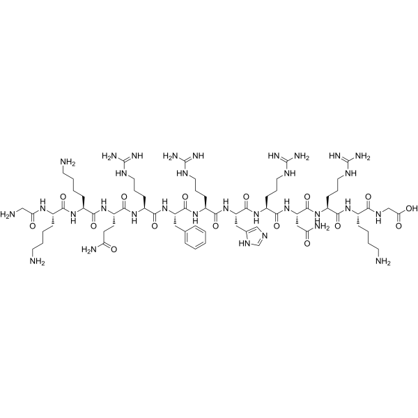
Vitronectin (367-378) corresponds to the 367-378 residue fragment of Vitronectin and is a heparin/glycosaminoglycan-binding peptide. Vitronectin (367-378) acts on cell surface glycosaminoglycans and does not directly bind integrins. Vitronectin (367-378) supports pluripotent stem cell adhesion, proliferation, long-term self-renewal, pluripotency markers, normal karyotype, and undifferentiated culture. Vitronectin (367-378) facilitates the differentiation of human pluripotent stem cells into definitive endoderm and mesoderm. Vitronectin (367-378) can be used in research related to stem cell fate regulation.
연구목적의 판매만을 진행합니다. 환자를 대상으로 한 판매는 하지 않습니다.
- Purity : 99.02%
- CAS No.: 1217344-74-5
- 화학식: C70H122N32O16
- 분자량:1667.92
-
보관:
Sealed storage, away from moisture.
Powder -80°C, 2 years , -20°C, 1 year* In solvent : -80°C, 6 months; -20°C, 1 month (sealed storage, away from moisture)

To place orders, for customer services and technical support, please contact: MedChemExpress USA
Tel: 609-228-6898 E-mail: [email protected] [email protected]


-
 Biological Activity
-
Chemical Information
-
용액&용해도
- Protocol
- 순도&문서
- References
-
 Help & FAQs




Biological Activity
제품 설명
In Vitro
Vitronectin (367-378) (500 ng/mL Noggin; 10 μM SB431542; 5 d) supported an adhesion rate of H9-derived ectoderm not exceeding 50%[1].
Vitronectin (367-378) alone cannot effectively support motor neuron differentiation of H9 cells, leading to cell aggregation and detachment, whereas the Vitronectin (367-378)/cRGD surface supports Map2+ and Map2+/Isl1+ motor neuron differentiation by day 15[1].
Vitronectin (367-378) promotes the differentiation of human pluripotent stem cells into definitive endoderm and mesoderm[1].
Vitronectin (367-378), compared with substrates such as Matrigel that can activate integrins, resulted in lower Akt phosphorylation levels, and the Smad/Akt signaling balance shifted toward mesendodermal differentiation[1].
Vitronectin (367-378) can be displayed on glass and streptavidin scaffolds, supporting human pluripotent stem cell adhesion and expansion[2].
Vitronectin (367-378) (GKKQRFRHRNRKG) supports the attachment and long-term self-renewal of multiple human embryonic stem cell and induced pluripotent stem cell lines; cells cultured on surfaces modified with this peptide maintain a normal karyotype, high expression of pluripotency markers (Oct-4, SSEA-4), and full trilineage differentiation potential[1].
Vitronectin (367-378) (peptide-functionalized surface; mTeSR medium; 6 days) mediates cell attachment by binding to cell surface glycosaminoglycans; treatment with soluble heparin or chondroitinase ABC weakens the adhesion of human embryonic stem cells[2].
Vitronectin (367-378) (combined with cyclic RGD peptide; streptavidin-modified surface; mTeSR medium; 1-2 months) supports human pluripotent stem cell attachment and long-term expansion; after colony formation, the ROCK inhibitor Y-27632 (HY-10071) can be withdrawn from the medium[2].
MedChemExpress (MCE) has not independently confirmed the accuracy of these methods. They are for reference only.


Chemical Information
-
CAS No. 1217344-74-5
-
Appearance Solid
-
분자량 1667.92
-
화학식 C70H122N32O16
-
Color White to off-white
-
Sequence Shortening
GKKQRFRHRNRKG
-
선적
Room temperature in continental US; may vary elsewhere.
-
보관
Sealed storage, away from moisture
Powder -80°C 2 years -20°C 1 year * In solvent : -80°C, 6 months; -20°C, 1 month (sealed storage, away from moisture)


용액&용해도
In Vitro:
H2O : 100 mg/mL (59.95 mM; Need ultrasonic)
Please refer to the solubility information to select the appropriate solvent. Once prepared, please aliquot and store the solution to prevent product inactivation from repeated freeze-thaw cycles.
Storage method and period of stock solution: -80°C, 6 months; -20°C, 1 month (sealed storage, away from moisture). When stored at -80°C, please use it within 6 months. When stored at -20°C, please use it within 1 month.
* Note: If you choose water as the stock solution, please dilute it to the working solution, then filter and sterilize it with a 0.22 μm filter before use.
Please refer to the solubility information to select the appropriate solvent. Once prepared, please aliquot and store the solution to prevent product inactivation from repeated freeze-thaw cycles.
Storage method and period of stock solution: -80°C, 6 months; -20°C, 1 month (sealed storage, away from moisture). When stored at -80°C, please use it within 6 months. When stored at -20°C, please use it within 1 month.
* Note: If you choose water as the stock solution, please dilute it to the working solution, then filter and sterilize it with a 0.22 μm filter before use.
Concentration (start) × Volume (start) = Concentration (final) × Volume (final)
In Vivo:
For the following dissolution methods, please prepare the working solution directly:
It is recommended to prepare fresh solutions and use them promptly within a short period of time.
The percentages shown for the solvents indicate their volumetric ratio in the final prepared solution. If precipitation or phase separation occurs during preparation, heat and/or sonication can be used to aid dissolution.
Add each solvent one by one: PBS
Solubility: 50 mg/mL (29.98 mM); Clear solution; Need ultrasonic


순도&문서
-
Data Sheet (275 KB)
-
SDS (252 KB)
- English - EN (252 KB)
- Français - FR (252 KB)
- Deutsch - DE (252 KB)
- Norwegian - NO (252 KB)
- Español - ES (252 KB)
- Swedish - SV (252 KB)
- Italian - IT (252 KB)
- Korean - KR (252 KB)
- Portuguese - PT (252 KB)
-
Handling Instructions (2659 KB)
References


Complete Stock Solution Preparation Table
Please refer to the solubility information to select the appropriate solvent. Once prepared, please aliquot and store the solution to prevent product inactivation from repeated freeze-thaw cycles.
Storage method and period of stock solution: -80°C, 6 months; -20°C, 1 month (sealed storage, away from moisture). When stored at -80°C, please use it within 6 months. When stored at -20°C, please use it within 1 month.
| Optional Solvent | Concentration Solvent Mass | 1 mg | 5 mg | 10 mg | 25 mg |
|---|---|---|---|---|---|
| H2O | 1 mM | 0.5995 mL | 2.9977 mL | 5.9955 mL | 14.9887 mL |
| 5 mM | 0.1199 mL | 0.5995 mL | 1.1991 mL | 2.9977 mL | |
| 10 mM | 0.0600 mL | 0.2998 mL | 0.5995 mL | 1.4989 mL | |
| 15 mM | 0.0400 mL | 0.1998 mL | 0.3997 mL | 0.9992 mL | |
| 20 mM | 0.0300 mL | 0.1499 mL | 0.2998 mL | 0.7494 mL | |
| 25 mM | 0.0240 mL | 0.1199 mL | 0.2398 mL | 0.5995 mL | |
| 30 mM | 0.0200 mL | 0.0999 mL | 0.1998 mL | 0.4996 mL | |
| 40 mM | 0.0150 mL | 0.0749 mL | 0.1499 mL | 0.3747 mL | |
| 50 mM | 0.0120 mL | 0.0600 mL | 0.1199 mL | 0.2998 mL |
* Note: If you choose water as the stock solution, please dilute it to the working solution, then filter and sterilize it with a 0.22 μm filter before use.


Vitronectin (367-378) Related Classifications
HY-P2452 Related Classifications
 Powered by Bioz