



Zampilimab

Based on 1 Customer Validation
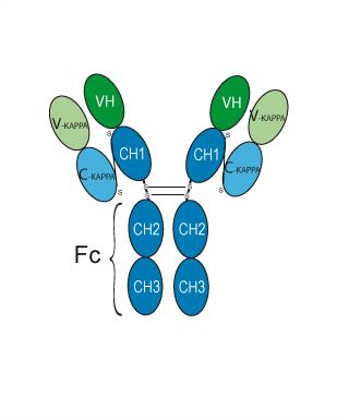
Zampilimab (UCB-7858) is a monoclonal antibody against transglutaminase 2 (TG2). Zampilimab inhibits TG2 crosslinking transamidation activity with an IC50 of 0.25 nM and a Kd of <50 pM. Zampilimab improves renal fibrosis.
For research use only. We do not sell to patients.
- Purity : ≥99.0%
- CAS No.: 2098280-42-1
- Molecular Weight:143.48 kDa
-
Storage:
Please store the product under the recommended conditions in the Certificate of Analysis.


To place orders, for customer services and technical support, please contact: MedChemExpress USA
Tel: 609-228-6898 E-mail: [email protected] [email protected]


-
 Biological Activity
-
Chemical Information
- Protocols
- Purity & Documentation
- References
-
 Help & FAQs


-
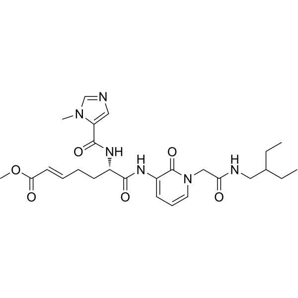
HY-19359
VilagletistatTransglutaminase 2 Inhibitor -

HY-P99003
Human IgG4 (S228P) kappa, Isotype ControlIgG4κ Isotype Control -
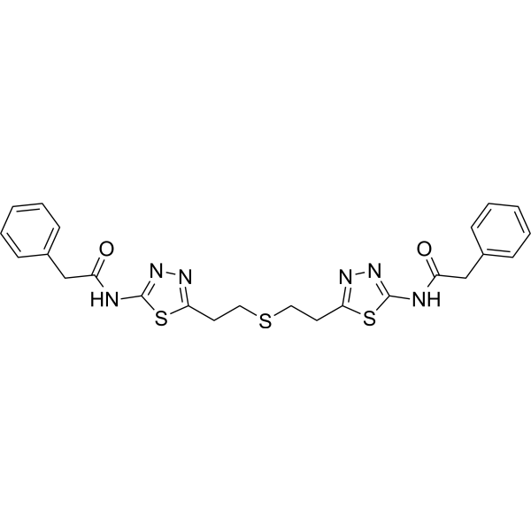
HY-12683
BPTESGlutaminase Inhibitor -
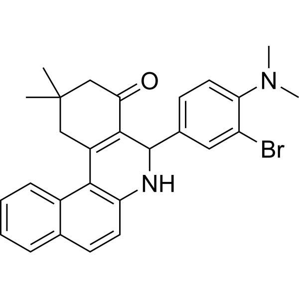
HY-12682
Glutaminase C-IN-1Glutaminase Inhibitor -
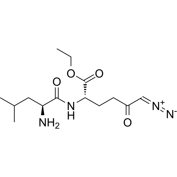
HY-122218
JHU-083Glutaminase Antagonist -
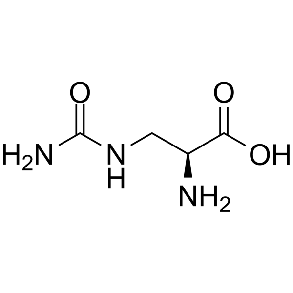
HY-121167
L-AlbizziinGlutamase Inhibitor -
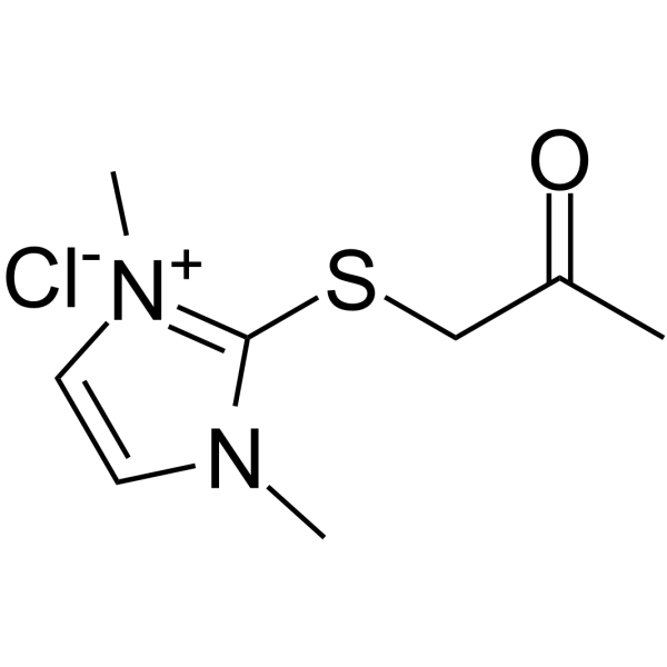
HY-117678
TG-2-IN-1TGM-2 Inhibitor -
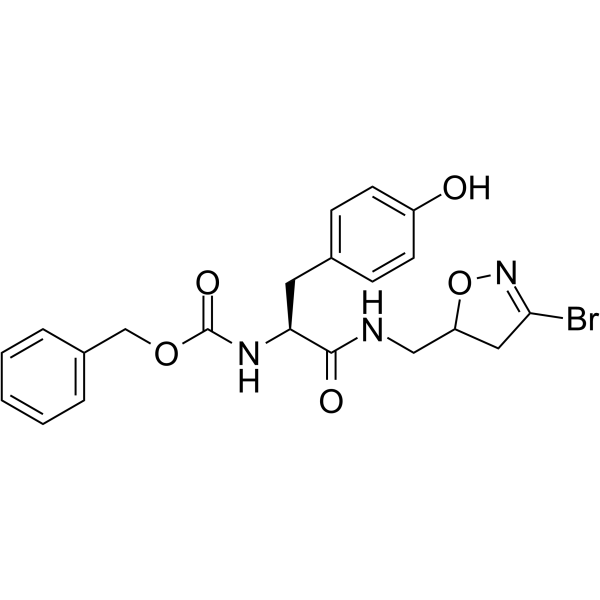
HY-123290
KCC009TG2 Inhibitor -
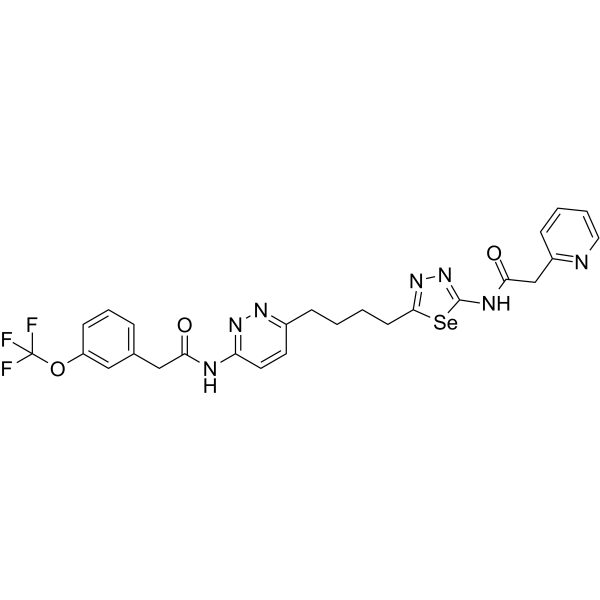
HY-114334
Glutaminase-IN-1KGA Inhibitor -
HY-W006187
2-Amino-2-(p-tolyl)acetic acidThiadiazole Intermediate -
HY-103671
IPN60090GLS1 Inhibitor -
HY-79583
Glutaminase-IN-3


Biological Activity
Description
Isotype
Human IgG4 kappa
Recommend Isotype Controls
Species Reactivity
Human
IC50 & Target
TGM2/Transglutaminase 2
In Vitro
Zampilimab inhibits TG2 crosslinking transamidation activity with an IC50 of 0.25 nM and Kd of <50 pM[3].
Zampilimab (mean IC50 ranging from 35 nM for collagen V through to 50 nM for fibronectin) inhibits the accumulation of multiple extracellular matrix proteins including collagens I, III, IV, V, and fibronectin in a primary human renal epithelial cell model of tubulointerstitial fibrosis[3].
Zampilimab (183-733 nM) slows wound closure in WI-38 cells and A549 cells[3].
MedChemExpress (MCE) has not independently confirmed the accuracy of these methods. They are for reference only.
In Vivo
MedChemExpress (MCE) has not independently confirmed the accuracy of these methods. They are for reference only.
Clinical Trial
| NCT Number | Sponsor | Condition | Start Date |
Phase

|
|---|---|---|---|---|
| NCT01329991 | Plexxikon| | 2011-05 | PHASE1 |
Gene ID
Accession
Conjugated
Unconjugated
Reconsititution
The product can be reconstituted/diluted with sterile PBS or saline.
Format
-
Human IgG4 kappa
Application
ELISA, FACS, Functional assay
Verified Bioactivity
-
 Immobilized TGM2/Transglutaminase 2 Protein, Human (sf9, His, solution, HY-P76114Y) can bind Zampilimab. The EC50 for this effect is 12.46 ng/mL.
 -
 Flow Cytometry analysis of Hela cells labelling TGM2/Transglutaminase 2 (red) with Zampilimab (anti-TGM2/Transglutaminase 2) (HY-P99512). Goat Anti-Human IgG (Alexa Fluor 488) (HY-P83776) at a dilution of 1/1000 was used as the secondary antibody. Blue-Human IgG4 kappa (HY-P99003). Black-Unlabelled control, cells without incubation with primary antibody.


Chemical Information
-
CAS No. 2098280-42-1
-
Appearance Liquid
-
Molecular Weight 143.48 kDa
-
Color Colorless to light yellow
-
SMILES
[Zampilimab]
-
Synonyms
UCB7858
-
Shipping
Shipping with dry ice.
-
Formulation
Please refer to the lot-specific COA for specific buffer information.
-
Storage
Please store the product under the recommended conditions in the Certificate of Analysis.


Purity & Documentation
-
Data Sheet (274 KB)
-
SDS (251 KB)
- English - EN (251 KB)
- Français - FR (251 KB)
- Deutsch - DE (251 KB)
- Norwegian - NO (251 KB)
- Español - ES (251 KB)
- Swedish - SV (251 KB)
- Italian - IT (251 KB)
- Korean - KR (251 KB)
- Portuguese - PT (251 KB)
-
Inhibitory Antibodies User Guide (603 KB)
References
[1]. Tempest R, et al. The Biological and Biomechanical Role of Transglutaminase-2 in the Tumour Microenvironment. Cancers (Basel). 2021 Jun 3;13(11):2788. [Content Brief]
[2]. Prat-Duran J, et al. Transglutaminase 2 as a novel target in chronic kidney disease - Methods, mechanisms and pharmacological inhibition. Pharmacol Ther. 2021 Jun;222:107787. [Content Brief]
[3]. Huang L, et al. The effect of TG2-inhibitory monoclonal antibody zampilimab on tissue fibrosis in human in vitro and primate in vivo models of chronic kidney disease. PLoS One. 2024 May 16;19(5):e0298864. [Content Brief]

Calculators
Concentration (start) × Volume (start) = Concentration (final) × Volume (final)


Zampilimab Related Classifications
HY-P99512 Related Classifications
 Powered by Bioz