



Flupirtine Maleate
Based on 1 publication(s) in Google Scholar
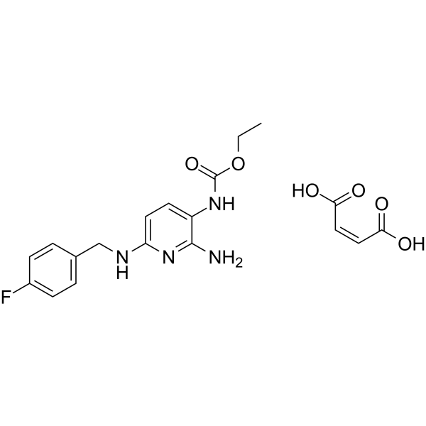
Flupirtine (D 9998) Maleate is an orally active, blood-brain barrier-crossing non-opioid analgesic and neuroprotective agent. Flupirtine Maleate is a neuronal potassium channel opener (Kv7 activator), a NMDA receptor antagonist and a GABA receptor activator. Flupirtine Maleate stabilizes blood-brain-barrier integrity, reduces oxidative stress and brain leukocyte infiltration, enhances angioneurogenesis, suppresses calcium influx, stabilizes neuronal resting membrane potential, and counteracts focal cerebral ischemia. Flupirtine Maleate exhibits analgesic, muscle relaxant properties, protects neurons from excitotoxic, ischemic, or cytokine-mediated death. Flupirtine Maleate functions as a non-opioid analgesic without antipyretic or antiphlogistic properties, shows no relevant affinity to opiate receptor. Flupirtine Maleate can be used for the research of focal cerebral ischemia, pain, Alzheimer’s disease, or multiple sclerosis.
For research use only. We do not sell to patients.
- Purity : 99.97%
- CAS No.: 75507-68-5
- Formula: C19H21FN4O6
- Molecular Weight:420.39
-
Storage:
4°C, sealed storage, away from moisture
* In solvent : -80°C, 6 months; -20°C, 1 month (sealed storage, away from moisture)

To place orders, for customer services and technical support, please contact: MedChemExpress USA
Tel: 609-228-6898 E-mail: [email protected] [email protected]


-
 Biological Activity
-
Chemical Information
-
Solvent & Solubility
- Protocols
- Purity & Documentation
- References
-
 Help & FAQs


-
HY-12651
Primaquine diphosphateAntimalarial Agent -
HY-15703
QNZ46NMDA Receptor Antagonist -

HY-13259
MG-132Proteasome Inhibitor -
HY-13555
β-LapachoneAutophagy Inducer -
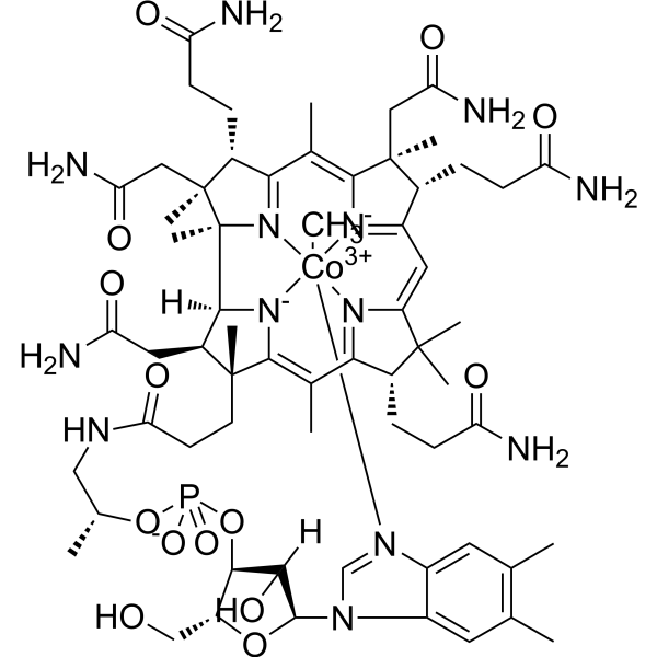
HY-B0586
MethylcobalaminVitamin B12 -
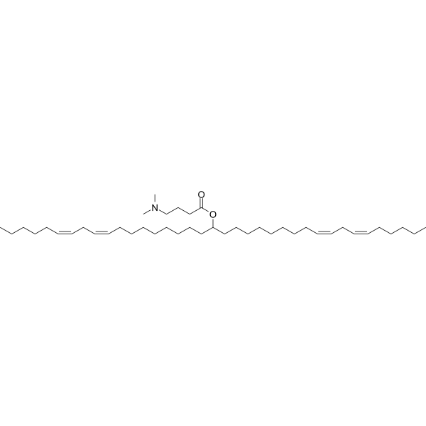
HY-112251
D-Lin-MC3-DMAsiRNA Delivery Vehicle -
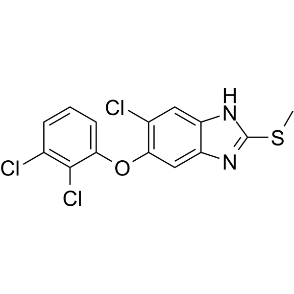
HY-B0621
TriclabendazoleParasite Inhibitor -
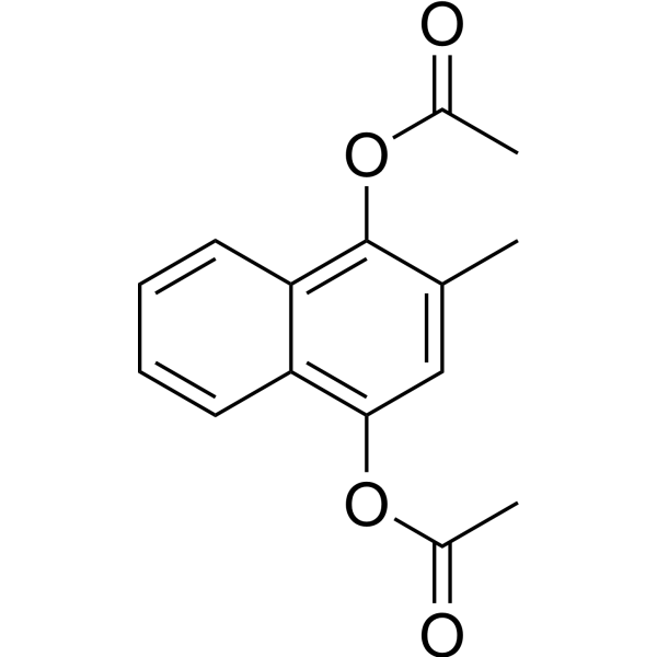
HY-B1508
Vitamin K4Endogenous Metabolite -
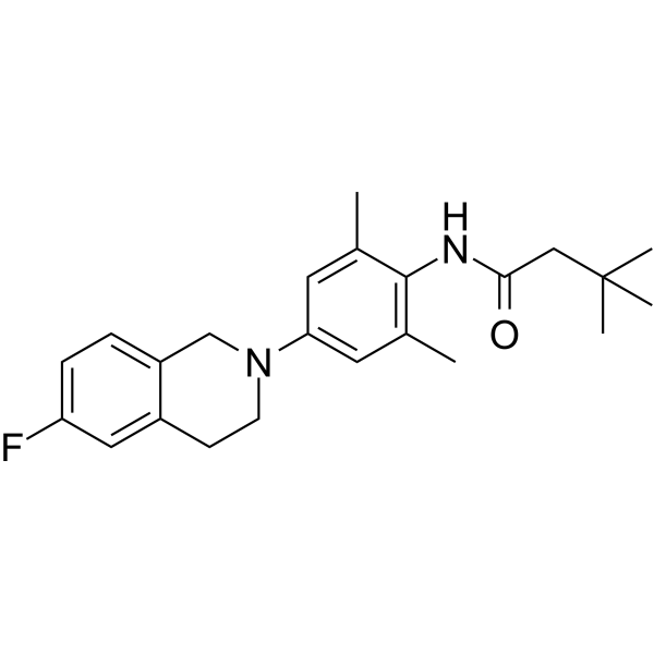
HY-139791
AzetukalnerKCNQ2/3 Activator -
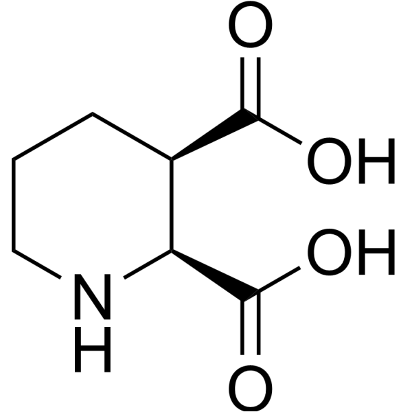
HY-121964
cis-Piperidine-2,3-dicarboxylic acidNMDA Agonist -
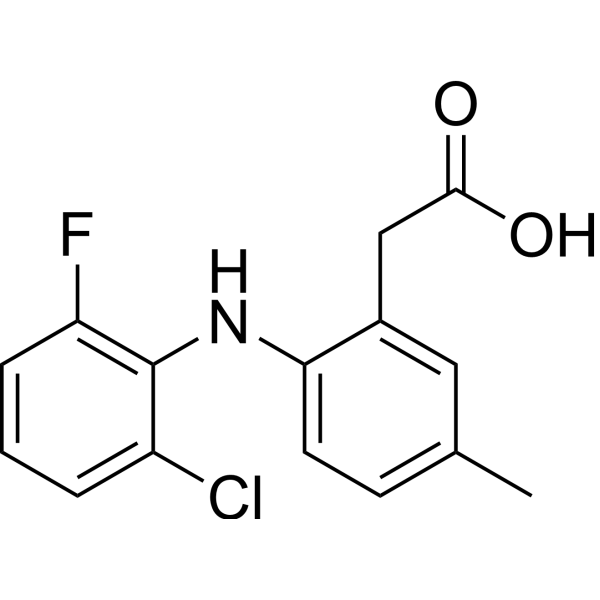
HY-13507
LumiracoxibCOX-2 Inhibitor -
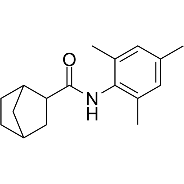
HY-101843
ML213Kv7.2/7.4 Channels Activator
Publications Citing Use of MedChemExpress (MCE) Flupirtine Maleate
MoreAll iGluR Isoforms
More

Biological Activity
Description
IC50 & Target
|
NMDA Receptor |
In Vitro
Flupirtine (0.1-100 μM for tsA cells; 3-30 μM for SCG neurons; 3-30 μM for hippocampal, DRG, DH neurons) Maleate enhances currents through KV7 channels in tsA 201 cells expressing KV7.2/7.3 subunits, rat SCG neurons, hippocampal neurons, DRG neurons, and DH neurons with EC50 values ranging from 4.4 to 6.1 μM[2].
Flupirtine (10, 30 μM) Maleate modulates GABAA receptors (enhancing low-concentration GABA currents) and NMDA receptors (inhibiting at 30 μM) but not TRPV1, non-NMDA glutamate, or glycine receptors in rat hippocampal neurons[2].
Flupirtine (30 μM) Maleate potentiates GABAA receptors in rat DRG, DH, and SCG neurons, with greater leftward shifts of GABA concentration-response curves in DRG and DH neurons than SCG neurons[2].
Flupirtine (0.1-100 μM) Maleate is more potent at enhancing GABAA receptor currents in rat DRG neurons (EC50 22 μM) than DH (EC50 53 μM) or hippocampal (EC50 65 μM) neurons, and therapeutic concentrations (3 μM) facilitate KV7 channels and GABAA receptors similarly in DRG/DH neurons[2].
Flupirtine (10-300 μM; 1.5 minutes) Maleate antagonizes NMDA-induced currents in cultured rat superior colliculus neurones with an IC50 of 182.1 μM for steady-state responses and 228.6 μM for peak responses[3].
Flupirtine (0.001-10 mM; 24 h) Maleate inhibits the growth of U373 MG cells with a GI50 of 0.47 mM, showing significant growth reduction at 1 and 10 mM after 24 h[4].
Flupirtine (1 mM; 24, 48 h) Maleate alters the cell cycle distribution of U373 MG cells, decreasing the percentage of cells in the G0-G1 phase compared to control after 24 and 48 h, with significant variations in cell cycle phases observed after 48 h[4].
MedChemExpress (MCE) has not independently confirmed the accuracy of these methods. They are for reference only. Further protocols information, click here.
-
Cell Line:U373 malignant glioma cell lines
-
Concentration:0.001, 0.01, 0.1, 1, and 10 mM
-
Incubation Time:24 hours
-
Result:Inhibited U373 MG cell growth with a GI50 of 0.47. Significantly reduced cell growth at high doses (1 and 10 mM) compared to low doses (0.001 to 0.1 mM) and control.
-
Cell Line:U373 malignant glioma cell lines
-
Concentration:1 mM
-
Incubation Time:24 h; 48 h
-
Result:Detected G0-G0 phase percentage of 45.48, Sub G0-G0 phase percentage of 2.49, S phase percentage of 24.47, and G₂-M phase percentage of 27.56 after 24 h treatment. Detected G0-G1 phase percentage of 56.39, Sub G0-G1 phase percentage of 1.82, S phase percentage of 18.99, and G2-M phase percentage of 22.80 after 48 h treatment. Observed significant variations in cell cycle phases after 48 h but not 24 h of treatment.
In Vivo
MedChemExpress (MCE) has not independently confirmed the accuracy of these methods. They are for reference only.
-
Animal Model:C57BL6 (male, 22-25 g, transient focal cerebral ischemia via left middle cerebral artery thread occlusion for 30 min)[1]
-
Dosage:1, 5, 10 mg/kg; 10 mg/kg (majority of experiments)
-
Administration:i.p.; single dose; during reperfusion or at 3, 6, 9, 12 h post-stroke (9 h post-stroke for majority of experiments)
-
Result:Reduced infarct volumes on day 2 post-stroke at 5 and 10 mg/kg (1 mg/kg had no effect); reduced infarct volumes, TUNEL+ cell counts, rt-PA-induced acute brain toxicity, Evans blue extravasation, oxidative stress (TBARS formation), intracerebral leukocyte infiltration, calpain activity, JNK and NF-κB activation, and proteasomal activity at 10 mg/kg (given up to 9 h post-stroke); increased STAT6 abundance, neuronal density (NeuN+ cells), CD31+ endothelial cells, Dcx+ immature neurons, and BrdU+/NeuN+ mature neurons on day 84; improved performance in rota rod, tight rope, corner turn, and foot fault tests up to day 84 at 10 mg/kg (given up to 9 h post-stroke).
Clinical Trial
| NCT Number | Sponsor | Condition | Start Date |
Phase

|
|---|---|---|---|---|
| NCT01329991 | Plexxikon| | 2011-05 | PHASE1 |


Chemical Information
-
CAS No. 75507-68-5
-
Appearance Solid
-
Molecular Weight 420.39
-
Formula C19H21FN4O6
-
Color White to off-white
-
SMILES
O=C(NC1=CC=C(N=C1N)NCC2=CC=C(C=C2)F)OCC.O=C(/C=C\C(O)=O)O
-
Synonyms
D 9998 Maleate
-
Shipping
Room temperature in continental US; may vary elsewhere.
-
Storage
4°C, sealed storage, away from moisture
* In solvent : -80°C, 6 months; -20°C, 1 month (sealed storage, away from moisture)


Publications (1)
-
Journal Impact Factor
-
Most Recent
-
Biochem Pharmacol
In vitro approach to elucidate the relevance of carboxylesterase 2 and N-acetyltransferase 2 to flupirtine-induced liver injury. [Abstract]2018 Sep:155:242-251. PMID: 30028988


Solvent & Solubility
In Vitro:
DMSO : 100 mg/mL (237.87 mM; Need ultrasonic; Hygroscopic DMSO has a significant impact on the solubility of product, please use newly opened DMSO)
H2O : 1.43 mg/mL (3.40 mM; ultrasonic and warming and heat to 60°C)
Please refer to the solubility information to select the appropriate solvent. Once prepared, please aliquot and store the solution to prevent product inactivation from repeated freeze-thaw cycles.
Storage method and period of stock solution: -80°C, 6 months; -20°C, 1 month (sealed storage, away from moisture). When stored at -80°C, please use it within 6 months. When stored at -20°C, please use it within 1 month.
* Note: If you choose water as the stock solution, please dilute it to the working solution, then filter and sterilize it with a 0.22 μm filter before use.
Please refer to the solubility information to select the appropriate solvent. Once prepared, please aliquot and store the solution to prevent product inactivation from repeated freeze-thaw cycles.
Storage method and period of stock solution: -80°C, 6 months; -20°C, 1 month (sealed storage, away from moisture). When stored at -80°C, please use it within 6 months. When stored at -20°C, please use it within 1 month.
* Note: If you choose water as the stock solution, please dilute it to the working solution, then filter and sterilize it with a 0.22 μm filter before use.
Concentration (start) × Volume (start) = Concentration (final) × Volume (final)
In Vivo:
Select the appropriate dissolution method based on your experimental animal and administration route.
- For the following dissolution methods, please ensure to first prepare a clear stock solution using an In Vitro approach and then sequentially add co-solvents:
- To ensure reliable experimental results, the clarified stock solution can be appropriately stored based on storage conditions. As for the working solution for In Vivo experiments, it is recommended to prepare freshly and use it on the same day.
- The percentages shown for the solvents indicate their volumetric ratio in the final prepared solution. If precipitation or phase separation occurs during preparation, heat and/or sonication can be used to aid dissolution.
Add each solvent one by one: 10% DMSO 40% PEG300 5% Tween-80 45% Saline
Solubility: ≥ 2.5 mg/mL (5.95 mM); Clear solution
This protocol yields a clear solution of ≥ 2.5 mg/mL (saturation unknown).
Taking 1 mL working solution as an example, add 100 μL DMSO stock solution (25.0 mg/mL) to 400 μL PEG300, and mix evenly; then add 50 μL Tween-80 and mix evenly; then add 450 μL Saline to adjust the volume to 1 mL.
Preparation of Saline: Dissolve 0.9 g sodium chloride in ddH₂O and dilute to 100 mL to obtain a clear Saline solution.
Add each solvent one by one: 10% DMSO 90% (20% SBE-β-CD in Saline)
Solubility: ≥ 2.5 mg/mL (5.95 mM); Clear solution
This protocol yields a clear solution of ≥ 2.5 mg/mL (saturation unknown).
Taking 1 mL working solution as an example, add 100 μL DMSO stock solution (25.0 mg/mL) to 900 μL 20% SBE-β-CD in Saline, and mix evenly.
Preparation of 20% SBE-β-CD in Saline (4°C, storage for one week): 2 g SBE-β-CD powder is dissolved in 10 mL Saline, completely dissolve until clear.
In Vivo Dissolution Calculator
Please enter the basic information of animal experiments:
-
-
-
-
Recommended: Prepare an additional quantity of animals to account for potential losses during experiments.
Please enter your animal formula composition:
-
%DMSO +
Recommended: Keep the proportion of DMSO in working solution below 2% if your animal is weak.
-
%+
-
+%Tween-80 + +
-
%Saline +
The co-solvents required include: DMSO, . All of co-solvents are available by MedChemExpress (MCE). , Tween 80. All of co-solvents are available by MedChemExpress (MCE).
Working solution concentration: 0.22 mg/mL
Method for preparing stock solution: mg drug dissolved in μL DMSO. Stock solution concentration: mg/mL. * In solvent : -80°C, 6 months; -20°C, 1 month (sealed storage, away from moisture)
1. Take μL DMSO stock solution;
2. Add μL .
μL , mix evenly;
3. Then add μL Tween 80, mix evenly;
4. Then add μL
Please ensure that the stock solution in the first step is dissolved to a clear state, and add co-solvents in sequence. You can use ultrasonic heating (ultrasonic cleaner, recommended frequency 20-40 kHz), vortexing, etc. to assist dissolution.

Protocols
-
ROS/oxidative-stress fluorescent staining
ROS/oxidative-stress fluorescent staining uses cell-permeant fluorogenic probes that become fluorescent after oxidation inside cells or tissues; commonly used examples include DCFH-DA/DCFDA for broad cellular oxidant detection, DHE for superoxide-related signal detection, MitoSOX for mitochondrial superoxide-related signal detection, and CellROX probes for oxidative-stress-associated fluorescence readouts. The assay detects probe oxidation rather than a single ROS species unless the probe and analysis method have been chemically validated for that species. DCFH-DA enters cells, is deacetylated by intracellular esterases to DCFH, and produces fluorescent DCF after oxidation, so the readout is used as an operational measure of total cellular oxidative stress rather than a species-specific ROS measurement. DHE and MitoSOX can report superoxide-related oxidation, but red fluorescence alone can include non-specific ethidium-like oxidation products; HPLC or optimized spectral approaches are
-
Fluorescent plasma-membrane potential dye assay
Fluorescent plasma-membrane potential dye assays measure changes in cell membrane potential using voltage-sensitive dyes whose fluorescence changes when cells depolarize or hyperpolarize. Anionic bis-oxonol dyes such as DiBAC4(3) enter depolarized cells more readily and show increased fluorescence after intracellular binding, while hyperpolarization reduces dye accumulation and fluorescence. FMP/FLIPR membrane-potential dyes are used for faster, homogeneous microplate assays of ion-channel or receptor-mediated membrane-potential changes.
-
Transepithelial/transendothelial electrical resistance assay
TEER measures electrical resistance across epithelial or endothelial monolayers cultured on permeable supports, and the readout reflects ionic conductance through the cell barrier, especially the paracellular pathway regulated by junctional integrity. TEER can be measured without destroying the monolayer and is commonly used before or during transport, permeability, barrier-disruption, and barrier-maturation experiments. TEER values are influenced by biological maturation and technical conditions; reported factors include temperature, medium formulation, passage number, electrode geometry, membrane properties, and junctional length during early monolayer maturation. Therefore, TEER should be interpreted with blank-insert subtraction, area normalization, repeated readings, and, when possible, orthogonal barrier readouts such as FITC-dextran flux or tight-junction staining.
-
Ca2+ Staining Technique
Ca2+ staining is an experimental technique that utilizes specific fluorescent probes (such as Fluo-4 AM, Fura-2, etc.) to qualitatively or quantitatively detect dynamic changes in intracellular Ca2+ concentrations; this is achieved by monitoring the changes in fluorescent signals generated when these probes bind to free intracellular calcium ions. The underlying principle relies primarily on the presence of chelating groups within the probe's molecular structure that possess high affinity for calcium ions.
-
Research Protocol for Inflammation-related Diseases
The NLRP3 inflammasome is a cytosolic innate immune signaling platform that integrates priming signals and danger-signal activation to promote caspase-1 activation, maturation of IL-1β and IL-18, and gasdermin D-mediated pyroptotic cell death. The core experimental logic is to determine whether inflammatory disease phenotypes are driven by increased NLRP3 expression, ASC-containing inflammasome assembly, caspase-1 cleavage, GSDMD cleavage, and extracellular release of IL-1β/IL-18 rather than by nonspecific cell injury alone. The pathway is strongly linked to inflammation-related disease phenotypes because monosodium urate crystals activate NALP3/NLRP3 inflammasome signaling in gout-like crystal inflammation, cholesterol crystals activate NLRP3 inflammasomes in atherogenesis models, and DSS-induced intestinal inflammation has been reported to involve NLRP3 inflammasome activity. However, experimental colitis studies also show context-dependent protective effects of NLRP3 inflammasome co


Purity & Documentation
-
Data Sheet (278 KB)
-
SDS (393 KB)
- English - EN (393 KB)
- Français - FR (393 KB)
- Deutsch - DE (393 KB)
- Norwegian - NO (393 KB)
- Español - ES (393 KB)
- Swedish - SV (393 KB)
- Italian - IT (393 KB)
- Korean - KR (393 KB)
- Portuguese - PT (393 KB)
-
Handling Instructions (2659 KB)
References
[1]. Jaeger HM, et al. The indirect NMDAR inhibitor flupirtine induces sustained post-ischemic recovery, neuroprotection and angioneurogenesis. Oncotarget. 2015;6(16):14033-14044. [Content Brief]
[2]. Klinger F, et al. Concomitant facilitation of GABAA receptors and KV7 channels by the non-opioid analgesic flupirtine. Br J Pharmacol. 2012;166(5):1631-1642. [Content Brief]
[3]. Kornhuber J, et al. Flupirtine shows functional NMDA receptor antagonism by enhancing Mg2+ block via activation of voltage independent potassium channels. Rapid communication. J Neural Transm (Vienna). 1999;106(9-10):857-67. [Content Brief]
[4]. Panchanathan E, et al. Effect of flupirtine on the growth and viability of U373 malignant glioma cells. Cancer Biol Med. 2013;10(3):142-147. [Content Brief]
[5]. Dörr J, et al. Disease Modification in Multiple Sclerosis by Flupirtine-Results of a Randomized Placebo Controlled Phase II Trial. Front Neurol. 2018;9:842. Published 2018 Oct 9. [Content Brief]


Complete Stock Solution Preparation Table
Please refer to the solubility information to select the appropriate solvent. Once prepared, please aliquot and store the solution to prevent product inactivation from repeated freeze-thaw cycles.
Storage method and period of stock solution: -80°C, 6 months; -20°C, 1 month (sealed storage, away from moisture). When stored at -80°C, please use it within 6 months. When stored at -20°C, please use it within 1 month.
| Optional Solvent | Concentration Solvent Mass | 1 mg | 5 mg | 10 mg | 25 mg |
|---|---|---|---|---|---|
| H2O / DMSO | 1 mM | 2.3787 mL | 11.8937 mL | 23.7874 mL | 59.4686 mL |
| DMSO | 5 mM | 0.4757 mL | 2.3787 mL | 4.7575 mL | 11.8937 mL |
| 10 mM | 0.2379 mL | 1.1894 mL | 2.3787 mL | 5.9469 mL | |
| 15 mM | 0.1586 mL | 0.7929 mL | 1.5858 mL | 3.9646 mL | |
| 20 mM | 0.1189 mL | 0.5947 mL | 1.1894 mL | 2.9734 mL | |
| 25 mM | 0.0951 mL | 0.4757 mL | 0.9515 mL | 2.3787 mL | |
| 30 mM | 0.0793 mL | 0.3965 mL | 0.7929 mL | 1.9823 mL | |
| 40 mM | 0.0595 mL | 0.2973 mL | 0.5947 mL | 1.4867 mL | |
| 50 mM | 0.0476 mL | 0.2379 mL | 0.4757 mL | 1.1894 mL | |
| 60 mM | 0.0396 mL | 0.1982 mL | 0.3965 mL | 0.9911 mL | |
| 80 mM | 0.0297 mL | 0.1487 mL | 0.2973 mL | 0.7434 mL | |
| 100 mM | 0.0238 mL | 0.1189 mL | 0.2379 mL | 0.5947 mL |
* Note: If you choose water as the stock solution, please dilute it to the working solution, then filter and sterilize it with a 0.22 μm filter before use.


Flupirtine Maleate Related Classifications
HY-17001 Related Classifications
 Powered by Bioz