



Magainin 2

Based on 1 Customer Validation
Magainin 2 (Magainin II) is an antimicrobial peptide (AMP) isolated from the skin of the African clawed frog Xenopus laevis. Magainin 2 displays antibiotic activity against numerous gram-negative and gram-positive bacteria. Magainin 2 also is active against protozoa. Magainin 2 exerts its cytotoxicity effects by preferential interactions with anionic phospholipids abundant in bacterial membranes.
For research use only. We do not sell to patients.
- Purity : 98.75%
- CAS No.: 108433-95-0
- Formula: C114H180N30O29S
- Molecular Weight:2466.90
-
Storage:
Sealed storage, away from moisture and light.
Powder -80°C, 2 years , -20°C, 1 year* In solvent : -80°C, 6 months; -20°C, 1 month (sealed storage, away from moisture and light)


To place orders, for customer services and technical support, please contact: MedChemExpress USA
Tel: 609-228-6898 E-mail: [email protected] [email protected]


-
 Biological Activity
-
Chemical Information
-
Solvent & Solubility
- Protocols
- Purity & Documentation
- References
-
 Help & FAQs


-

HY-P0233
MelittinPLA2 Activator -

HY-P0269
Magainin 1Antimicrobial Peptide -

HY-P0263
DermaseptinAntimicrobial Agent -

HY-P0261
IndolicidinAntimicrobial Peptide -
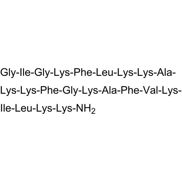
HY-105088
PexigananAntimicrobial Peptide -

HY-P1539
Cecropin ABacterial Inhibitor -
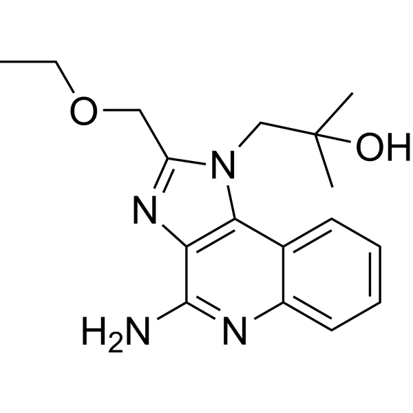
HY-13740
ResiquimodTLR7/8 Agonist -
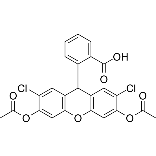
HY-D0940
H2DCFDAROS-Sensitive Probe -

HY-P0092
Cecropin BCytochrome P450 Inhibitor -

HY-P1222
LL-37, humanAntimicrobial Peptide -

HY-P4855
CRAMP (mouse)Antibacterial Peptide -

HY-P2317
Cecropin P1, porcineAntibacterial Peptide
All Antibiotic Isoforms
More

Biological Activity
Description
In Vitro
Magainin 2 exhibits bactericidal effects and induces morphological changes in Escherichia coli regarding early apoptosis. Magainin 2 induces the expression of a bacterial protein with affinity for the caspase substrate and effects the expression of RecA as a caspase-like protein[1]. Magainin 2 kill bacteria by permeabilizing the cell membranes without exhibiting significant toxicity against mammalian cells. The main target of the peptide is considered to be the lipid matrix of the membranes. Application of 10 μg /mL magainin 2 to Paramecium caudatum, a protozoan, in pond water caused an osmotic swelling of the cell and a subsequent cell burst, suggesting that the peptide could perturb membrane functions responsible for osmotic balance[2]. Magainin 2 permeabilizes bacterial and mammalian membranes in significantly different ways. The peptide forms pores with a diameter of about 2.8 nm (less than 6.6 nm) in B. megaterium, and translocates into the cytosol. In contrast, the peptide significantly perturbes the membrane of CHO-K1 cells, permitting the entry of a large molecule (larger than 23 nm) into the cytosol, accompanied by membrane budding and lipid flip-flop, mainly accumulating in mitochondria and nuclei[3].
MedChemExpress (MCE) has not independently confirmed the accuracy of these methods. They are for reference only. Further protocols information, click here.


Chemical Information
-
CAS No. 108433-95-0
-
Appearance Solid
-
Molecular Weight 2466.90
-
Formula C114H180N30O29S
-
Color White to off-white
-
Synonyms
Magainin II
-
Sequence
Gly-Ile-Gly-Lys-Phe-Leu-His-Ser-Ala-Lys-Lys-Phe-Gly-Lys-Ala-Phe-Val-Gly-Glu-Ile-Met-Asn-Ser
-
Sequence Shortening
GIGKFLHSAKKFGKAFVGEIMNS
-
Structure Classification
-
Initial Source
the skin of the African clawed frog
-
Shipping
Room temperature in continental US; may vary elsewhere.
-
Storage
Sealed storage, away from moisture and light
Powder -80°C 2 years -20°C 1 year * In solvent : -80°C, 6 months; -20°C, 1 month (sealed storage, away from moisture and light)


Solvent & Solubility
In Vitro:
H2O : 50 mg/mL (20.27 mM; Need ultrasonic)
Please refer to the solubility information to select the appropriate solvent. Once prepared, please aliquot and store the solution to prevent product inactivation from repeated freeze-thaw cycles.
Storage method and period of stock solution: -80°C, 6 months; -20°C, 1 month (sealed storage, away from moisture and light). When stored at -80°C, please use it within 6 months. When stored at -20°C, please use it within 1 month.
* Note: If you choose water as the stock solution, please dilute it to the working solution, then filter and sterilize it with a 0.22 μm filter before use.
Please refer to the solubility information to select the appropriate solvent. Once prepared, please aliquot and store the solution to prevent product inactivation from repeated freeze-thaw cycles.
Storage method and period of stock solution: -80°C, 6 months; -20°C, 1 month (sealed storage, away from moisture and light). When stored at -80°C, please use it within 6 months. When stored at -20°C, please use it within 1 month.
* Note: If you choose water as the stock solution, please dilute it to the working solution, then filter and sterilize it with a 0.22 μm filter before use.
Concentration (start) × Volume (start) = Concentration (final) × Volume (final)

Protocols
-
Research Protocol for Infectious Diseases
Infectious-disease experiments test how pathogens interact with host barriers, innate immune receptors, inflammatory signaling, pathogen replication, and tissue injury; pattern-recognition receptors such as TLRs, RIG-I-like receptors, NOD-like receptors, and inflammasomes detect microbial molecules and activate NF-κB, interferon, and cytokine responses. The central hypothesis is that infection severity reflects the balance between pathogen burden and host response: protective inflammation restricts pathogen growth, whereas excessive or mislocalized inflammation contributes to tissue damage and disease phenotype. Unresolved questions include which host pathways are protective versus pathogenic, why some infection models fail to translate to human disease, and which combined readouts best predict clinically relevant infection outcomes.
-
Cell Cytotoxicity Assay
Cytotoxicity assays are usually based on the assessment of cell membrane damage, which can also be indirectly detected by measuring cell viability. Detection methods include MTT assay, CKK-8 assay, LDH assay and ATP assay, etc.
-
Mammalian live/dead viability and cytotoxicity staining
Live/dead viability and cytotoxicity staining assays are based on the simultaneous detection of intracellular esterase activity in metabolically active (viable) cells and membrane integrity loss in non-viable cells. In commonly used dual-staining approaches, membrane-permeant fluorogenic substrates are converted by intracellular esterases into fluorescent products in live cells, while impermeant DNA-binding dyes selectively enter cells with compromised plasma membranes and label nucleic acids in dead or dying cells, enabling discrimination between viable and non-viable populations by fluorescence microscopy or flow cytometry.
-
Gram Staining of Tissue Sections
Gram staining of tissue sections is a histochemical technique used to differentiate Gram-positive and Gram-negative bacteria within histological specimens based on differences in bacterial cell wall structure and dye retention, adapted from classical bacteriological Gram staining into tissue-compatible “histological Gram stain” variants. In tissue applications, modifications of the Brown-Hopps and Brown-Brenn methods are commonly used to improve differentiation of microorganisms embedded within host connective tissue and to reduce overstaining or loss of Gram-negative signal, which are known limitations of earlier approaches. The principle relies on crystal violet-iodine complex retention in Gram-positive organisms and subsequent decolorization and counterstaining steps that allow contrast visualization of Gram-negative organisms against tissue background.
-
Cell Viability Determination by MTT Colorimetric Assay
The following protocol uses the MTT colorimetric assay as a classic literature-established method for assessing cell viability/metabolic activity in cultured mammalian cells. MTT[3-(4,5-dimethylthiazol-2-yl)-2,5-diphenyltetrazolium bromide] is reduced by metabolically active cells to a colored formazan product; the amount of formazan is quantified spectrophotometrically and provides an indirect measure of metabolically active viable cells. Importantly, MTT reduction reflects cellular oxidoreductase/metabolic activity rather than an absolute direct count of living cells, so changes in cellular metabolism can alter the signal independently of cell number.


Purity & Documentation
-
Data Sheet (271 KB)
-
SDS (252 KB)
- English - EN (252 KB)
- Français - FR (252 KB)
- Deutsch - DE (252 KB)
- Norwegian - NO (252 KB)
- Español - ES (252 KB)
- Swedish - SV (252 KB)
- Italian - IT (252 KB)
- Korean - KR (252 KB)
- Portuguese - PT (252 KB)
-
Handling Instructions (2659 KB)
References
[1]. M Zasloff, et al. Magainins, a class of antimicrobial peptides from Xenopus skin: isolation, characterization of two active forms, and partial cDNA sequence of a precursor. Proc Natl Acad Sci U S A. 1987 Aug;84(15):5449-53. [Content Brief]
[2]. K Matsuzaki, et al. Magainins as paradigm for the mode of action of pore forming polypeptides. Biochim Biophys Acta. 1998 Nov 10;1376(3):391-400. [Content Brief]


Complete Stock Solution Preparation Table
Please refer to the solubility information to select the appropriate solvent. Once prepared, please aliquot and store the solution to prevent product inactivation from repeated freeze-thaw cycles.
Storage method and period of stock solution: -80°C, 6 months; -20°C, 1 month (sealed storage, away from moisture and light). When stored at -80°C, please use it within 6 months. When stored at -20°C, please use it within 1 month.
| Optional Solvent | Concentration Solvent Mass | 1 mg | 5 mg | 10 mg | 25 mg |
|---|---|---|---|---|---|
| H2O | 1 mM | 0.4054 mL | 2.0268 mL | 4.0537 mL | 10.1342 mL |
| 5 mM | 0.0811 mL | 0.4054 mL | 0.8107 mL | 2.0268 mL | |
| 10 mM | 0.0405 mL | 0.2027 mL | 0.4054 mL | 1.0134 mL | |
| 15 mM | 0.0270 mL | 0.1351 mL | 0.2702 mL | 0.6756 mL | |
| 20 mM | 0.0203 mL | 0.1013 mL | 0.2027 mL | 0.5067 mL |
* Note: If you choose water as the stock solution, please dilute it to the working solution, then filter and sterilize it with a 0.22 μm filter before use.


Magainin 2 Related Classifications
HY-P0270 Related Classifications
 Powered by Bioz