



Buntanetap
Based on 1 publication(s) in Google Scholar
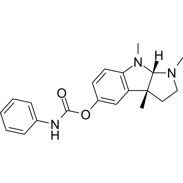
Buntanetap ((+)-Phenserine) is a selective acetylcholinesterase inhibitor. Buntanetap is a multiple neurotoxic protein translation inhibitor with oral activity, including amyloid precursor protein (APP), α-synuclein (αSYN) and huntingtin protein (HTT). Buntanetap reduces the production of β-amyloid precursor protein by blocking its mRNA translation. Buntanetap has anti-inflammatory effects and can be used in the study of Alzheimer's disease and Parkinson's disease.
For research use only. We do not sell to patients.
- Purity : 99.71%
- CAS No.: 116839-68-0
- Formula: C20H23N3O2
- Molecular Weight:337.42
-
Storage:Powder -20°C, 3 years , 4°C, 2 years ; In solvent -80°C, 6 months , -20°C, 1 month

To place orders, for customer services and technical support, please contact: MedChemExpress USA
Tel: 609-228-6898 E-mail: [email protected] [email protected]


-
 Biological Activity
-
Chemical Information
-
Solvent & Solubility
- Protocols
- Purity & Documentation
- References
-
 Help & FAQs


-
HY-17368
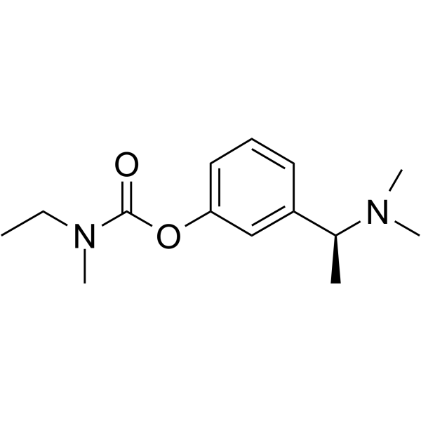
RivastigmineCholinesterase Inhibitor -
HY-10162
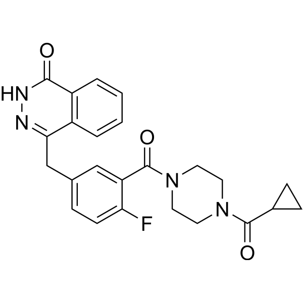
OlaparibPARP Inhibitor -
HY-101855
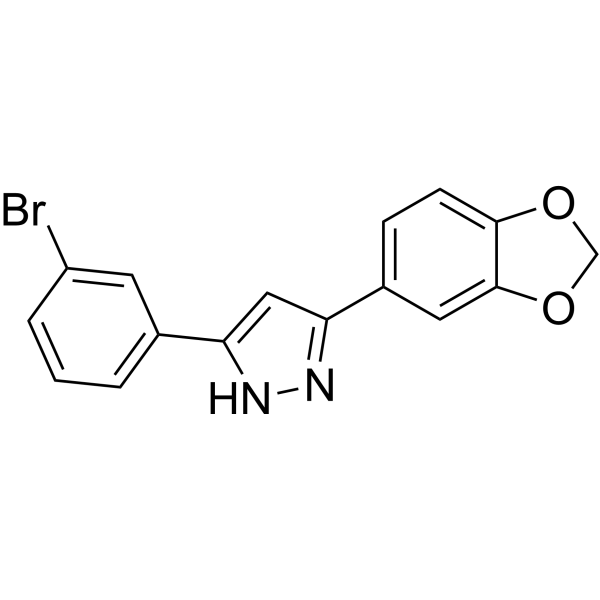
EmrusolminOligomeric Aggregation Inhibitor -
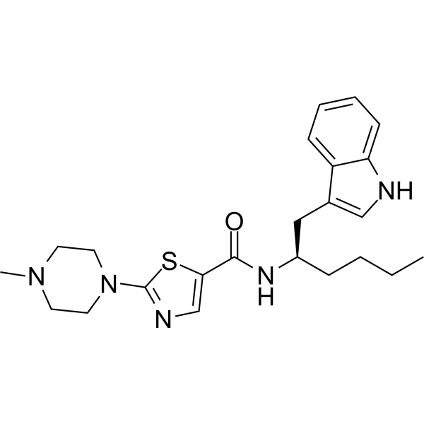
HY-145580
Minzasolminα-Synuclein Oligomerization Inhibitor -
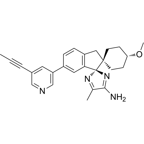
HY-100740
LanabecestatBACE1 Inhibitor -
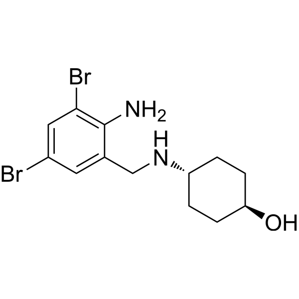
HY-B1039
AmbroxolBromhexine Metabolite -
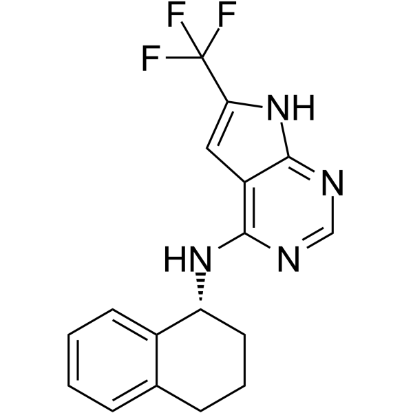
HY-152943
MTK458Mitophagy Activator -
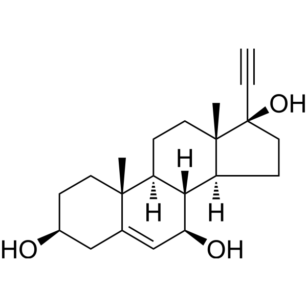
HY-108039
BezisterimNF-κB Inhibitor -
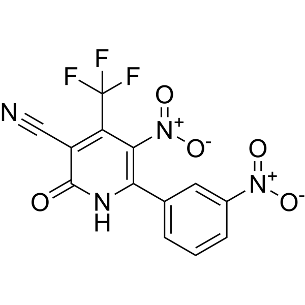
HY-124876
SynuClean-Dα-synuclein Aggregation Inhibitor -
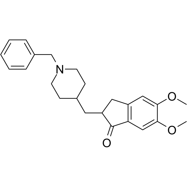
HY-14566
DonepezilSpecific AChE Inhibitor -
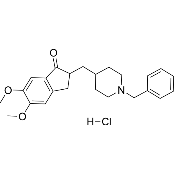
HY-B0034
Donepezil HydrochlorideAChE Inhibitor -
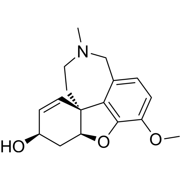
HY-76299
GalanthamineCholinesterase (ChE) Inhibitor
Publications Citing Use of MedChemExpress (MCE) Buntanetap
MoreAll α-synuclein Isoforms
More

Biological Activity
Description
IC50 & Target
|
α-synuclein |
In Vitro
Buntanetap (1-25 μM; 24 h) significantly reduces the expression of APP, αSYN, and HTT in SH-SY5Y cells[2].
MedChemExpress (MCE) has not independently confirmed the accuracy of these methods. They are for reference only. Further protocols information, click here.


Chemical Information
-
CAS No. 116839-68-0
-
Appearance Solid
-
Molecular Weight 337.42
-
Formula C20H23N3O2
-
Color Light brown to brown
-
SMILES
CN1[C@@](N(C)CC2)([H])[C@@]2(C)C3=C1C=CC(OC(NC4=CC=CC=C4)=O)=C3
-
Synonyms
(+)-Phenserine; ANVS401
-
Shipping
Room temperature in continental US; may vary elsewhere.
-
Storage
Powder -20°C 3 years 4°C 2 years In solvent -80°C 6 months -20°C 1 month


Publications (1)
-
Journal Impact Factor
-
Most Recent
-
Biochemistry (Mosc)
A Conjugate of Aminoadamantane and Tetrahydro-γ-Carboline Inhibits Accumulation of Mutant α-Synuclein A53T in the Cellular Model of Proteinopathy. [Abstract]2026 May;91(5):789-799. PMID: 42219390


Solvent & Solubility
In Vitro:
DMSO : 230 mg/mL (681.64 mM; Need ultrasonic; Hygroscopic DMSO has a significant impact on the solubility of product, please use newly opened DMSO)
Please refer to the solubility information to select the appropriate solvent. Once prepared, please aliquot and store the solution to prevent product inactivation from repeated freeze-thaw cycles.
Storage method and period of stock solution: -80°C, 6 months; -20°C, 1 month. When stored at -80°C, please use it within 6 months. When stored at -20°C, please use it within 1 month.
Please refer to the solubility information to select the appropriate solvent. Once prepared, please aliquot and store the solution to prevent product inactivation from repeated freeze-thaw cycles.
Storage method and period of stock solution: -80°C, 6 months; -20°C, 1 month. When stored at -80°C, please use it within 6 months. When stored at -20°C, please use it within 1 month.
Concentration (start) × Volume (start) = Concentration (final) × Volume (final)
In Vivo:
Select the appropriate dissolution method based on your experimental animal and administration route.
- For the following dissolution methods, please ensure to first prepare a clear stock solution using an In Vitro approach and then sequentially add co-solvents:
- To ensure reliable experimental results, the clarified stock solution can be appropriately stored based on storage conditions. As for the working solution for In Vivo experiments, it is recommended to prepare freshly and use it on the same day.
- The percentages shown for the solvents indicate their volumetric ratio in the final prepared solution. If precipitation or phase separation occurs during preparation, heat and/or sonication can be used to aid dissolution.
Add each solvent one by one: 10% DMSO 40% PEG300 5% Tween-80 45% Saline
Solubility: ≥ 5 mg/mL (14.82 mM); Clear solution
This protocol yields a clear solution of ≥ 5 mg/mL (saturation unknown).
Taking 1 mL working solution as an example, add 100 μL DMSO stock solution (50.0 mg/mL) to 400 μL PEG300, and mix evenly; then add 50 μL Tween-80 and mix evenly; then add 450 μL Saline to adjust the volume to 1 mL.
Preparation of Saline: Dissolve 0.9 g sodium chloride in ddH₂O and dilute to 100 mL to obtain a clear Saline solution.
Add each solvent one by one: 10% DMSO 90% (20% SBE-β-CD in Saline)
Solubility: ≥ 5 mg/mL (14.82 mM); Clear solution
This protocol yields a clear solution of ≥ 5 mg/mL (saturation unknown).
Taking 1 mL working solution as an example, add 100 μL DMSO stock solution (50.0 mg/mL) to 900 μL 20% SBE-β-CD in Saline, and mix evenly.
Preparation of 20% SBE-β-CD in Saline (4°C, storage for one week): 2 g SBE-β-CD powder is dissolved in 10 mL Saline, completely dissolve until clear.
In Vivo Dissolution Calculator
Please enter the basic information of animal experiments:
-
-
-
-
Recommended: Prepare an additional quantity of animals to account for potential losses during experiments.
Please enter your animal formula composition:
-
%DMSO +
Recommended: Keep the proportion of DMSO in working solution below 2% if your animal is weak.
-
%+
-
+%Tween-80 + +
-
%Saline +
The co-solvents required include: DMSO, . All of co-solvents are available by MedChemExpress (MCE). , Tween 80. All of co-solvents are available by MedChemExpress (MCE).
Working solution concentration: 0.22 mg/mL
Method for preparing stock solution: mg drug dissolved in μL DMSO. Stock solution concentration: mg/mL.
1. Take μL DMSO stock solution;
2. Add μL .
μL , mix evenly;
3. Then add μL Tween 80, mix evenly;
4. Then add μL
Please ensure that the stock solution in the first step is dissolved to a clear state, and add co-solvents in sequence. You can use ultrasonic heating (ultrasonic cleaner, recommended frequency 20-40 kHz), vortexing, etc. to assist dissolution.

Protocols
-
Neurotoxicity Study
This protocol assesses in vitro neurotoxicity by combining neuronal viability, mitochondrial/metabolic activity, neurite outgrowth, and optional neuronal network function readouts. Calcein-AM or resazurin/PrestoBlue readouts estimate viable or metabolically active cells; βIII-tubulin immunofluorescence detects neuronal morphology and neurite networks; TMRE detects mitochondrial membrane potential; and MEA recordings detect functional changes in neuronal network activity.
-
RNA extraction experimental
By lysing cells, releasing RNA, and removing impurities such as proteins and DNA, high-purity RNA products are finally obtained. The commonly used traditional method is the guanidine isothiocyanate/phenol/chloroform method (Trizol), which is suitable for a variety of animal materials including animal tissues, microorganisms, cultured cells, etc., and most plant materials.
-
Human pluripotent stem cell midbrain dopaminergic neuron differentiation
Human pluripotent stem cells are directed toward midbrain dopaminergic neurons by first inducing a neural floor-plate-like progenitor state, then patterning cells with ventralizing SHH signaling and midbrain/WNT-FGF cues, and finally maturing progenitors into neurons expressing dopaminergic markers such as TH, NURR1/NR4A2, PITX3, DAT/SLC6A3, VMAT2/SLC18A2, GIRK2/KCNJ6, FOXA2, LMX1A, and EN1. The main readouts are loss of pluripotency, acquisition of FOXA2+/LMX1A+ midbrain floor-plate progenitors, emergence of βIII-tubulin+/MAP2+ neurons, and production of TH+ dopaminergic neurons with molecular, dopamine-release, and electrophysiological features of midbrain dopaminergic identity.
-
Research Protocol for Inflammation-related Diseases
The NLRP3 inflammasome is a cytosolic innate immune signaling platform that integrates priming signals and danger-signal activation to promote caspase-1 activation, maturation of IL-1β and IL-18, and gasdermin D-mediated pyroptotic cell death. The core experimental logic is to determine whether inflammatory disease phenotypes are driven by increased NLRP3 expression, ASC-containing inflammasome assembly, caspase-1 cleavage, GSDMD cleavage, and extracellular release of IL-1β/IL-18 rather than by nonspecific cell injury alone. The pathway is strongly linked to inflammation-related disease phenotypes because monosodium urate crystals activate NALP3/NLRP3 inflammasome signaling in gout-like crystal inflammation, cholesterol crystals activate NLRP3 inflammasomes in atherogenesis models, and DSS-induced intestinal inflammation has been reported to involve NLRP3 inflammasome activity. However, experimental colitis studies also show context-dependent protective effects of NLRP3 inflammasome co
-
Amyloid: Congo Red Amyloid Staining
Congo red amyloid staining is a histochemical method used to detect extracellular amyloid deposits in tissue sections based on the affinity of Congo red dye for β-pleated sheet-rich protein aggregates. When bound to amyloid, Congo red produces characteristic apple-green birefringence under polarized light microscopy, which is widely regarded as a diagnostic feature of amyloid deposition in histopathology. The diagnostic principle relies on the combination of dye binding (congophilia) and optical anisotropy under polarized illumination, which distinguishes amyloid from most non-amyloid eosinophilic extracellular deposits in routine histological evaluation. Amyloid identification by Congo red staining remains a cornerstone in diagnostic pathology despite the availability of adjunct methods such as immunohistochemistry and mass spectrometry, particularly because of its ability to localize deposits directly within tissue architecture. The specificity of Congo red-positive deposits is incre
-
Alzheimer’s Disease Modeling
Alzheimer’s Disease (AD) is a neurodegenerative disorder characterized by a progressive decline in cognitive functions and loss of specific types of neurons and synapses. Alzheimer's symptoms can be simulated in mice by injecting drugs (such as Aβ) or genetically modified.


Purity & Documentation
-
Data Sheet (276 KB)
-
SDS (254 KB)
- English - EN (254 KB)
- Français - FR (254 KB)
- Deutsch - DE (254 KB)
- Norwegian - NO (254 KB)
- Español - ES (254 KB)
- Swedish - SV (254 KB)
- Italian - IT (254 KB)
- Korean - KR (254 KB)
- Portuguese - PT (254 KB)
-
Handling Instructions (2659 KB)
References
[1]. Fang C, et al. Buntanetap, a Novel Translational Inhibitor of Multiple Neurotoxic Proteins, Proves to Be Safe and Promising in Both Alzheimer's and Parkinson's Patients. J Prev Alzheimers Dis. 2023;10(1):25-33. [Content Brief]
[2]. Chen XQ, et al. Posiphen Reduces the Levels of Huntingtin Protein through Translation Suppression. Pharmaceutics. 2021 Dec 7;13(12):2109. [Content Brief]


Complete Stock Solution Preparation Table
Please refer to the solubility information to select the appropriate solvent. Once prepared, please aliquot and store the solution to prevent product inactivation from repeated freeze-thaw cycles.
Storage method and period of stock solution: -80°C, 6 months; -20°C, 1 month. When stored at -80°C, please use it within 6 months. When stored at -20°C, please use it within 1 month.
| Optional Solvent | Concentration Solvent Mass | 1 mg | 5 mg | 10 mg | 25 mg |
|---|---|---|---|---|---|
| DMSO | 1 mM | 2.9637 mL | 14.8183 mL | 29.6367 mL | 74.0916 mL |
| 5 mM | 0.5927 mL | 2.9637 mL | 5.9273 mL | 14.8183 mL | |
| 10 mM | 0.2964 mL | 1.4818 mL | 2.9637 mL | 7.4092 mL | |
| 15 mM | 0.1976 mL | 0.9879 mL | 1.9758 mL | 4.9394 mL | |
| 20 mM | 0.1482 mL | 0.7409 mL | 1.4818 mL | 3.7046 mL | |
| 25 mM | 0.1185 mL | 0.5927 mL | 1.1855 mL | 2.9637 mL | |
| 30 mM | 0.0988 mL | 0.4939 mL | 0.9879 mL | 2.4697 mL | |
| 40 mM | 0.0741 mL | 0.3705 mL | 0.7409 mL | 1.8523 mL | |
| 50 mM | 0.0593 mL | 0.2964 mL | 0.5927 mL | 1.4818 mL | |
| 60 mM | 0.0494 mL | 0.2470 mL | 0.4939 mL | 1.2349 mL | |
| 80 mM | 0.0370 mL | 0.1852 mL | 0.3705 mL | 0.9261 mL | |
| 100 mM | 0.0296 mL | 0.1482 mL | 0.2964 mL | 0.7409 mL |


Buntanetap Related Classifications
HY-16009 Related Classifications
 Powered by Bioz