
A031
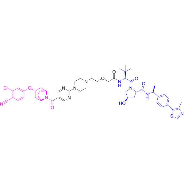
A031 is a PROTAC degrader targeting the androgen receptor. A031 induces time-dependent degradation of androgen receptor protein. A031 exerts tumor growth inhibitory effects in a zebrafish model xenografted with human prostate cancer cells. A031 can be used in studies related to prostate cancer.
(Pink: Androgen Receptor ligand (HY-175937); Blue: VHL and VHL ligand (HY-112078); Black: linker).
For research use only. We do not sell to patients.
- CAS No.: 2682255-44-1
- Formula: C50H61ClN10O7S
- Molecular Weight:981.60
-
Storage:
Please store the product under the recommended conditions in the Certificate of Analysis.
All PROTACs Isoforms
More
Biological Activity
Description
In Vitro
A031 (0.125-1.0 μM; 4 days) potently inhibits AR-positive human prostate cancer VCaP cell viability with an IC50 of less than 0.25 μM and achieves a 69.56% inhibition rate at 1.0 μM after 4 days of treatment[1].
A031 (2.0 μM; 2-4.5 h) induces time-dependent degradation of AR protein in AR-positive human prostate cancer VCaP cells, with near-complete degradation observed after 4.5 h of treatment with 2.0 μM A031[1].
A031 (2.0 μM) acts as a bona fide AR degrader in AR-positive human prostate cancer VCaP cells, as its AR-degrading activity is blocked by proteasome and NEDD8 pathway inhibitors but not by VHL-3 pre-treatment[1].
MedChemExpress (MCE) has not independently confirmed the accuracy of these methods. They are for reference only. Further protocols information, click here.
-
Cell Line:AR-positive human prostate cancer VCaP cells
-
Concentration:0.125 μM; 0.25 μM; 0.5 μM; 1.0 μM
-
Incubation Time:4 days
-
Result:Inhibited VCaP cell viability in a dose-dependent manner.
Achieved a cell inhibition rate of 69.56% at 1.0 μM.
Showed a good inhibition rate even at 0.125 μM.
Had an IC50 for cell inhibition of less than 0.25 μM.
-
Cell Line:AR-positive human prostate cancer VCaP cells
-
Concentration:2.0 μM
-
Incubation Time:2 h, 3.5 h, 4.5 h
-
Result:Reduced AR protein levels within 2 h of treatment.
Almost completely degraded AR protein after 4.5 h of treatment.
Parmacokinetics
| Species | Dose | Route | T1/2 | Cmax | AUClast | AUCinf | Vz | CL |
|---|---|---|---|---|---|---|---|---|
| Rat[1] | 1 mg/kg | i.v. | 0.57 h | 14366.2 ng/mL | 1773.8 ng·h/mL | 1773.8 ng·h/mL | 0.47 L/kg | 0.57 L/h/kg |
In Vivo
A031 (6.25-100 μM; static exposure; 2 days) has a minimal toxic concentration of 25 μM in wild-type AB zebrafish, showing no mortality or toxicity at concentrations up to this level[1].
MedChemExpress (MCE) has not independently confirmed the accuracy of these methods. They are for reference only.
-
Animal Model:wild-type AB strain (2 days post-fertilization)[1]
-
Dosage:2.8 μM; 8.3 μM; 25 μM
-
Administration:static exposure; 2 days
-
Result:Achieved a 28% tumor growth inhibition rate with a tumor fluorescence intensity of 379577 pixels.
Achieved a 55% tumor growth inhibition rate with a tumor fluorescence intensity of 240013 pixels.
Achieved a 61% tumor growth inhibition rate with a tumor fluorescence intensity of 208489 pixels.
-
Animal Model:wild-type AB strain (3 days post-fertilization)[1]
-
Dosage:6.25 μM; 12.5 μM; 25 μM; 50 μM; 100 μM
-
Administration:static exposure; 5 days
-
Result:Caused 0% mortality and no toxic phenotypes at 6.25 μM, 12.5 μM, and 25 μM.
Caused obvious precipitation at 50 μM and 100 μM, with mortality data not reported.
Determined a minimal toxic concentration (MTC) of 25 μM.
Chemical Information
-
CAS No. 2682255-44-1
-
Molecular Weight 981.60
-
Formula C50H61ClN10O7S
-
SMILES
O=C(N([C@@H]1C2)[C@H](CC1)C[C@@H]2OC3=CC(Cl)=C(C=C3)C#N)C4=CN=C(N5CCN(CC5)CCOCC(N[C@@H](C(C)(C)C)C(N6[C@@H](C[C@H](C6)O)C(N[C@H](C7=CC=C(C8=C(N=CS8)C)C=C7)C)=O)=O)=O)N=C4
-
Shipping
Room temperature in continental US; may vary elsewhere.
-
Storage
Please store the product under the recommended conditions in the Certificate of Analysis.
Protocols
-
Research Protocol for Endocrine Diseases
Endocrine diseases often arise from disrupted hormone production, hormone signaling, or target-tissue responsiveness; for diabetes-focused endocrine disease models, insulin signaling regulates glucose uptake, hepatic glucose output, lipid metabolism, and β-cell compensation. Type 2 diabetes develops through interacting defects in insulin resistance, β-cell dysfunction, adipose inflammation, hepatic glucose overproduction, altered incretin signaling, and ectopic lipid metabolism. A major unresolved question is whether endocrine dysfunction is driven primarily by target-tissue insulin resistance, intrinsic β-cell failure, immune/inflammatory stress, or combined multi-organ failure that differs by disease stage.
-
How to Select a Suitable Non-Mouse Animal Model
Selecting a suitable non-mouse animal model is a structured decision based on the research question, required anatomy or physiology, disease mechanism, endpoint feasibility, translational relevance, and ethical justification. Non-mouse models are preferred when mice cannot reproduce key human-relevant features, such as organ size, surgical anatomy, cardiovascular physiology, neuroanatomy, immune features, pharmacology, toxicology, or long-term clinical procedures. Candidate species may include rats, rabbits, guinea pigs, ferrets, zebrafish, pigs, sheep, goats, dogs, cats, horses, and non-human primates, but each species must be justified by its specific scientific advantage rather than convenience or tradition. Unresolved questions include how to quantify translational superiority across species, how to balance increased biological relevance against higher ethical burden, and when human-derived systems or new approach methodologies should replace animal use.
-
Patient-Derived Xenograft (PDX)
Patient-derived xenograft (PDX) models are generated by engrafting primary human tumor tissue directly into immunodeficient mice, allowing in vivo propagation of patient tumor biology without initial in vitro adaptation. These models are used to preserve key histopathological and molecular characteristics of the original tumor and enable assessment of tumor growth dynamics and therapeutic response in a living organism. The biological readout is tumor engraftment and subsequent growth in the murine host, which reflects the ability of human tumor cells to survive, vascularize, and expand in an immunocompromised microenvironment.
-
Patient-Derived Orthotopic Xenograft (PDOX)
Patient-derived orthotopic xenograft (PDOX) modeling implants fresh patient tumor tissue or patient-derived tumor cells into the anatomically corresponding organ or tissue site of immunodeficient mice, usually by surgical orthotopic implantation, to preserve patient tumor histology, local microenvironmental context, invasion, metastatic behavior, and treatment-response features better than subcutaneous implantation. PDOX readouts include tumor engraftment, orthotopic tumor growth, local invasion, metastasis, recurrence after resection, histologic similarity to the donor tumor, biomarker retention, molecular concordance, survival, and response or resistance to therapy. PDOX models are used for preclinical drug testing and individualized therapy evaluation, but engraftment success varies by tumor type and specimen quality.
-
Subcutaneous Cell-Line-Derived Xenograft
Subcutaneous cell-line-derived xenograft (CDX) models are established by implanting cultured human cancer cell lines into immunodeficient mice, where the injected cells form localized tumors that can be monitored in vivo as a measure of tumorigenic potential, growth kinetics, and treatment response. These models are widely used in oncology research because they allow reproducible tumor formation and enable comparative assessment of tumor growth between different cell lines or genetic manipulations in a controlled in vivo microenvironment. Subcutaneous implantation of cancer cells in immunodeficient mice is a standard approach for evaluating tumor growth behavior and therapeutic response across multiple cancer types, including prostate, esophageal, pancreatic, and colon cancer models.
-
Orthotopic Cell-Line Xenograft
Orthotopic cell-line xenograft models involve implantation of human cancer cell lines into the anatomically corresponding organ of immunodeficient mice to reproduce tumor growth within a native microenvironment, enabling more clinically relevant tumor behavior compared with subcutaneous models. These models are widely used because orthotopic placement better recapitulates tumor progression, including invasion and metastatic spread, which are often underrepresented in heterotopic implantation systems. Compared with conventional xenografts, orthotopic implantation is described as more technically complex but provides improved simulation of tumor-microenvironment interactions and metastatic behavior, making it particularly valuable for translational oncology research. Surgical orthotopic implantation approaches have been emphasized as enabling faithful reproduction of clinical cancer features, including metastasis and disease progression patterns that align with the tumor’s organ of origi
-
How to Choose the Right Model Animal
Choosing the right model animal is a validity-driven decision in which the species, strain, sex, age, genetic background, disease-induction method, outcome measures, and welfare burden must match the scientific question rather than laboratory tradition or convenience. A model should be selected by judging face validity, construct validity, and predictive validity: whether it resembles the human phenotype, whether it reproduces relevant mechanisms, and whether results are likely to predict human biology or treatment response. Animal studies often fail to translate because of species differences, weak disease resemblance, poor experimental design, inadequate reporting, publication bias, and underuse of randomization, blinding, and sample-size justification. Unresolved questions include how to rank competing models objectively, how much human-disease complexity must be reproduced for a given objective, and when non-animal systems such as organoids, ex vivo tissue, or computational models
Purity & Documentation
References
Calculators
Concentration (start) × Volume (start) = Concentration (final) × Volume (final)

