



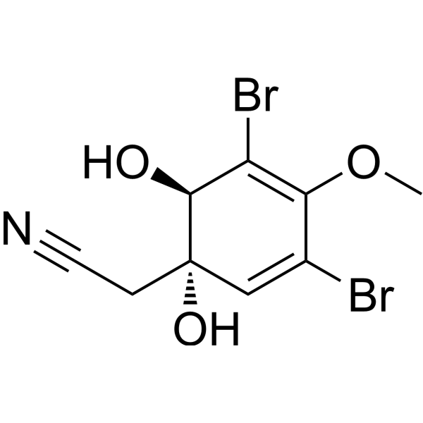
Aeroplysinin 1
Based on 1 publication(s) in Google Scholar
Aeroplysinin 1 ((+)-Aeroplysinin-1), a secondary metabolite isolated from marine sponges, shows potent antibiotic effects on Gram-positive bacteria and exerts antiviral activity against HIV-1 (IC50=14.6 μM). Aeroplysinin 1 has anti-inflammatory, anti-angiogenic and anti-tumor activities. Aeroplysinin 1 induces apoptosis in endothelial cells.
For research use only. We do not sell to patients.
- Purity : 95.0%
- CAS No.: 28656-91-9
- Formula: C9H9Br2NO3
- Molecular Weight:338.98
-
Storage:Powder -20°C, 3 years ; In solvent -80°C, 6 months , -20°C, 1 month

To place orders, for customer services and technical support, please contact: MedChemExpress USA
Tel: 609-228-6898 E-mail: [email protected] [email protected]


-
 Biological Activity
-
Chemical Information
-
Solvent & Solubility
- Protocols
- Purity & Documentation
- References
-
 Help & FAQs


Publications Citing Use of MedChemExpress (MCE) Aeroplysinin 1
More

Biological Activity
Description
IC50 & Target
[1]|
Bacterial |
HIV-1 14.6 μM (IC50) |
Apoptosis |
Cellular Effect
|
Cell Line

|
Type | Value | Description | References |
|---|---|---|---|---|
| DU-145 | IC50 |
0.54 μM
Compound: 16; Apl-1
|
Antiproliferative activity against human DU-145 cells assessed as inhibition of cell proliferation measured after 2 to 4 hrs incubation by MTT assay
Antiproliferative activity against human DU-145 cells assessed as inhibition of cell proliferation measured after 2 to 4 hrs incubation by MTT assay
|
[PMID: 39270448] |
| HeLa S3 | IC50 |
18.8 μM
Compound: 1, aeroplysinin-1
|
Cytotoxicity against human HeLaS3 cells after 2 hrs by MTT assay
Cytotoxicity against human HeLaS3 cells after 2 hrs by MTT assay
|
[PMID: 8786366] |
| HeLa S3 | IC50 |
2 μM
Compound: 1, aeroplysinin-1
|
Dose enhancement factor, ratio of IC50 for human HeLaS3 cells after 4 days to IC50 for buthionine sulfoximine-pretreated human HeLaS3 cells after 4 days by MTT assay
Dose enhancement factor, ratio of IC50 for human HeLaS3 cells after 4 days to IC50 for buthionine sulfoximine-pretreated human HeLaS3 cells after 4 days by MTT assay
|
[PMID: 8786366] |
| HeLa S3 | IC50 |
2.8 μM
Compound: 1, aeroplysinin-1
|
Cytotoxicity against buthionine sulfoximine-pretreated human HeLaS3 cells after 4 days by MTT assay
Cytotoxicity against buthionine sulfoximine-pretreated human HeLaS3 cells after 4 days by MTT assay
|
[PMID: 8786366] |
| HeLa S3 | IC50 |
27.5 μM
Compound: 1, aeroplysinin-1
|
Cytotoxicity against human HeLaS3 cells after 2 weeks by clonogenic assay
Cytotoxicity against human HeLaS3 cells after 2 weeks by clonogenic assay
|
[PMID: 8786366] |
| HeLa S3 | IC50 |
3.5 μM
Compound: 1, aeroplysinin-1
|
Dose enhancement factor, ratio of IC50 for human HeLaS3 cells after 2 hrs to IC50 for buthionine sulfoximine-pretreated human HeLaS3 cells after 2 hrs by MTT assay
Dose enhancement factor, ratio of IC50 for human HeLaS3 cells after 2 hrs to IC50 for buthionine sulfoximine-pretreated human HeLaS3 cells after 2 hrs by MTT assay
|
[PMID: 8786366] |
| HeLa S3 | IC50 |
5.4 μM
Compound: 1, aeroplysinin-1
|
Cytotoxicity against buthionine sulfoximine-pretreated human HeLaS3 cells after 2 hrs by MTT assay
Cytotoxicity against buthionine sulfoximine-pretreated human HeLaS3 cells after 2 hrs by MTT assay
|
[PMID: 8786366] |
| HeLa S3 | IC50 |
5.6 μM
Compound: 1, aeroplysinin-1
|
Cytotoxicity against human HeLaS3 cells after 4 days by MTT assay
Cytotoxicity against human HeLaS3 cells after 4 days by MTT assay
|
[PMID: 8786366] |
| PC-3 | IC50 |
0.12 μM
Compound: 16; Apl-1
|
Antiproliferative activity against human PC-3 cells assessed as inhibition of cell proliferation measured after 2 to 4 hrs incubation by MTT assay
Antiproliferative activity against human PC-3 cells assessed as inhibition of cell proliferation measured after 2 to 4 hrs incubation by MTT assay
|
[PMID: 39270448] |
In Vitro
Aeroplysinin 1 shows anti-proliferative effect against tumor cells (HT-1080, HTC-116, HeLa, THP-1, NOMO-1 and HL-60 cells), with IC50s ranging from 2.3 to 17 μM[1].
Aeroplysinin-1 also exhibits an antiviral activity toward HIV-1 caused by inhibition of its reverse transcriptase activity[1].
Aeroplysinin 1 inhibits P. phosphoreum, C. wailesii, P. minimum and HIV with IC50s of 3.5, 5.6, 7.0 and 14.6 μM[1].
Aeroplysinin 1 inhibits human endothelial cells (EVLC-2, HMEC, RF-24, and HUVEC cells), with IC50s ranging from 2.6 to 4.7 μM[2].
(+)-Aeroplysinin-1 (0.25-0.5 μM) blocks the EGF-dependent proliferation of both MCF-7 and ZR-75-1 human breast cancer cells and inhibits the ligand-induced endocytosis of the EGF receptor in vitro[3].
MedChemExpress (MCE) has not independently confirmed the accuracy of these methods. They are for reference only. Further protocols information, click here.


Chemical Information
-
CAS No. 28656-91-9
-
Appearance Solid
-
Molecular Weight 338.98
-
Formula C9H9Br2NO3
-
Color White to off-white
-
SMILES
N#CC[C@]1(O)C=C(Br)C(OC)=C(Br)[C@@H]1O
-
Synonyms
(+)-Aeroplysinin-1
-
Initial Source
-
Shipping
Room temperature in continental US; may vary elsewhere.
-
Storage
Powder -20°C 3 years In solvent -80°C 6 months -20°C 1 month


Publications (1)
-
Journal Impact Factor
-
Most Recent


Solvent & Solubility
In Vitro:
DMSO : 3.39 mg/mL (10.00 mM; Need ultrasonic and warming; Hygroscopic DMSO has a significant impact on the solubility of product, please use newly opened DMSO)
Please refer to the solubility information to select the appropriate solvent. Once prepared, please aliquot and store the solution to prevent product inactivation from repeated freeze-thaw cycles.
Storage method and period of stock solution: -80°C, 6 months; -20°C, 1 month. When stored at -80°C, please use it within 6 months. When stored at -20°C, please use it within 1 month.
Please refer to the solubility information to select the appropriate solvent. Once prepared, please aliquot and store the solution to prevent product inactivation from repeated freeze-thaw cycles.
Storage method and period of stock solution: -80°C, 6 months; -20°C, 1 month. When stored at -80°C, please use it within 6 months. When stored at -20°C, please use it within 1 month.
Concentration (start) × Volume (start) = Concentration (final) × Volume (final)

Protocols
-
Research Protocol for Infectious Diseases
Infectious-disease experiments test how pathogens interact with host barriers, innate immune receptors, inflammatory signaling, pathogen replication, and tissue injury; pattern-recognition receptors such as TLRs, RIG-I-like receptors, NOD-like receptors, and inflammasomes detect microbial molecules and activate NF-κB, interferon, and cytokine responses. The central hypothesis is that infection severity reflects the balance between pathogen burden and host response: protective inflammation restricts pathogen growth, whereas excessive or mislocalized inflammation contributes to tissue damage and disease phenotype. Unresolved questions include which host pathways are protective versus pathogenic, why some infection models fail to translate to human disease, and which combined readouts best predict clinically relevant infection outcomes.
-
Apoptosis
Apoptosis, also called programmed cell death, is generally characterized by distinct morphological characteristics.
-
TUNEL staining for apoptotic DNA fragmentation
TUNEL staining detects DNA strand breaks by using terminal deoxynucleotidyl transferase to add labeled nucleotides to exposed 3′-OH DNA termini, generating either microscopic staining in fixed cells or tissue sections, or fluorescence/cytometric signal in cell suspensions. TUNEL positivity reflects DNA fragmentation but should not be interpreted alone as definitive apoptosis, because TUNEL can also label necrotic, autolytic, mechanically damaged, or DNA-repair-associated DNA breaks.
-
Cell Cytotoxicity Assay
Cytotoxicity assays are usually based on the assessment of cell membrane damage, which can also be indirectly detected by measuring cell viability. Detection methods include MTT assay, CKK-8 assay, LDH assay and ATP assay, etc.
-
Annexin V plus membrane-impermeant dye apoptosis staining
Annexin V-based apoptosis assays rely on the detection of phosphatidylserine (PS) externalization from the inner leaflet of the plasma membrane to the outer leaflet, an early biochemical hallmark of apoptosis. Fluorescently labeled Annexin V binds PS in a calcium-dependent manner, enabling identification of early apoptotic cells by flow cytometry or fluorescence microscopy. When combined with a membrane-impermeant DNA-binding dye (e. g. , propidium iodide), this approach allows discrimination between viable (Annexin V−/dye−), early apoptotic (Annexin V+/dye−), and late apoptotic or necrotic (Annexin V+/dye+) cell populations by assessing membrane integrity and PS exposure.
-
Endothelial Tube Formation Assay
Endothelial tube formation assay evaluates the ability of endothelial cells to attach, migrate, align, and organize into capillary-like networks when cultured on gelled basement membrane extract or Matrigel; the readout is the morphology and quantity of tube-like networks, which reflects an in vitro endothelial morphogenesis step related to angiogenesis. Basement membrane extract/Matrigel provides laminin-rich extracellular matrix cues that support endothelial differentiation into capillary-like structures, but it can contain biologically active growth factors, so growth-factor-reduced matrix is preferred when testing defined angiogenic stimulators or inhibitors.
-
Apoptosis Solutions
Apoptosis is a regulated, generally non-lytic cell-death pathway that removes unwanted, damaged, infected, or abnormal cells through coordinated morphological changes, caspase activation, DNA fragmentation, and membrane remodeling. The intrinsic apoptosis pathway is controlled mainly by mitochondrial outer membrane permeabilization, BCL-2 family proteins, cytochrome c release, apoptosome formation, caspase-9 activation, and downstream executioner caspase-3/7 activation. The extrinsic apoptosis pathway is initiated by death receptors such as Fas, TNFR, and TRAIL receptors, which recruit adaptor proteins and activate caspase-8 before engaging executioner caspases or mitochondrial amplification through BID cleavage. Apoptosis is linked to many phenotypes, including cancer cell killing, tissue homeostasis, immune regulation, neurodegeneration, infection response, and treatment-induced cytotoxicity; unresolved questions include how apoptosis interacts with necroptosis, pyroptosis, ferroptos
-
Gram Staining of Tissue Sections
Gram staining of tissue sections is a histochemical technique used to differentiate Gram-positive and Gram-negative bacteria within histological specimens based on differences in bacterial cell wall structure and dye retention, adapted from classical bacteriological Gram staining into tissue-compatible “histological Gram stain” variants. In tissue applications, modifications of the Brown-Hopps and Brown-Brenn methods are commonly used to improve differentiation of microorganisms embedded within host connective tissue and to reduce overstaining or loss of Gram-negative signal, which are known limitations of earlier approaches. The principle relies on crystal violet-iodine complex retention in Gram-positive organisms and subsequent decolorization and counterstaining steps that allow contrast visualization of Gram-negative organisms against tissue background.
-
Research Protocol for Inflammation-related Diseases
The NLRP3 inflammasome is a cytosolic innate immune signaling platform that integrates priming signals and danger-signal activation to promote caspase-1 activation, maturation of IL-1β and IL-18, and gasdermin D-mediated pyroptotic cell death. The core experimental logic is to determine whether inflammatory disease phenotypes are driven by increased NLRP3 expression, ASC-containing inflammasome assembly, caspase-1 cleavage, GSDMD cleavage, and extracellular release of IL-1β/IL-18 rather than by nonspecific cell injury alone. The pathway is strongly linked to inflammation-related disease phenotypes because monosodium urate crystals activate NALP3/NLRP3 inflammasome signaling in gout-like crystal inflammation, cholesterol crystals activate NLRP3 inflammasomes in atherogenesis models, and DSS-induced intestinal inflammation has been reported to involve NLRP3 inflammasome activity. However, experimental colitis studies also show context-dependent protective effects of NLRP3 inflammasome co


Purity & Documentation
-
Data Sheet (276 KB)
-
SDS (252 KB)
- English - EN (252 KB)
- Français - FR (252 KB)
- Deutsch - DE (252 KB)
- Norwegian - NO (252 KB)
- Español - ES (252 KB)
- Swedish - SV (252 KB)
- Italian - IT (252 KB)
- Korean - KR (252 KB)
- Portuguese - PT (252 KB)
-
Handling Instructions (2659 KB)
References
[1]. García-Vilas JA, et al. Aeroplysinin-1, a Sponge-Derived Multi-Targeted Bioactive Marine Drug. Mar Drugs. 2015;14(1):1. Published 2015 Dec 22. [Content Brief]
[2]. Martínez-Poveda B, et al. The brominated compound aeroplysinin-1 inhibits proliferation and the expression of key pro- inflammatory molecules in human endothelial and monocyte cells. PLoS One. 2013;8(1):e55203. [Content Brief]
[3]. Kreuter MH, et al. Inhibition of intrinsic protein tyrosine kinase activity of EGF-receptor kinase complex from human breast cancer cells by the marine sponge metabolite (+)-aeroplysinin-1. Comp Biochem Physiol B. 1990;97(1):151‐158. [Content Brief]


Complete Stock Solution Preparation Table
Please refer to the solubility information to select the appropriate solvent. Once prepared, please aliquot and store the solution to prevent product inactivation from repeated freeze-thaw cycles.
Storage method and period of stock solution: -80°C, 6 months; -20°C, 1 month. When stored at -80°C, please use it within 6 months. When stored at -20°C, please use it within 1 month.
| Optional Solvent | Concentration Solvent Mass | 1 mg | 5 mg | 10 mg | 25 mg |
|---|---|---|---|---|---|
| DMSO | 1 mM | 2.9500 mL | 14.7501 mL | 29.5003 mL | 73.7507 mL |
| 5 mM | 0.5900 mL | 2.9500 mL | 5.9001 mL | 14.7501 mL |


Aeroplysinin 1 Related Classifications
HY-19827 Related Classifications
 Powered by Bioz