



AKT-IN-3

Based on 1 Customer Validation
AKT-IN-3 (compound E22) is a potent, orally active low hERG blocking Akt inhibitor, with 1.4 nM, 1.2 nM and 1.7 nM for Akt1, Akt2 and Akt3, respectively. AKT-IN-3 (compound E22) also exhibits good inhibitory activity against other AGC family kinases, such as PKA, PKC, ROCK1, RSK1, P70S6K, and SGK. AKT-IN-3 (compound E22) induces apoptosis and inhibits metastasis of cancer cells.
For research use only. We do not sell to patients.
- Purity : 98.12%
- CAS No.: 2374740-21-1
- Formula: C23H23Cl2F2N5O3
- Molecular Weight:526.36
-
Storage:Powder -20°C, 3 years , 4°C, 2 years ; In solvent -80°C, 6 months , -20°C, 1 month

To place orders, for customer services and technical support, please contact: MedChemExpress USA
Tel: 609-228-6898 E-mail: [email protected] [email protected]


-
 Biological Activity
-
Chemical Information
-
Solvent & Solubility
- Protocols
- Purity & Documentation
- References
-
 Help & FAQs


-
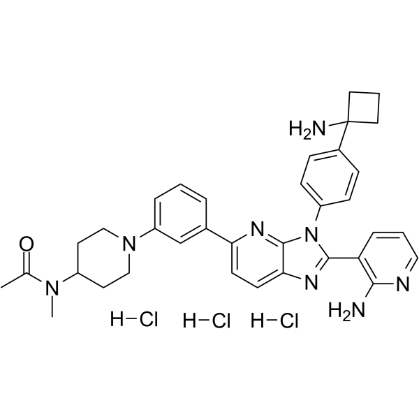
HY-137458A
Vevorisertib trihydrochlorideAKT Inhibitor -
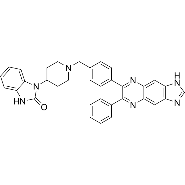
HY-10355
AKT inhibitor VIIIAKT Inhibitor -
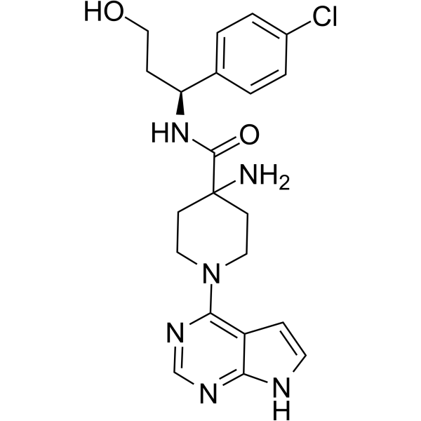
HY-15431
CapivasertibAkt Inhibitor -
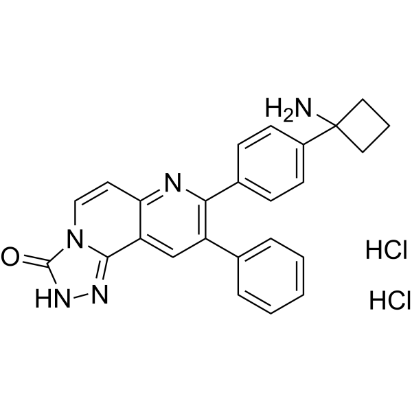
HY-10358
MK-2206 dihydrochlorideAllosteric Akt Inhibitor -
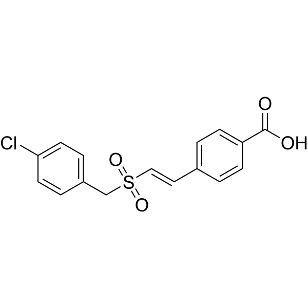
HY-101625
RecilisibRadioprotectant -
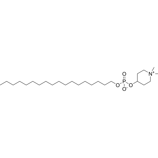
HY-50909
PerifosineAkt Inhibitor -
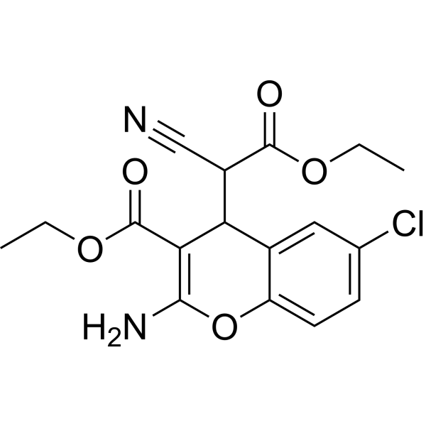
HY-18749
SC79Akt Activator -
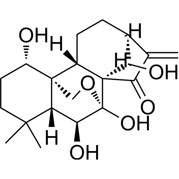
HY-N0004
OridoninBacterial Inhibitor -
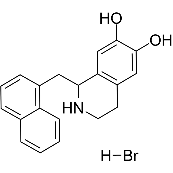
HY-15477
YS-49PI3K/Akt Activator -
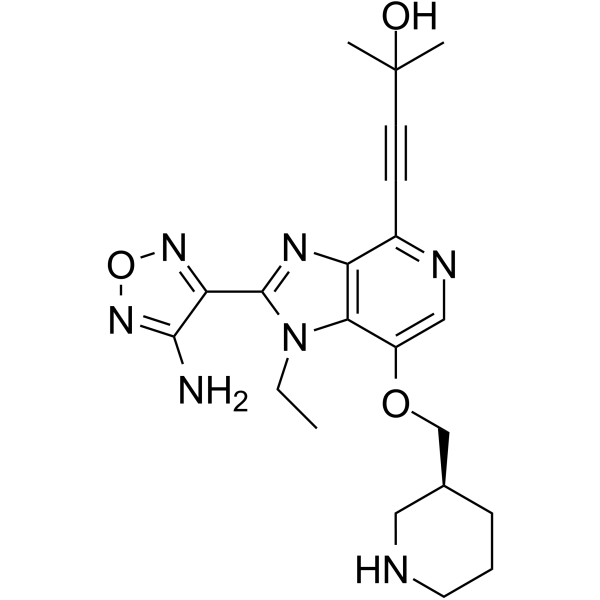
HY-10249
GSK-690693Akt Inhibitor -
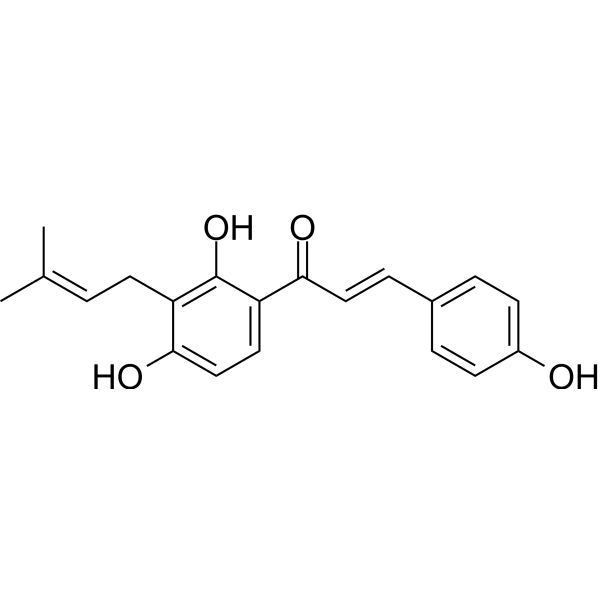
HY-13065
IsobavachalconeAkt Inhibitor -
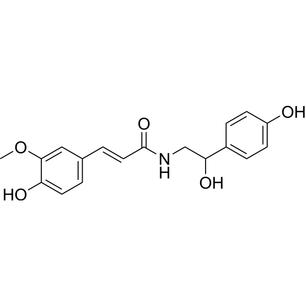
HY-N2232
N-FeruloyloctopamineNatural Product


Biological Activity
Description
IC50 & Target
[1]|
Akt1 1.4 nM (IC50) |
Akt2 1.2 nM (IC50) |
Akt3 1.7 nM (IC50) |
PKA 0.3 nM (IC50) |
P70S6K 8.9 (IC50) |
Cellular Effect
|
Cell Line

|
Type | Value | Description | References |
|---|---|---|---|---|
| HCT-116 | IC50 |
1.1 μM
Compound: E22
|
Antiproliferative activity against human HCT116 cells after 72 hrs by SRB assay
Antiproliferative activity against human HCT116 cells after 72 hrs by SRB assay
|
[PMID: 31298542] |
| LNCaP | IC50 |
0.05 μM
Compound: E22
|
Antiproliferative activity against human LNCAP cells
Antiproliferative activity against human LNCAP cells
|
[PMID: 31298542] |
| LNCaP | IC50 |
44 nM
Compound: E22
|
Inhibition of Akt in human LNCaP cells assessed as reduction in PRAS40 phosphorylation at T246 residue after 1 hr by ELISA
Inhibition of Akt in human LNCaP cells assessed as reduction in PRAS40 phosphorylation at T246 residue after 1 hr by ELISA
|
[PMID: 31298542] |
| OVCAR-8 | IC50 |
0.8 μM
Compound: E22
|
Antiproliferative activity against human OVCAR8 cells
Antiproliferative activity against human OVCAR8 cells
|
[PMID: 31298542] |


Chemical Information
-
CAS No. 2374740-21-1
-
Appearance Solid
-
Molecular Weight 526.36
-
Formula C23H23Cl2F2N5O3
-
Color White to off-white
-
SMILES
CNC(C[C@H]1NC[C@@H](NC(C2=CC(C3=C(Cl)C=NN3C)=C(Cl)O2)=O)[C@H](C4=CC(F)=C(F)C=C4)C1)=O
-
Shipping
Room temperature in continental US; may vary elsewhere.
-
Storage
Powder -20°C 3 years 4°C 2 years In solvent -80°C 6 months -20°C 1 month


Solvent & Solubility
In Vitro:
DMSO : ≥ 100 mg/mL (189.98 mM; Hygroscopic DMSO has a significant impact on the solubility of product, please use newly opened DMSO)
* "≥" means soluble, but saturation unknown.
Please refer to the solubility information to select the appropriate solvent. Once prepared, please aliquot and store the solution to prevent product inactivation from repeated freeze-thaw cycles.
Storage method and period of stock solution: -80°C, 6 months; -20°C, 1 month. When stored at -80°C, please use it within 6 months. When stored at -20°C, please use it within 1 month.
Please refer to the solubility information to select the appropriate solvent. Once prepared, please aliquot and store the solution to prevent product inactivation from repeated freeze-thaw cycles.
Storage method and period of stock solution: -80°C, 6 months; -20°C, 1 month. When stored at -80°C, please use it within 6 months. When stored at -20°C, please use it within 1 month.
Concentration (start) × Volume (start) = Concentration (final) × Volume (final)

Protocols
-
Detection of Apoptosis
Apoptosis, also called programmed cell death, is generally characterized by distinct morphological characteristics.
-
TUNEL staining for apoptotic DNA fragmentation
TUNEL staining detects DNA strand breaks by using terminal deoxynucleotidyl transferase to add labeled nucleotides to exposed 3′-OH DNA termini, generating either microscopic staining in fixed cells or tissue sections, or fluorescence/cytometric signal in cell suspensions. TUNEL positivity reflects DNA fragmentation but should not be interpreted alone as definitive apoptosis, because TUNEL can also label necrotic, autolytic, mechanically damaged, or DNA-repair-associated DNA breaks.
-
Annexin V plus membrane-impermeant dye apoptosis staining
Annexin V-based apoptosis assays rely on the detection of phosphatidylserine (PS) externalization from the inner leaflet of the plasma membrane to the outer leaflet, an early biochemical hallmark of apoptosis. Fluorescently labeled Annexin V binds PS in a calcium-dependent manner, enabling identification of early apoptotic cells by flow cytometry or fluorescence microscopy. When combined with a membrane-impermeant DNA-binding dye (e. g. , propidium iodide), this approach allows discrimination between viable (Annexin V−/dye−), early apoptotic (Annexin V+/dye−), and late apoptotic or necrotic (Annexin V+/dye+) cell populations by assessing membrane integrity and PS exposure.
-
Apoptosis Solutions
Apoptosis is a regulated, generally non-lytic cell-death pathway that removes unwanted, damaged, infected, or abnormal cells through coordinated morphological changes, caspase activation, DNA fragmentation, and membrane remodeling. The intrinsic apoptosis pathway is controlled mainly by mitochondrial outer membrane permeabilization, BCL-2 family proteins, cytochrome c release, apoptosome formation, caspase-9 activation, and downstream executioner caspase-3/7 activation. The extrinsic apoptosis pathway is initiated by death receptors such as Fas, TNFR, and TRAIL receptors, which recruit adaptor proteins and activate caspase-8 before engaging executioner caspases or mitochondrial amplification through BID cleavage. Apoptosis is linked to many phenotypes, including cancer cell killing, tissue homeostasis, immune regulation, neurodegeneration, infection response, and treatment-induced cytotoxicity; unresolved questions include how apoptosis interacts with necroptosis, pyroptosis, ferroptos
-
Orthotopic Cell-Line Xenograft
Orthotopic cell-line xenograft models involve implantation of human cancer cell lines into the anatomically corresponding organ of immunodeficient mice to reproduce tumor growth within a native microenvironment, enabling more clinically relevant tumor behavior compared with subcutaneous models. These models are widely used because orthotopic placement better recapitulates tumor progression, including invasion and metastatic spread, which are often underrepresented in heterotopic implantation systems. Compared with conventional xenografts, orthotopic implantation is described as more technically complex but provides improved simulation of tumor-microenvironment interactions and metastatic behavior, making it particularly valuable for translational oncology research. Surgical orthotopic implantation approaches have been emphasized as enabling faithful reproduction of clinical cancer features, including metastasis and disease progression patterns that align with the tumor’s organ of origi


Purity & Documentation
-
Data Sheet (274 KB)
-
SDS (251 KB)
- English - EN (251 KB)
- Français - FR (251 KB)
- Deutsch - DE (251 KB)
- Norwegian - NO (251 KB)
- Español - ES (251 KB)
- Swedish - SV (251 KB)
- Italian - IT (251 KB)
- Korean - KR (251 KB)
- Portuguese - PT (251 KB)
-
Handling Instructions (2659 KB)
References


Complete Stock Solution Preparation Table
Please refer to the solubility information to select the appropriate solvent. Once prepared, please aliquot and store the solution to prevent product inactivation from repeated freeze-thaw cycles.
Storage method and period of stock solution: -80°C, 6 months; -20°C, 1 month. When stored at -80°C, please use it within 6 months. When stored at -20°C, please use it within 1 month.
| Optional Solvent | Concentration Solvent Mass | 1 mg | 5 mg | 10 mg | 25 mg |
|---|---|---|---|---|---|
| DMSO | 1 mM | 1.8998 mL | 9.4992 mL | 18.9984 mL | 47.4960 mL |
| 5 mM | 0.3800 mL | 1.8998 mL | 3.7997 mL | 9.4992 mL | |
| 10 mM | 0.1900 mL | 0.9499 mL | 1.8998 mL | 4.7496 mL | |
| 15 mM | 0.1267 mL | 0.6333 mL | 1.2666 mL | 3.1664 mL | |
| 20 mM | 0.0950 mL | 0.4750 mL | 0.9499 mL | 2.3748 mL | |
| 25 mM | 0.0760 mL | 0.3800 mL | 0.7599 mL | 1.8998 mL | |
| 30 mM | 0.0633 mL | 0.3166 mL | 0.6333 mL | 1.5832 mL | |
| 40 mM | 0.0475 mL | 0.2375 mL | 0.4750 mL | 1.1874 mL | |
| 50 mM | 0.0380 mL | 0.1900 mL | 0.3800 mL | 0.9499 mL | |
| 60 mM | 0.0317 mL | 0.1583 mL | 0.3166 mL | 0.7916 mL | |
| 80 mM | 0.0237 mL | 0.1187 mL | 0.2375 mL | 0.5937 mL | |
| 100 mM | 0.0190 mL | 0.0950 mL | 0.1900 mL | 0.4750 mL |


AKT-IN-3 Related Classifications
HY-126257 Related Classifications
 Powered by Bioz