



Aloperine
Based on 8 publication(s) in Google Scholar
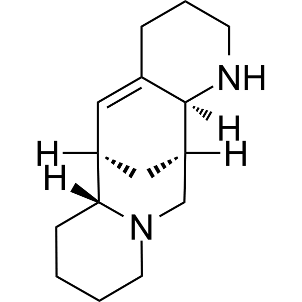
Aloperine is an alkaloid in sophora plants such as Sophora alopecuroides L, which has shown anti-cancer, anti-inflammatory and anti-virus properties. Aloperine is widely used to treat patients with allergic contact dermatitis eczema and other skin inflammation in China. Aloperine induces apoptosis and autophagy in HL-60 cells.
For research use only. We do not sell to patients.
- Purity : 99.80%
- CAS No.: 56293-29-9
- Formula: C15H24N2
- Molecular Weight:232.37
-
Storage:
4°C, sealed storage, away from moisture
* In solvent : -80°C, 6 months; -20°C, 1 month (sealed storage, away from moisture)

To place orders, for customer services and technical support, please contact: MedChemExpress USA
Tel: 609-228-6898 E-mail: [email protected] [email protected]


-
 Biological Activity
-
Chemical Information
-
Solvent & Solubility
- Protocols
- Purity & Documentation
- References
-
 Help & FAQs


-
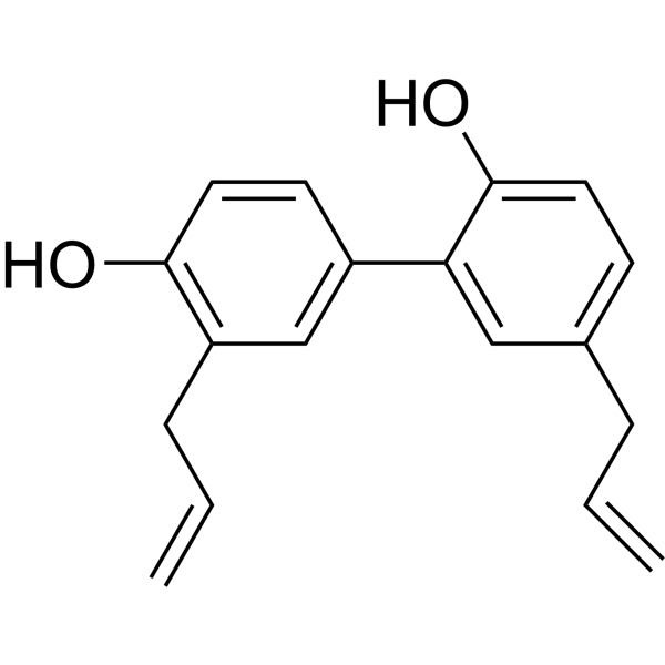
HY-N0003
HonokiolAutophagy Inducer -
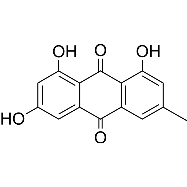
HY-14393
EmodinNatural Anthraquinone -
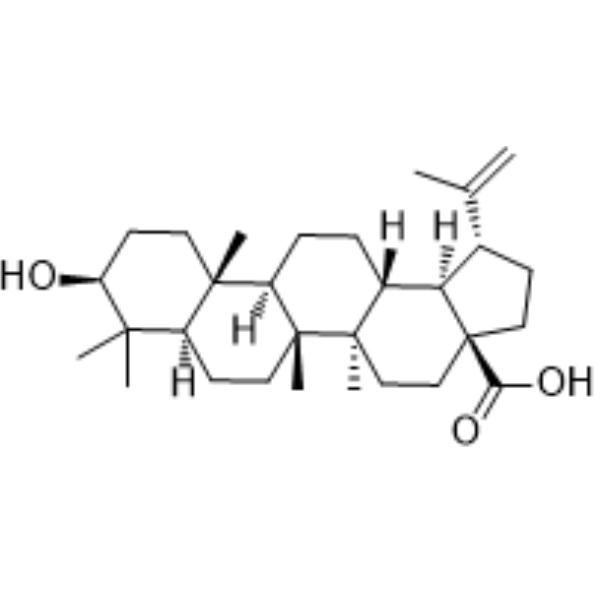
HY-10529
Betulinic acidApoptosis Inducer -
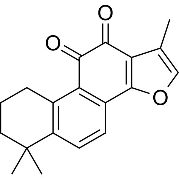
HY-N0135
Tanshinone IIAVEGFR Inhibitor -
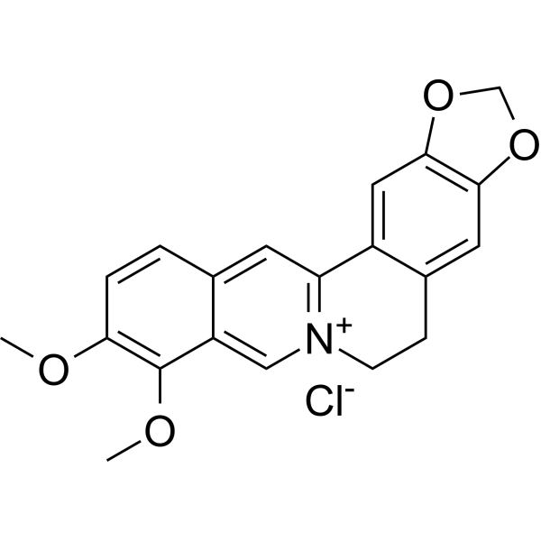
HY-18258
Berberine chlorideAntibiotic -
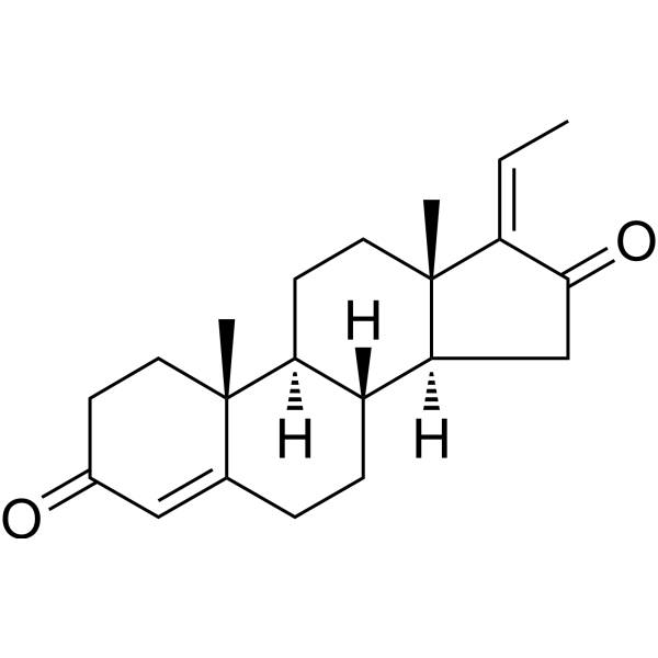
HY-107738
GuggulsteroneSterol -
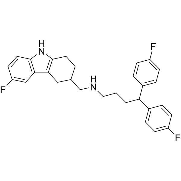
HY-123872
MSC1094308p97/VPS4B Inhibitor -

HY-13516R
Aloperine (Standard)Autophagy Inducer -
HY-17504
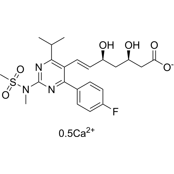
Rosuvastatin CalciumHMGCR Inhibitor -
HY-10221
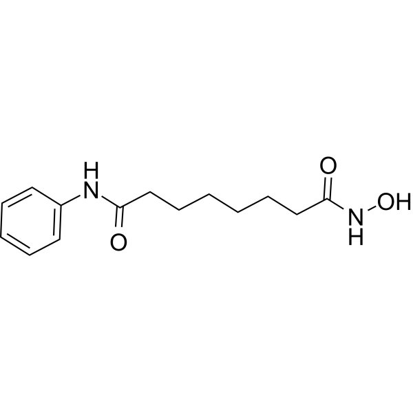
VorinostatHDAC Inhibitor -
HY-15142
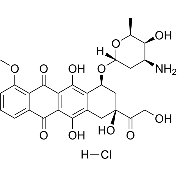
Doxorubicin hydrochlorideTopoisomerase Inhibitor -
HY-B1370
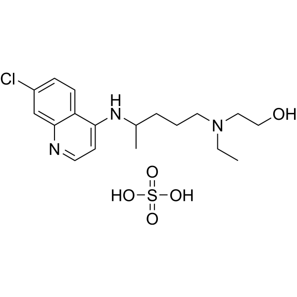
Hydroxychloroquine sulfateToll-like Receptor (TLR) Inhibitor
Publications Citing Use of MedChemExpress (MCE) Aloperine
More- Mol Biomed. 2023 Nov 17;4(1):42. [Abstract]
- Biomolecules. 2026 Jun;16(6):791. [Abstract]
- Int Immunopharmacol. 2025 Sep 23:166:115590. [Abstract]
- Int J Mol Sci. 2024 Aug 21;25(16):9083. [Abstract]
- Int Immunopharmacol. 2021 Aug:97:107720. [Abstract]
- Am J Physiol Cell Physiol. 2023 Oct 1;325(4):C1058-C1072. [Abstract]
- Vet Microbiol. 2026 May:316:110992. [Abstract]
- Res Sq. 2026 Apr 13.
 Customer Validation & Images

-
Cell Proliferation/Viability Assay
-
Cell Imaging/Staining
-
Flow Cytometry
-
WB
-
RT-PCR


Biological Activity
Description
Cellular Effect
|
Cell Line

|
Type | Value | Description | References |
|---|---|---|---|---|
| HEK-293T | CC50 |
>200 μM
Compound: Aloperine
|
Cytotoxicity against HEK293T cells assessed as reduction in cell viability
Cytotoxicity against HEK293T cells assessed as reduction in cell viability
|
[PMID: 29494844] |
| MDCK | CC50 |
>80 μM
Compound: 1
|
Cytotoxicity against MDCK cells assessed as reduction in cell viability after 48 hrs by CellTiter-Glo assay
Cytotoxicity against MDCK cells assessed as reduction in cell viability after 48 hrs by CellTiter-Glo assay
|
[PMID: 25147619] |
| MDCK | CC50 |
>80 μM
Compound: 1
|
Cytotoxicity in MDCK cells assessed as reduction of cell viability incubated for 2 days by CytoTox-Glo cytotoxicity assay
Cytotoxicity in MDCK cells assessed as reduction of cell viability incubated for 2 days by CytoTox-Glo cytotoxicity assay
|
[PMID: 29152054] |
| MDCK | EC50 |
14.5 μM
Compound: 1
|
Antiviral activity against Influenza A virus (A/Puerto Rico/8/34(H1N1)) infected in MDCK cells assessed as inhibition of virus-induced cytocidal activity after 48 hrs by CellTiter-Glo assay
Antiviral activity against Influenza A virus (A/Puerto Rico/8/34(H1N1)) infected in MDCK cells assessed as inhibition of virus-induced cytocidal activity after 48 hrs by CellTiter-Glo assay
|
[PMID: 25147619] |
| MT4 | CC50 |
>86 μM
Compound: 1
|
Cytotoxicity in human MT4 cells assessed as reduction of cell viability incubated for 3 days by CytoTox-Glo cytotoxicity assay
Cytotoxicity in human MT4 cells assessed as reduction of cell viability incubated for 3 days by CytoTox-Glo cytotoxicity assay
|
[PMID: 29152054] |
| MT4 | CC50 |
>86.2 μM
Compound: 1
|
Cytotoxicity against human MT4 cells assessed as reduction in cell viability after 3 days by CellTiter-Glo luminescent assay
Cytotoxicity against human MT4 cells assessed as reduction in cell viability after 3 days by CellTiter-Glo luminescent assay
|
[PMID: 26985308] |
| MT4 | EC50 |
1.75 μM
Compound: 1
|
Antiviral activity against HIV1 NL4-3 infected in human MT4 cells assessed as reduction in viral replication measured on day 3 post infection by Nano-Glo luciferase reporter gene assay
Antiviral activity against HIV1 NL4-3 infected in human MT4 cells assessed as reduction in viral replication measured on day 3 post infection by Nano-Glo luciferase reporter gene assay
|
[PMID: 26985308] |
In Vitro
Aloperine (1-20 μM; 24 hours) gives growth-inhibitory IC50 values in cancer cells ranges from 0.04 to 1.36 mM, the IC50 values in HL-60, U937, K562, EC109, A549 and HepG2 cells are 0.04, 0.27, 0.36, 1.11, 1.18 and 1.36 mM,respectively[1]. Aloperine (1-20 μM; 24 hours) induces apoptosis and decreases bcl-2 expression in HL-60 cells[1]. Aloperine (20–100 μM; 18 hours) induces autophagy and formation of acidic vacuole in HL-60 cells[1].
MedChemExpress (MCE) has not independently confirmed the accuracy of these methods. They are for reference only. Further protocols information, click here.


Chemical Information
-
CAS No. 56293-29-9
-
Appearance Solid
-
Molecular Weight 232.37
-
Formula C15H24N2
-
Color White to off-white
-
SMILES
[H][C@]1(C2)[C@](CCCC3)([H])N3C[C@]2([H])[C@@]4([H])C(CCCN4)=C1
-
Structure Classification
-
Initial Source
-
Shipping
Room temperature in continental US; may vary elsewhere.
-
Storage
4°C, sealed storage, away from moisture
* In solvent : -80°C, 6 months; -20°C, 1 month (sealed storage, away from moisture)


Publications (8)
-
Journal Impact Factor
-
Most Recent
-
Mol Biomed
Aloperine targets lysosomes to inhibit late autophagy and induces cell death through apoptosis and paraptosis in glioblastoma. [Abstract]2023 Nov 17;4(1):42. PMID: 37975957
Aloperine purchased from MedChemExpress. Usage Cited in: Mol Biomed. 2023 Nov 17;4(1):42. [Abstract]
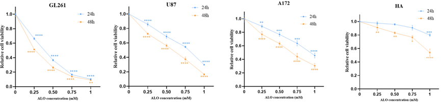
Cell viability of GL261, U87, A172, and HA exposed to Aloperine (ALO) (0.25, 0.5, 0.75, 1 mM) for different concentrations and time courses by cck-8 test.
Aloperine purchased from MedChemExpress. Usage Cited in: Mol Biomed. 2023 Nov 17;4(1):42. [Abstract]
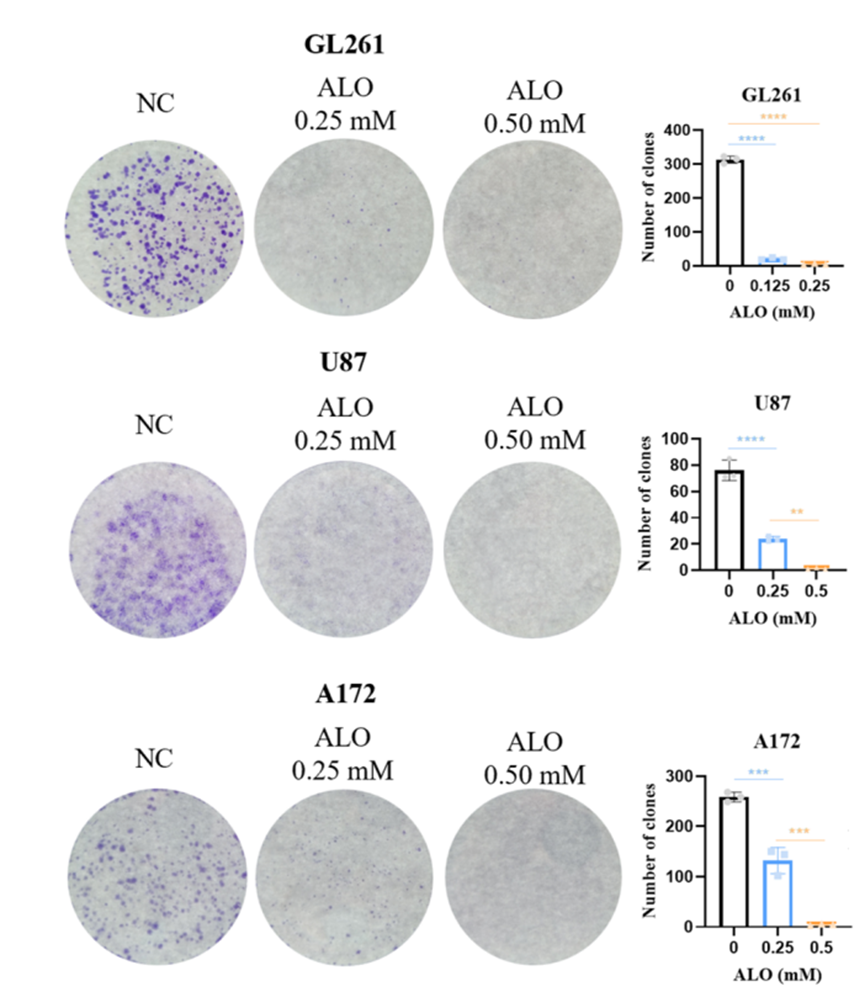
Clone forming ability of GL261, U87, and A172 single cell after Aloperine (ALO) (0.25, 0.5 mM) treatment by clone forming assay.
Aloperine purchased from MedChemExpress. Usage Cited in: Mol Biomed. 2023 Nov 17;4(1):42. [Abstract]
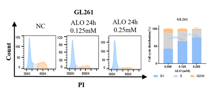
Cell cycle distribution of GL261 and U87 exposed to Aloperine (ALO) (0.125, 0.25 mM) by flow cytometry.
Aloperine purchased from MedChemExpress. Usage Cited in: Mol Biomed. 2023 Nov 17;4(1):42. [Abstract]
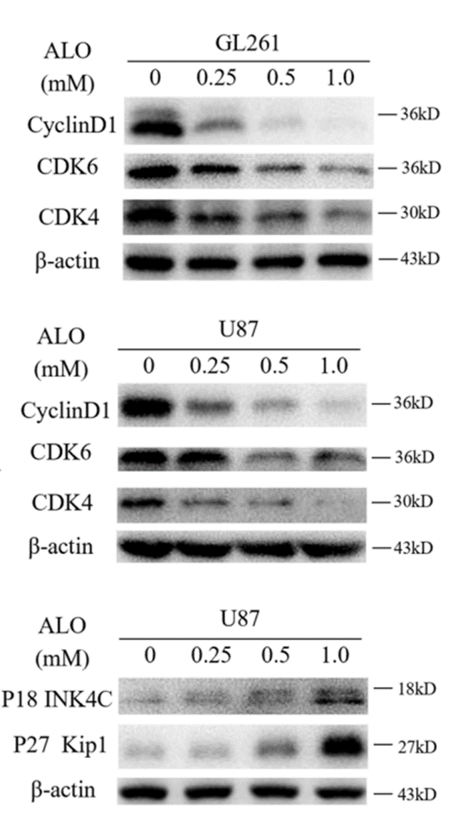
Expression of G1-phase cell cycle related proteins and cell cycle inhibitors with Aloperine (ALO) (0.25, 0.5, 1.0 mM) in GL261 and U87, tested by western blot.
Aloperine purchased from MedChemExpress. Usage Cited in: Mol Biomed. 2023 Nov 17;4(1):42. [Abstract]
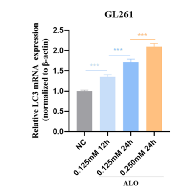
Transcriptional expression of LC3B, after Aloperine (ALO) (0.125, 0.250 mM, 24 h) treatment in GL261 tested by qPCR.
-
Biomolecules
Aloperine Suppresses the Tumorigenicity of Esophageal Squamous Cell Carcinoma by Targeting the AP-1/IL-6/STAT3 Signaling Axis. [Abstract]2026 Jun;16(6):791. PMID: 42352257 -
Int Immunopharmacol
Aloperine exerts anti-tumor effect and activates the tumor cell-intrinsic STING pathway in gallbladder cancer. [Abstract]2025 Sep 23:166:115590. PMID: 40991999 -
Int J Mol Sci
2024 Aug 21;25(16):9083. PMID: 39201769 -
Int Immunopharmacol
Aloperine improves osteoporosis in ovariectomized mice by inhibiting RANKL-induced NF-κB, ERK and JNK approaches. [Abstract]2021 Aug:97:107720. PMID: 33945918 -
Am J Physiol Cell Physiol
Aloperine Protects Pulmonary Hypertension via Triggering PPARγ Signaling and Inhibiting Calcium Regulatory Pathway in Pulmonary Arterial Smooth Muscle Cells. [Abstract]2023 Oct 1;325(4):C1058-C1072. PMID: 37661916 -
Vet Microbiol
The Chinese medicine monomer Schisandrin C inhibits PRRSV infection by regulating the OGT-PI3K/AKT/mTOR signaling pathway. [Abstract]2026 May:316:110992. PMID: 41865607 -


Solvent & Solubility
In Vitro:
DMSO : 5.56 mg/mL (23.93 mM; ultrasonic and warming and heat to 60°C; Hygroscopic DMSO has a significant impact on the solubility of product, please use newly opened DMSO)
Please refer to the solubility information to select the appropriate solvent. Once prepared, please aliquot and store the solution to prevent product inactivation from repeated freeze-thaw cycles.
Storage method and period of stock solution: -80°C, 6 months; -20°C, 1 month (sealed storage, away from moisture). When stored at -80°C, please use it within 6 months. When stored at -20°C, please use it within 1 month.
Please refer to the solubility information to select the appropriate solvent. Once prepared, please aliquot and store the solution to prevent product inactivation from repeated freeze-thaw cycles.
Storage method and period of stock solution: -80°C, 6 months; -20°C, 1 month (sealed storage, away from moisture). When stored at -80°C, please use it within 6 months. When stored at -20°C, please use it within 1 month.
Concentration (start) × Volume (start) = Concentration (final) × Volume (final)
In Vivo:
Select the appropriate dissolution method based on your experimental animal and administration route.
- For the following dissolution methods, please ensure to first prepare a clear stock solution using an In Vitro approach and then sequentially add co-solvents:
- To ensure reliable experimental results, the clarified stock solution can be appropriately stored based on storage conditions. As for the working solution for In Vivo experiments, it is recommended to prepare freshly and use it on the same day.
- The percentages shown for the solvents indicate their volumetric ratio in the final prepared solution. If precipitation or phase separation occurs during preparation, heat and/or sonication can be used to aid dissolution.
Add each solvent one by one: 10% DMSO 40% PEG300 5% Tween-80 45% Saline
Solubility: ≥ 2.08 mg/mL (8.95 mM); Clear solution
This protocol yields a clear solution of ≥ 2.08 mg/mL (saturation unknown).
Taking 1 mL working solution as an example, add 100 μL DMSO stock solution (20.8 mg/mL) to 400 μL PEG300, and mix evenly; then add 50 μL Tween-80 and mix evenly; then add 450 μL Saline to adjust the volume to 1 mL.
Preparation of Saline: Dissolve 0.9 g sodium chloride in ddH₂O and dilute to 100 mL to obtain a clear Saline solution.
Add each solvent one by one: 10% DMSO 90% (20% SBE-β-CD in Saline)
Solubility: ≥ 2.08 mg/mL (8.95 mM); Clear solution
This protocol yields a clear solution of ≥ 2.08 mg/mL (saturation unknown).
Taking 1 mL working solution as an example, add 100 μL DMSO stock solution (20.8 mg/mL) to 900 μL 20% SBE-β-CD in Saline, and mix evenly.
Preparation of 20% SBE-β-CD in Saline (4°C, storage for one week): 2 g SBE-β-CD powder is dissolved in 10 mL Saline, completely dissolve until clear.
In Vivo Dissolution Calculator
Please enter the basic information of animal experiments:
-
-
-
-
Recommended: Prepare an additional quantity of animals to account for potential losses during experiments.
Please enter your animal formula composition:
-
%DMSO +
Recommended: Keep the proportion of DMSO in working solution below 2% if your animal is weak.
-
%+
-
+%Tween-80 + +
-
%Saline +
The co-solvents required include: DMSO, . All of co-solvents are available by MedChemExpress (MCE). , Tween 80. All of co-solvents are available by MedChemExpress (MCE).
Working solution concentration: 0.22 mg/mL
Method for preparing stock solution: mg drug dissolved in μL DMSO. Stock solution concentration: mg/mL. * In solvent : -80°C, 6 months; -20°C, 1 month (sealed storage, away from moisture)
1. Take μL DMSO stock solution;
2. Add μL .
μL , mix evenly;
3. Then add μL Tween 80, mix evenly;
4. Then add μL
Please ensure that the stock solution in the first step is dissolved to a clear state, and add co-solvents in sequence. You can use ultrasonic heating (ultrasonic cleaner, recommended frequency 20-40 kHz), vortexing, etc. to assist dissolution.

Protocols
-
Apoptosis
Apoptosis, also called programmed cell death, is generally characterized by distinct morphological characteristics.
-
TUNEL staining for apoptotic DNA fragmentation
TUNEL staining detects DNA strand breaks by using terminal deoxynucleotidyl transferase to add labeled nucleotides to exposed 3′-OH DNA termini, generating either microscopic staining in fixed cells or tissue sections, or fluorescence/cytometric signal in cell suspensions. TUNEL positivity reflects DNA fragmentation but should not be interpreted alone as definitive apoptosis, because TUNEL can also label necrotic, autolytic, mechanically damaged, or DNA-repair-associated DNA breaks.
-
Imiquimod-Induced Psoriasiform Dermatitis
Imiquimod (IMQ)-induced psoriasiform dermatitis is a widely used murine model in which topical application of IMQ, a Toll-like receptor 7 (TLR7) agonist, triggers innate immune activation in the skin and induces a psoriasis-like inflammatory cascade characterized by epidermal hyperplasia, immune cell infiltration, and cytokine production dominated by the IL-23/IL-17 axis. This inflammatory response is mediated through activation of dendritic cells and downstream induction of IL-23, IL-17A, IL-22, and related pro-inflammatory mediators, recapitulating key features of human plaque psoriasis and enabling mechanistic and therapeutic studies. The model is commonly induced using Aldara (5% IMQ cream) applied topically to murine skin, resulting in rapid onset of erythema, scaling, and thickening that can be quantified as disease severity indices and validated histologically.
-
Annexin V plus membrane-impermeant dye apoptosis staining
Annexin V-based apoptosis assays rely on the detection of phosphatidylserine (PS) externalization from the inner leaflet of the plasma membrane to the outer leaflet, an early biochemical hallmark of apoptosis. Fluorescently labeled Annexin V binds PS in a calcium-dependent manner, enabling identification of early apoptotic cells by flow cytometry or fluorescence microscopy. When combined with a membrane-impermeant DNA-binding dye (e. g. , propidium iodide), this approach allows discrimination between viable (Annexin V−/dye−), early apoptotic (Annexin V+/dye−), and late apoptotic or necrotic (Annexin V+/dye+) cell populations by assessing membrane integrity and PS exposure.
-
TPA/Croton Oil Ear Edema and Dermatitis
The TPA (12-O-tetradecanoylphorbol-13-acetate) and croton oil-induced mouse ear edema model is a well-established acute cutaneous inflammation system used to evaluate topical anti-inflammatory activity by measuring edema formation, neutrophil infiltration, vascular permeability, and cytokine-mediated skin responses in vivo. The inflammatory response is triggered by topical application of phorbol esters (TPA) or croton oil constituents, leading to rapid activation of protein kinase C signaling, leukocyte recruitment, and increased vascular permeability, which can be quantified by ear thickness, weight, dye extravasation, and biochemical markers such as myeloperoxidase (MPO) activity and pro-inflammatory mediators in ear tissue homogenates. This model is widely used for screening anti-inflammatory agents, where reductions in edema and inflammatory biomarkers reflect suppression of acute dermal inflammation and immune cell infiltration. Histological evaluation typically confirms epidermal
-
Autophagy
Autophagy is a process in which eukaryotic cells use lysosomes to degrade their own cytoplasmic proteins and damaged organelles under the regulation of autophagy related gene (Atg). Microtubule-associated proteins light chain 3 (LC3) is recognized as autophagy marker, which transfers from cytoplasmic LC3 (LC3-I) to membrane type (LC3-II). LC3-II/I ratio could be detected by Western Blot and fluorescence microscopy.
-
Contact Hypersensitivity Dermatitis
Contact hypersensitivity (CHS) dermatitis is a T cell-mediated delayed-type (Type IV) immune reaction in which low-molecular-weight haptens applied to the skin bind host proteins to form complete antigens, triggering sensitization followed by a secondary inflammatory response upon re-exposure (elicitation phase), which is commonly quantified by ear swelling as a readout of skin inflammation in murine models. This model is widely used to study allergic contact dermatitis because it is antigen-specific, reproducible, and reflects key immunological events including dendritic cell activation, T cell priming in draining lymph nodes, and effector T cell-driven tissue inflammation. DNFB- and oxazolone-induced CHS models are standard systems for evaluating both acute and chronic T cell-dependent skin inflammation and for testing immunomodulatory interventions.
-
Apoptosis Solutions
Apoptosis is a regulated, generally non-lytic cell-death pathway that removes unwanted, damaged, infected, or abnormal cells through coordinated morphological changes, caspase activation, DNA fragmentation, and membrane remodeling. The intrinsic apoptosis pathway is controlled mainly by mitochondrial outer membrane permeabilization, BCL-2 family proteins, cytochrome c release, apoptosome formation, caspase-9 activation, and downstream executioner caspase-3/7 activation. The extrinsic apoptosis pathway is initiated by death receptors such as Fas, TNFR, and TRAIL receptors, which recruit adaptor proteins and activate caspase-8 before engaging executioner caspases or mitochondrial amplification through BID cleavage. Apoptosis is linked to many phenotypes, including cancer cell killing, tissue homeostasis, immune regulation, neurodegeneration, infection response, and treatment-induced cytotoxicity; unresolved questions include how apoptosis interacts with necroptosis, pyroptosis, ferroptos
-
Research Protocol for Inflammation-related Diseases
The NLRP3 inflammasome is a cytosolic innate immune signaling platform that integrates priming signals and danger-signal activation to promote caspase-1 activation, maturation of IL-1β and IL-18, and gasdermin D-mediated pyroptotic cell death. The core experimental logic is to determine whether inflammatory disease phenotypes are driven by increased NLRP3 expression, ASC-containing inflammasome assembly, caspase-1 cleavage, GSDMD cleavage, and extracellular release of IL-1β/IL-18 rather than by nonspecific cell injury alone. The pathway is strongly linked to inflammation-related disease phenotypes because monosodium urate crystals activate NALP3/NLRP3 inflammasome signaling in gout-like crystal inflammation, cholesterol crystals activate NLRP3 inflammasomes in atherogenesis models, and DSS-induced intestinal inflammation has been reported to involve NLRP3 inflammasome activity. However, experimental colitis studies also show context-dependent protective effects of NLRP3 inflammasome co
-
Lysosome and acidic-vesicle live-cell staining
Lysosome and acidic-vesicle live-cell staining detects acidic intracellular compartments by using membrane-permeant acidotropic probes that accumulate in low-pH vesicles, including lysosomes, late endosomes, autolysosomes, and acidic phagosomes. LysoTracker staining is commonly used as an intensity-based readout of acidic lysosomal compartment abundance or enlargement, while acridine orange produces green fluorescence in less concentrated compartments and red fluorescence after concentration-dependent accumulation in acidic vesicular organelles. Loss or reduction of acridine-orange red signal can be used as a readout of lysosomal membrane permeabilization or reduced acidic-vesicle integrity. This protocol is designed for live cultured cells and can be adapted for fluorescence microscopy, high-content imaging, plate-reader readout, or flow cytometry when the selected literature supports the readout. Because these dyes report acidotropic accumulation rather than lysosome identity alone,
-
Macroautophagy Solutions
Macroautophagy is a conserved lysosome-dependent degradation pathway in which cytoplasmic material is sequestered into double-membrane autophagosomes and delivered to lysosomes for degradation and recycling. The pathway supports cellular homeostasis during nutrient limitation, organelle stress, protein-aggregate accumulation, infection, differentiation, and tissue remodeling by coupling cargo sequestration, autophagosome maturation, lysosomal fusion, and degradation of cargo-derived macromolecules. The core molecular sequence includes initiation by nutrient- and stress-regulated autophagy machinery, autophagosome nucleation, LC3/ATG8-family conjugation to autophagosomal membranes, cargo selection through receptors such as SQSTM1/p62, autophagosome-lysosome fusion, and lysosomal degradation. LC3 was identified as a mammalian homolog of yeast Atg8 that localizes to autophagosomal membranes after processing, and p62/SQSTM1 was shown to connect ubiquitinated cargo with autophagic degradati


Purity & Documentation
-
Data Sheet (277 KB)
-
SDS (392 KB)
- English - EN (392 KB)
- Français - FR (392 KB)
- Deutsch - DE (392 KB)
- Norwegian - NO (392 KB)
- Español - ES (392 KB)
- Swedish - SV (392 KB)
- Italian - IT (392 KB)
- Korean - KR (392 KB)
- Portuguese - PT (392 KB)
-
Handling Instructions (2659 KB)
References
[1]. Lin Z, et al. In vitro anti-tumour activities of quinolizidine alkaloids derived from Sophora flavescens Ait. Basic Clin Pharmacol Toxicol. 2011 May;108(5):304-9. [Content Brief]
[2]. Yuan XY, et al. Effects and mechanisms of aloperine on 2, 4-dinitrofluorobenzene-induced allergic contact dermatitis in BALB/c mice. Eur J Pharmacol. 2010 Mar 10;629(1-3):147-52. [Content Brief]


Complete Stock Solution Preparation Table
Please refer to the solubility information to select the appropriate solvent. Once prepared, please aliquot and store the solution to prevent product inactivation from repeated freeze-thaw cycles.
Storage method and period of stock solution: -80°C, 6 months; -20°C, 1 month (sealed storage, away from moisture). When stored at -80°C, please use it within 6 months. When stored at -20°C, please use it within 1 month.
| Optional Solvent | Concentration Solvent Mass | 1 mg | 5 mg | 10 mg | 25 mg |
|---|---|---|---|---|---|
| DMSO | 1 mM | 4.3035 mL | 21.5173 mL | 43.0346 mL | 107.5866 mL |
| 5 mM | 0.8607 mL | 4.3035 mL | 8.6069 mL | 21.5173 mL | |
| 10 mM | 0.4303 mL | 2.1517 mL | 4.3035 mL | 10.7587 mL | |
| 15 mM | 0.2869 mL | 1.4345 mL | 2.8690 mL | 7.1724 mL | |
| 20 mM | 0.2152 mL | 1.0759 mL | 2.1517 mL | 5.3793 mL |


Aloperine Related Classifications
HY-13516 Related Classifications
 Powered by Bioz