



ARD-1676
Based on 1 publication(s) in Google Scholar
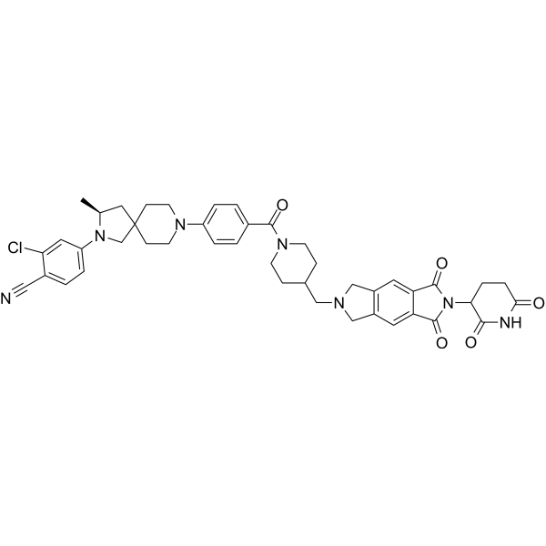
ARD-1676 is an orally active androgen receptor (AR) PROTAC degrader. ARD-1676 induces proteasomal degradation of AR, inhibits AR-regulated gene expression, suppresses cell growth, reduces AR protein levels and inhibits tumor growth in in vivo models. ARD-1676 can be used in studies related to AR+ human prostate cancer and spinal bulbar muscular atrophy.
(Pink: Androgen Receptor ligand (HY-150878); Blue: Cereblon ligand (HY-W248665); Black: linker).
For research use only. We do not sell to patients.
- CAS No.: 2632305-36-1
- Formula: C44H46ClN7O5
- Molecular Weight:788.33
-
Storage:
4°C, protect from light
* In solvent : -80°C, 6 months; -20°C, 1 month (protect from light)

To place orders, for customer services and technical support, please contact: MedChemExpress USA
Tel: 609-228-6898 E-mail: [email protected] [email protected]


-
 Biological Activity
-
Chemical Information
-
Solvent & Solubility
- Protocols
- Purity & Documentation
- References
-
 Help & FAQs


Publications Citing Use of MedChemExpress (MCE) ARD-1676
MoreAll PROTACs Isoforms
More

Biological Activity
Description
Cellular Effect
|
Cell Line

|
Type | Value | Description | References |
|---|---|---|---|---|
| LNCaP | IC50 |
2.8 nM
Compound: 39; ARD-1676
|
Growth inhibition in human LNCaP cells assessed as reduction in cell viability
Growth inhibition in human LNCaP cells assessed as reduction in cell viability
|
[PMID: 37683104] |
| VCaP | IC50 |
11.5 nM
Compound: 39; ARD-1676
|
Growth inhibition in human VCaP cells
Growth inhibition in human VCaP cells
|
[PMID: 37683104] |
In Vitro
ARD-1676 (4 days) inhibits the growth of VCaP human prostate cancer cells, with an IC50 of 11.5 nM after 4 days of co-incubation with 0.1 nM R1881[1].
ARD-1676 (4 days) inhibits the growth of human prostate cancer LNCaP cells, with an IC50 of 2.8 nM after 4 days of co-incubation with 0.1 nM R1881[1].
ARD-1676 (10-100 nM; 1-24 h) induces rapid degradation of AR in human prostate cancer cell lines VCaP and LNCaP. At concentrations of 10 nM and 100 nM, the degradation rate exceeds 50% within 1 h, and reaches the maximum degradation level at 3-6 h[1].
ARD-1676 (0.03-30 nM; 24 h) potently inhibits AR-regulated KLK3 and TMPRSS2 gene expression in VCaP and LNCaP human prostate cancer cells[1].
ARD-1676 (1 nM-10 μM; 24 h) potently and efficiently degrades most clinically relevant AR mutants (including wild-type, point, and deletion mutants) in HEK293 cells, but exhibits low activity against ARL702H and ARS889G mutants[1].
ARD-1676 (0.1-1000 nM; 15 min-48 h) potently and rapidly induces proteasomal degradation of polyglutamine AR112Q in Tet-on PC12 cells. Its effect takes effect as early as 15 min and persists for up to 48 h, and it also effectively degrades both cytoplasmic and nuclear pools of this misfolded protein[2].
ARD-1676 (100 nM; 24 h) potently reduces AR protein levels in control and SBMA patient-derived iMNs without altering AR mRNA expression or inducing cytotoxicity[2].
MedChemExpress (MCE) has not independently confirmed the accuracy of these methods. They are for reference only. Further protocols information, click here.
-
Cell Line:HEK293 cells overexpressing clinically relevant AR mutants (wild-type, K388R, Δ388-390, L702H, V716M, W742C, H875Y, F877L, T878A, S889G, Δ873-879)
-
Concentration:1, 10, 100 nM, 1 and 10 μM
-
Incubation Time:24 h (in the presence of 50 μg/mL cycloheximide)
-
Result:Potently degraded wild-type, K388R, V716M, W742C, H875Y, F877L, and T878A AR mutants at concentrations as low as 1 nM.
Effectively depleted Δ388-390 and Δ873-879 deletion mutants.
Showed reduced efficacy against L702H and S889G mutants relative to other variants.
-
Cell Line:VCaP and LNCaP AR+ human prostate cancer cell lines
-
Concentration:0.03, 0.1, 0.3, 1, 3, 10 and 30 nM (in the presence of 0.1 nM R1881)
-
Incubation Time:24 h
-
Result:Reduced KLK3 and TMPRSS2 mRNA levels by > 50% at 1 nM in VCaP cells and 3 nM in LNCaP cells, demonstrating ~100-fold greater potency than enzalutamide for suppressing AR-regulated gene expression.
Parmacokinetics
| Species | Dose | Route | T1/2 | AUC0-t | Vss | CL | Tmax | Cmax | F |
|---|---|---|---|---|---|---|---|---|---|
| Mice[1] | 2 mg/kg | i.v. | 4.3 h | 50538 ng·h/mL | 0.23 L/kg | 0.04 L/h/kg | / | / | / |
| Mice[1] | 5 mg/kg | p.o. | 4.4 h | 85243 ng·h/mL | / | / | 2.0 h | 9124 ng/mL | 67 % |
| Rat[1] | 2 mg/kg | i.v. | 4.5 h | 5492 ng·h/mL | 1.7 L/kg | 0.36 L/h/kg | / | / | / |
| Rat[1] | 5 mg/kg | p.o. | 5.6 h | 6016 ng·h/mL | / | / | 4.0 h | 871 ng/mL | 44 % |
| Dog[1] | 1 mg/kg | i.v. | 9.9 h | 4857 ng·h/mL | 1.74 L/kg | 0.19 L/h/kg | / | / | / |
| Dog[1] | 10 mg/kg | p.o. | 27.4 h | 15170 ng·h/mL | / | / | 3.0 h | 1031 ng/mL | 31 % |
| Cynomolgus Monkey[1] | 1 mg/kg | i.v. | 10 h | 5171 ng·h/mL | 2.07 L/kg | 0.2 L/h/kg | / | / | / |
| Cynomolgus Monkey[1] | 2 mg/kg | p.o. | 9.62 h | 10302 ng·h/mL | / | / | 3.33 h | 1520 ng/mL | 99 % |
In Vivo
ARD-1676 (30 mg/kg; p.o.; single administration) achieves in vivo target engagement in AR113Q SBMA mice by significantly reducing polyQ AR protein levels in skeletal muscle, without inducing acute toxicity[2].
MedChemExpress (MCE) has not independently confirmed the accuracy of these methods. They are for reference only.
-
Animal Model:SCID mice (male)[1]
-
Dosage:10 mg/kg (TGI); 20 mg/kg (TGI); 40 mg/kg (TGI); 12.5 mg/kg (AR protein reduction)
-
Administration:p.o.; once daily for 45 days (for TGI); single dose (for AR protein reduction)
-
Result:Reduced AR protein levels in tumor tissue by 96% at 6 h and 93% at 24 h.
Achieved tumor growth inhibition (TGI) of 50% at 10 mg/kg, 68% at 20 mg/kg, and 85% at 40 mg/kg.
Caused no animal weight loss or signs of toxicity during treatment.
-
Animal Model:AR113Q knock-in (male, 8-10 weeks old)[2]
-
Dosage:30 mg/kg
-
Administration:p.o.; single dose
-
Result:Reduced polyQ androgen receptor (AR) protein levels significantly in levator ani/bulbocavernosus muscle compared to vehicle control.
Showed no change in Ar mRNA expression in tibialis anterior muscle.
Detected no significant change in AR protein levels in lumbar spinal cord.
Observed no alterations in serum liver or kidney function biomarkers, indicating no acute toxicity.


Chemical Information
-
CAS No. 2632305-36-1
-
Appearance Solid
-
Molecular Weight 788.33
-
Formula C44H46ClN7O5
-
Color White to light yellow
-
SMILES
C[C@H]1CC2(CN1C3=CC=C(C(Cl)=C3)C#N)CCN(CC2)C4=CC=C(C=C4)C(N5CCC(CC5)CN6CC7=C(C6)C=C8C(N(C(C8=C7)=O)C9CCC(NC9=O)=O)=O)=O
-
Shipping
Room temperature in continental US; may vary elsewhere.
-
Storage
4°C, protect from light
* In solvent : -80°C, 6 months; -20°C, 1 month (protect from light)


Publications (1)
-
Journal Impact Factor
-
Most Recent
-
Anal Chem
Hydrogen/Deuterium Exchange for Chiral Stability Assessment in Acidic Methine-Containing Compounds. [Abstract]2025 Dec 2;97(47):26097-26107. PMID: 41243541


Solvent & Solubility
In Vitro:
DMSO : 100 mg/mL (126.85 mM; Need ultrasonic; Hygroscopic DMSO has a significant impact on the solubility of product, please use newly opened DMSO)
Please refer to the solubility information to select the appropriate solvent. Once prepared, please aliquot and store the solution to prevent product inactivation from repeated freeze-thaw cycles.
Storage method and period of stock solution: -80°C, 6 months; -20°C, 1 month (protect from light). When stored at -80°C, please use it within 6 months. When stored at -20°C, please use it within 1 month.
Please refer to the solubility information to select the appropriate solvent. Once prepared, please aliquot and store the solution to prevent product inactivation from repeated freeze-thaw cycles.
Storage method and period of stock solution: -80°C, 6 months; -20°C, 1 month (protect from light). When stored at -80°C, please use it within 6 months. When stored at -20°C, please use it within 1 month.
Concentration (start) × Volume (start) = Concentration (final) × Volume (final)
In Vivo:
Select the appropriate dissolution method based on your experimental animal and administration route.
- For the following dissolution methods, please ensure to first prepare a clear stock solution using an In Vitro approach and then sequentially add co-solvents:
- To ensure reliable experimental results, the clarified stock solution can be appropriately stored based on storage conditions. As for the working solution for In Vivo experiments, it is recommended to prepare freshly and use it on the same day.
- The percentages shown for the solvents indicate their volumetric ratio in the final prepared solution. If precipitation or phase separation occurs during preparation, heat and/or sonication can be used to aid dissolution.
Add each solvent one by one: 10% DMSO 40% PEG300 5% Tween-80 45% Saline
Solubility: ≥ 2.5 mg/mL (3.17 mM); Clear solution
This protocol yields a clear solution of ≥ 2.5 mg/mL (saturation unknown).
Taking 1 mL working solution as an example, add 100 μL DMSO stock solution (25.0 mg/mL) to 400 μL PEG300, and mix evenly; then add 50 μL Tween-80 and mix evenly; then add 450 μL Saline to adjust the volume to 1 mL.
Preparation of Saline: Dissolve 0.9 g sodium chloride in ddH₂O and dilute to 100 mL to obtain a clear Saline solution.
Add each solvent one by one: 10% DMSO 90% (20% SBE-β-CD in Saline)
Solubility: ≥ 2.5 mg/mL (3.17 mM); Clear solution
This protocol yields a clear solution of ≥ 2.5 mg/mL (saturation unknown).
Taking 1 mL working solution as an example, add 100 μL DMSO stock solution (25.0 mg/mL) to 900 μL 20% SBE-β-CD in Saline, and mix evenly.
Preparation of 20% SBE-β-CD in Saline (4°C, storage for one week): 2 g SBE-β-CD powder is dissolved in 10 mL Saline, completely dissolve until clear.
In Vivo Dissolution Calculator
Please enter the basic information of animal experiments:
-
-
-
-
Recommended: Prepare an additional quantity of animals to account for potential losses during experiments.
Please enter your animal formula composition:
-
%DMSO +
Recommended: Keep the proportion of DMSO in working solution below 2% if your animal is weak.
-
%+
-
+%Tween-80 + +
-
%Saline +
The co-solvents required include: DMSO, . All of co-solvents are available by MedChemExpress (MCE). , Tween 80. All of co-solvents are available by MedChemExpress (MCE).
Working solution concentration: 0.22 mg/mL
Method for preparing stock solution: mg drug dissolved in μL DMSO. Stock solution concentration: mg/mL. * In solvent : -80°C, 6 months; -20°C, 1 month (protect from light)
1. Take μL DMSO stock solution;
2. Add μL .
μL , mix evenly;
3. Then add μL Tween 80, mix evenly;
4. Then add μL
Please ensure that the stock solution in the first step is dissolved to a clear state, and add co-solvents in sequence. You can use ultrasonic heating (ultrasonic cleaner, recommended frequency 20-40 kHz), vortexing, etc. to assist dissolution.

Protocols
-
RT-PCR
Reverse transcription technology uses RNA as a template to synthesize DNA. RT-PCR is simple, specific and sensitive, and can be used to detect gene expression levels and expression differences in cells; detect RNA virus content; clone cDNA sequences of specific genes.
-
RNA extraction experimental
By lysing cells, releasing RNA, and removing impurities such as proteins and DNA, high-purity RNA products are finally obtained. The commonly used traditional method is the guanidine isothiocyanate/phenol/chloroform method (Trizol), which is suitable for a variety of animal materials including animal tissues, microorganisms, cultured cells, etc., and most plant materials.
-
Somatic Cell Culture
A method of simulating the in vivo environment in vitro to maintain the cell growth, differentation and main functions.
-
Patient-Derived Orthotopic Xenograft (PDOX)
Patient-derived orthotopic xenograft (PDOX) modeling implants fresh patient tumor tissue or patient-derived tumor cells into the anatomically corresponding organ or tissue site of immunodeficient mice, usually by surgical orthotopic implantation, to preserve patient tumor histology, local microenvironmental context, invasion, metastatic behavior, and treatment-response features better than subcutaneous implantation. PDOX readouts include tumor engraftment, orthotopic tumor growth, local invasion, metastasis, recurrence after resection, histologic similarity to the donor tumor, biomarker retention, molecular concordance, survival, and response or resistance to therapy. PDOX models are used for preclinical drug testing and individualized therapy evaluation, but engraftment success varies by tumor type and specimen quality.
-
CCK-8/WST-8 Cell Proliferation Assay
The CCK-8/WST-8 assay is based on the reduction of the water-soluble tetrazolium salt WST-8 to a water-soluble formazan product by cellular dehydrogenases in metabolically active cells, where the generated formazan amount is proportional to the number of living cells and is quantified by measuring absorbance in the visible range, providing a colorimetric readout for cell viability and proliferation assessment. This class of tetrazolium-based assays improves upon earlier MTT-based systems by producing a water-soluble formazan, eliminating the need for organic solubilization steps and enabling direct spectrophotometric measurement in culture medium.
-
Cell Counting-Based Growth Curve Assay
Cell counting-based growth curve assays quantify cell proliferation by directly measuring changes in viable cell number over time using manual or automated counting methods such as hemocytometer-based counting or instrument-assisted cell enumeration, enabling construction of growth curves that reflect population expansion dynamics in response to culture conditions. A widely used approach is trypan blue exclusion with hemocytometer counting, where membrane-compromised (non-viable) cells take up the dye, allowing discrimination between viable and non-viable cells while simultaneously enabling total cell number quantification. Repeated sampling across time points allows estimation of proliferation rate, growth phases, and comparative growth kinetics between experimental conditions.
-
Real Time qPCR (Q-PCR)
Real-time quantitative PCR (qPCR) quantifies an amplifiable nucleic-acid target by monitoring fluorescence during PCR cycling rather than measuring product only after amplification. The increase in fluorescence tracks accumulation of PCR product, and the quantification cycle (Cq; historically also Ct/CP) is related to the initial amount of target: samples containing more starting target generally reach the defined fluorescence threshold in fewer cycles.
-
Cell Viability Determination by MTT Colorimetric Assay
The following protocol uses the MTT colorimetric assay as a classic literature-established method for assessing cell viability/metabolic activity in cultured mammalian cells. MTT[3-(4,5-dimethylthiazol-2-yl)-2,5-diphenyltetrazolium bromide] is reduced by metabolically active cells to a colored formazan product; the amount of formazan is quantified spectrophotometrically and provides an indirect measure of metabolically active viable cells. Importantly, MTT reduction reflects cellular oxidoreductase/metabolic activity rather than an absolute direct count of living cells, so changes in cellular metabolism can alter the signal independently of cell number.
-
Protocol for Cell Counting and Cell Density Analysis
Cell counting and cell-density analysis estimate the number of cells in a known volume or field area. Manual hemocytometer counting uses a chamber of defined geometry to convert counted cells into cells/mL, while automated counters and image-analysis workflows detect cell objects from optical, brightfield, fluorescence, impedance, or digital-image features. Trypan blue viability counting is based on dye exclusion: viable cells with intact membranes exclude dye, while non-viable cells with compromised membranes stain blue. The readout is total cell density, viable-cell density, dead-cell density, and percent viability. Cell density can also be estimated from microscopy images by counting objects per image area, from flow cytometry using calibrated volume or reference particles, or from in situ microscopy in bioreactors after calibration against reference methods such as hemocytometer or flow cytometry.
-
Research Protocol for Endocrine Diseases
Endocrine diseases often arise from disrupted hormone production, hormone signaling, or target-tissue responsiveness; for diabetes-focused endocrine disease models, insulin signaling regulates glucose uptake, hepatic glucose output, lipid metabolism, and β-cell compensation. Type 2 diabetes develops through interacting defects in insulin resistance, β-cell dysfunction, adipose inflammation, hepatic glucose overproduction, altered incretin signaling, and ectopic lipid metabolism. A major unresolved question is whether endocrine dysfunction is driven primarily by target-tissue insulin resistance, intrinsic β-cell failure, immune/inflammatory stress, or combined multi-organ failure that differs by disease stage.
-
Patient-Derived Xenograft (PDX)
Patient-derived xenograft (PDX) models are generated by engrafting primary human tumor tissue directly into immunodeficient mice, allowing in vivo propagation of patient tumor biology without initial in vitro adaptation. These models are used to preserve key histopathological and molecular characteristics of the original tumor and enable assessment of tumor growth dynamics and therapeutic response in a living organism. The biological readout is tumor engraftment and subsequent growth in the murine host, which reflects the ability of human tumor cells to survive, vascularize, and expand in an immunocompromised microenvironment.
-
MTT Cell Proliferation Assay
The MTT assay is a colorimetric endpoint assay for estimating viable cell number, cell growth, cytotoxicity, or cell activation in cultured mammalian cells. Living cells reduce the yellow tetrazolium salt MTT into purple/blue formazan, while dead cells do not generate the same signal; the resulting color can be quantified with a multiwell spectrophotometer. MTT reduction is commonly interpreted as a readout of metabolic activity that often correlates with viable cell number, but it should not be treated as a direct cell-counting method unless the assay is optimized for the cell type and experimental condition. Studies show that MTT reduction can involve mitochondrial and non-mitochondrial reducing systems, and formazan may accumulate in intracellular lipid droplets rather than simply marking mitochondria.
-
Subcutaneous Cell-Line-Derived Xenograft
Subcutaneous cell-line-derived xenograft (CDX) models are established by implanting cultured human cancer cell lines into immunodeficient mice, where the injected cells form localized tumors that can be monitored in vivo as a measure of tumorigenic potential, growth kinetics, and treatment response. These models are widely used in oncology research because they allow reproducible tumor formation and enable comparative assessment of tumor growth between different cell lines or genetic manipulations in a controlled in vivo microenvironment. Subcutaneous implantation of cancer cells in immunodeficient mice is a standard approach for evaluating tumor growth behavior and therapeutic response across multiple cancer types, including prostate, esophageal, pancreatic, and colon cancer models.
-
Orthotopic Cell-Line Xenograft
Orthotopic cell-line xenograft models involve implantation of human cancer cell lines into the anatomically corresponding organ of immunodeficient mice to reproduce tumor growth within a native microenvironment, enabling more clinically relevant tumor behavior compared with subcutaneous models. These models are widely used because orthotopic placement better recapitulates tumor progression, including invasion and metastatic spread, which are often underrepresented in heterotopic implantation systems. Compared with conventional xenografts, orthotopic implantation is described as more technically complex but provides improved simulation of tumor-microenvironment interactions and metastatic behavior, making it particularly valuable for translational oncology research. Surgical orthotopic implantation approaches have been emphasized as enabling faithful reproduction of clinical cancer features, including metastasis and disease progression patterns that align with the tumor’s organ of origi


Purity & Documentation
-
Data Sheet (284 KB)
-
SDS (252 KB)
- English - EN (252 KB)
- Français - FR (252 KB)
- Deutsch - DE (252 KB)
- Norwegian - NO (252 KB)
- Español - ES (252 KB)
- Swedish - SV (252 KB)
- Italian - IT (252 KB)
- Korean - KR (252 KB)
- Portuguese - PT (252 KB)
-
Handling Instructions (2659 KB)
References
[1]. Xiang W, et al. Discovery of ARD-1676 as a Highly Potent and Orally Efficacious AR PROTAC Degrader with a Broad Activity against AR Mutants for the Treatment of AR + Human Prostate Cancer. Journal of medicinal chemistry. 2023 Sep 28;66(18):13280-13303. [Content Brief]
[2]. Sangotra A, et al. PROTACs therapeutically target the polyglutamine androgen receptor in spinal and bulbar muscular atrophy models. Neurotherapeutics : the journal of the American Society for Experimental NeuroTherapeutics. 2025 Oct;22(6):e00732. [Content Brief]


Complete Stock Solution Preparation Table
Please refer to the solubility information to select the appropriate solvent. Once prepared, please aliquot and store the solution to prevent product inactivation from repeated freeze-thaw cycles.
Storage method and period of stock solution: -80°C, 6 months; -20°C, 1 month (protect from light). When stored at -80°C, please use it within 6 months. When stored at -20°C, please use it within 1 month.
| Optional Solvent | Concentration Solvent Mass | 1 mg | 5 mg | 10 mg | 25 mg |
|---|---|---|---|---|---|
| DMSO | 1 mM | 1.2685 mL | 6.3425 mL | 12.6850 mL | 31.7126 mL |
| 5 mM | 0.2537 mL | 1.2685 mL | 2.5370 mL | 6.3425 mL | |
| 10 mM | 0.1269 mL | 0.6343 mL | 1.2685 mL | 3.1713 mL | |
| 15 mM | 0.0846 mL | 0.4228 mL | 0.8457 mL | 2.1142 mL | |
| 20 mM | 0.0634 mL | 0.3171 mL | 0.6343 mL | 1.5856 mL | |
| 25 mM | 0.0507 mL | 0.2537 mL | 0.5074 mL | 1.2685 mL | |
| 30 mM | 0.0423 mL | 0.2114 mL | 0.4228 mL | 1.0571 mL | |
| 40 mM | 0.0317 mL | 0.1586 mL | 0.3171 mL | 0.7928 mL | |
| 50 mM | 0.0254 mL | 0.1269 mL | 0.2537 mL | 0.6343 mL | |
| 60 mM | 0.0211 mL | 0.1057 mL | 0.2114 mL | 0.5285 mL | |
| 80 mM | 0.0159 mL | 0.0793 mL | 0.1586 mL | 0.3964 mL | |
| 100 mM | 0.0127 mL | 0.0634 mL | 0.1269 mL | 0.3171 mL |


ARD-1676 Related Classifications
HY-156111 Related Classifications
 Powered by Bioz
Keywords
- ARD-1676
- 2632305-36-1
- ARD1676
- ARD 1676
- PROTACs
- Androgen Receptor
- MDA-Pca-2b human prostate cancer cells
- cereblon
- LNCaP human prostate cancer cells
- SBMA patient-derived iSKMs
- VCaP human prostate cancer cells
- androgen receptor
- spinal and bulbar muscular atrophy
- Tet-on PC12 cells
- SBMA patient-derived iMNs
- HEK293 cells
- Inhibitor
- inhibitor
- inhibit