



AZ10397767
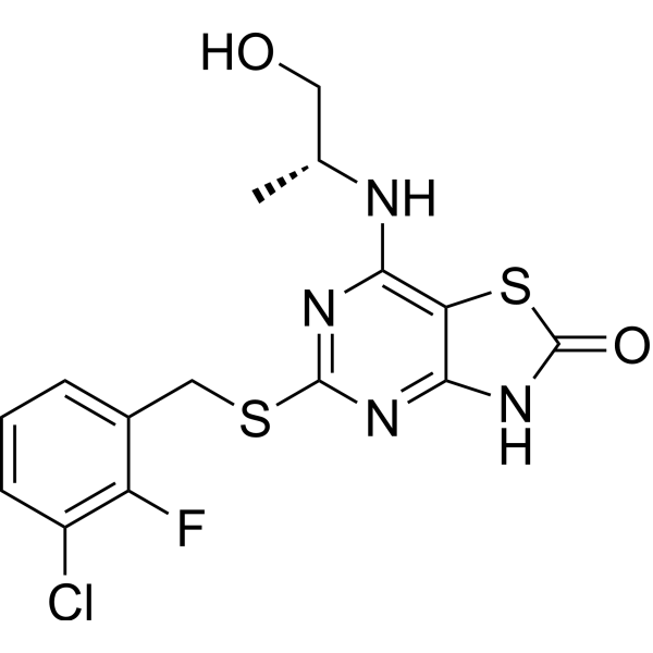
AZ10397767 is an orally active, selective CXCR2 receptor antagonist with an IC50 of 1 nM. AZ10397767 attenuates the Oxaliplatin (HY-17371)-induced NF-κB transcriptional activity and potentiates Oxaliplatin-induced apoptosis in androgen-independent prostate cancer (AIPC) cells. AZ10397767 significantly inhibits neutrophil recruitment into tumors which then adversely affects tumor growth in vitro and in vivo.
For research use only. We do not sell to patients.
- CAS No.: 333742-63-5
- Formula: C15H14ClFN4O2S2
- Molecular Weight:400.88
-
Storage:
Please store the product under the recommended conditions in the Certificate of Analysis.

To place orders, for customer services and technical support, please contact: MedChemExpress USA
Tel: 609-228-6898 E-mail: [email protected] [email protected]


-
 Biological Activity
-
Chemical Information
- Protocols
- Purity & Documentation
- References
-
 Help & FAQs


-
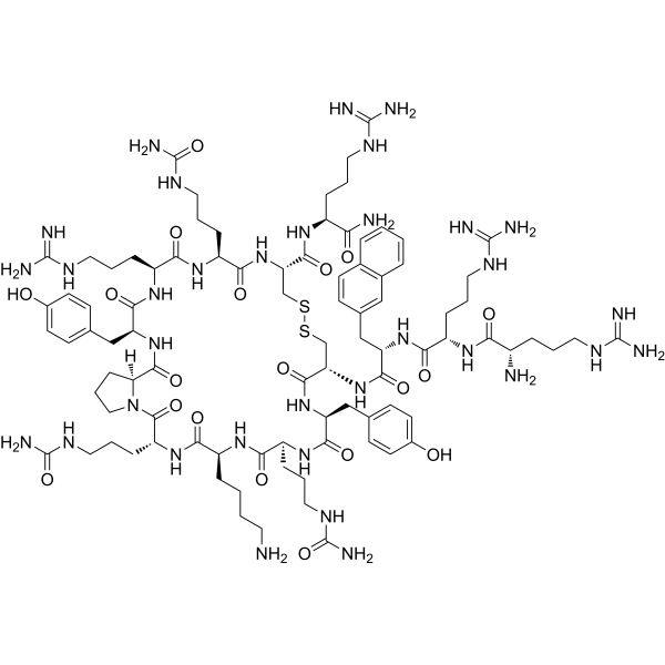
HY-P1102
TC14012CXCR4 Antagonist -
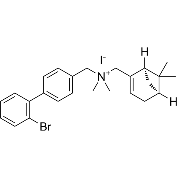
HY-110269
VUF 11222CXCR3 Agonist -
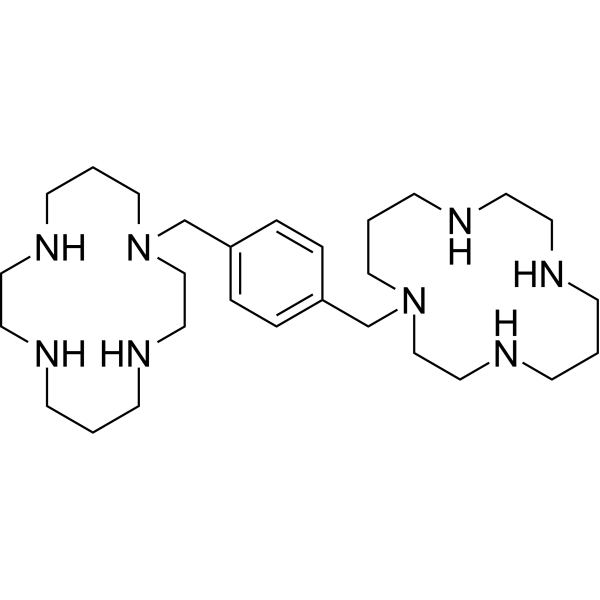
HY-10046
PlerixaforCXCR4 Antagonist -
HY-16711
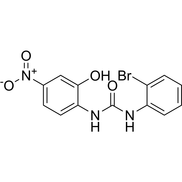
SB225002CXCR2 Antagonist -
HY-15319
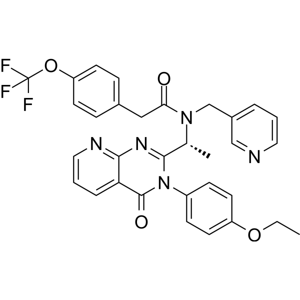
AMG 487CXCR3 Antagonist -
HY-15251
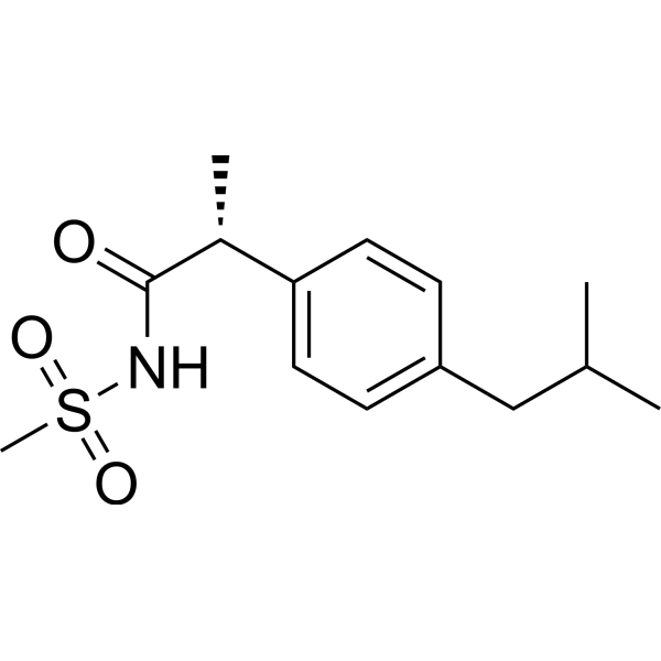
ReparixinCXCR1/2 Inhibitor -
HY-50912
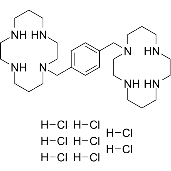
Plerixafor octahydrochlorideCXCR4 Antagonist -
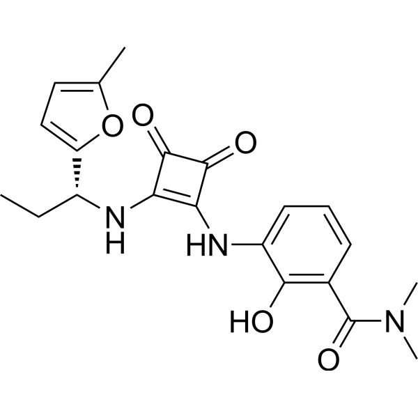
HY-10198
NavarixinCXCR1/2 Antagonist -
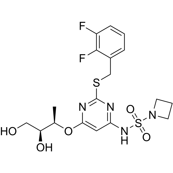
HY-19855
AZD-5069CXCR2 Antagonist -
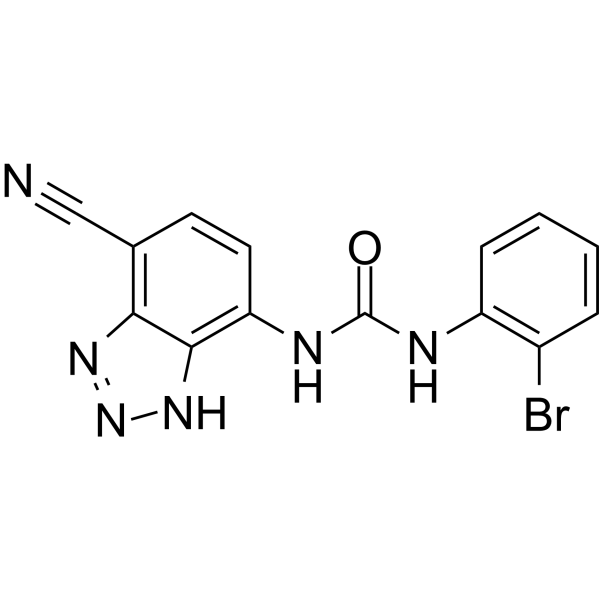
HY-50688
SB-265610CXCR2 Antagonist -
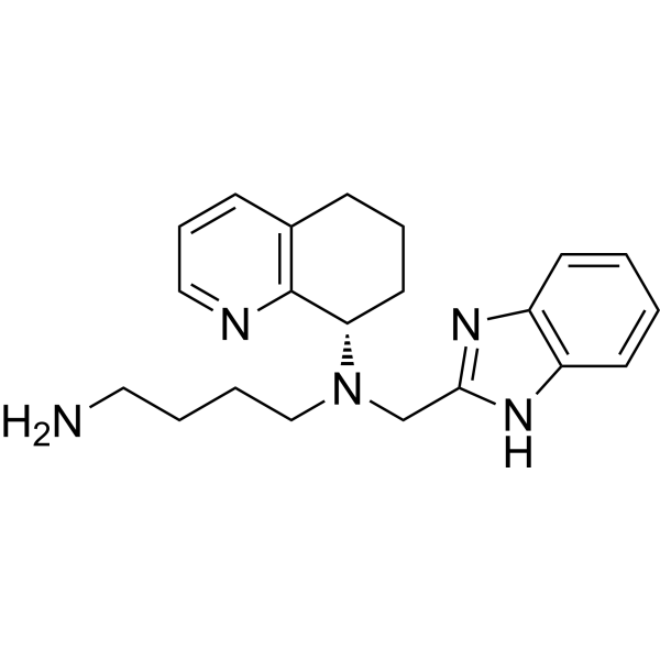
HY-50101
MavorixaforCXCR4 Antagonist -
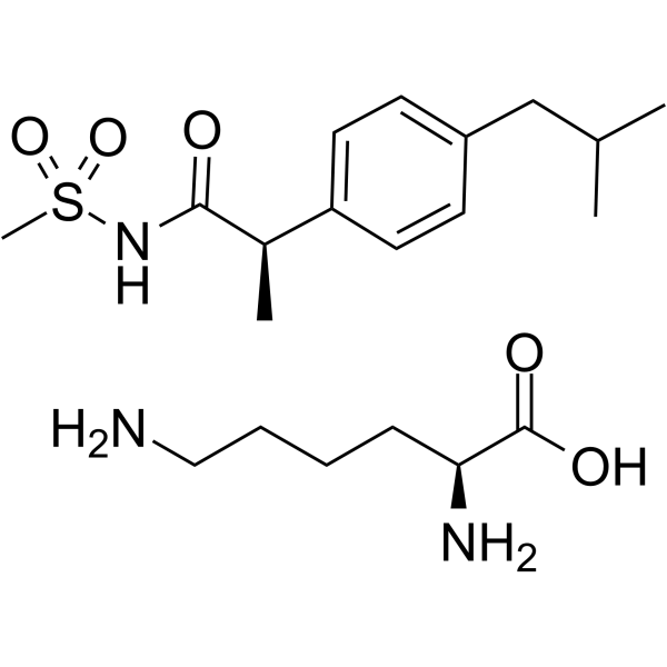
HY-15252
Reparixin L-lysine saltCXCR Inhibitor


Biological Activity
Description
IC50 & Target
[1]|
CXCR2 1 nM (IC50) |
Cellular Effect
|
Cell Line

|
Type | Value | Description | References |
|---|---|---|---|---|
| HEK293 | IC50 |
1 nM
Compound: 30a
|
Displacement of [125I]IL8 from human recombinant CXCR2 expressed in HEK293 cells by SPA assay
Displacement of [125I]IL8 from human recombinant CXCR2 expressed in HEK293 cells by SPA assay
|
[PMID: 18240390] |
In Vitro
AZ10397767 (20 nM; 48 h) abrogates the IL-8-induced (3 nM) increase in proliferation, reducing cell number to below basal levels[2].
AZ10397767 (20 nM; 72 h) increases Oxaliplatin (HY-17371) cytotoxicity, and potentiates Oxaliplatin-induced apoptosis in AIPC cells. AZ10397767 by itself fails to induce apoptosis in either PC3 or DU145 cells[3].
AZ10397767 (20 nM; 24 h) attenuates the Oxaliplatin-induced NF-κB transcriptional activity and the increases in mRNA transcript levels for each of the CXC-chemokines (CXCL8 and CXCL1) and antiapoptotic genes (Bcl-2 and survivin) in the PC3 and DU145 cells[3].
MedChemExpress (MCE) has not independently confirmed the accuracy of these methods. They are for reference only. Further protocols information, click here.
-
Cell Line:LNCaP cells and 22Rv1 cells
-
Concentration:20 nM
-
Incubation Time:48 h
-
Result:Abrogated the IL-8-induced (3 nM) increase in proliferation, reducing cell number to below basal levels.
-
Cell Line:PC3 or DU145 cells
-
Concentration:20 nM
-
Incubation Time:72 h
-
Result:Coadministration with 0.1 or 1 μM Oxaliplatin resulted in a marked increase in the sub-G0/G1 cell population in either cell line.
Potentiates Oxaliplatin-induced apoptosis in AIPC cells.
-
Cell Line:PC3 or DU145 cells
-
Concentration:20 nM
-
Incubation Time:24 h
-
Result:Attenuated the Oxaliplatin (1 μM)-induced NF-κB transcriptional activity and the increases in mRNA transcript levels for each of the CXC-chemokines (CXCL8 and CXCL1) and antiapoptotic genes (Bcl-2 and survivin) in the PC3 and DU145 cells.
In Vivo
AZ10397767 (compound 30a) has a CL of 4 ml/min/kg in rat[1].
MedChemExpress (MCE) has not independently confirmed the accuracy of these methods. They are for reference only.
-
Animal Model:SCID mice with A549 cells[4]
-
Dosage:100 mg/kg
-
Administration:Orally; twice daily; for 22 days
-
Result:Tumors were 36% smaller than their control counterparts.
Significantly (p < 0.01) reduced the number of tumor-infiltrating neutrophils compared to mice receiving vehicle control.


Chemical Information
-
CAS No. 333742-63-5
-
Molecular Weight 400.88
-
Formula C15H14ClFN4O2S2
-
SMILES
O=C1SC2=C(N[C@H](C)CO)N=C(SCC3=CC=CC(Cl)=C3F)N=C2N1
-
Shipping
Room temperature in continental US; may vary elsewhere.
-
Storage
Please store the product under the recommended conditions in the Certificate of Analysis.

Protocols
-
Annexin V plus membrane-impermeant dye apoptosis staining
Annexin V-based apoptosis assays rely on the detection of phosphatidylserine (PS) externalization from the inner leaflet of the plasma membrane to the outer leaflet, an early biochemical hallmark of apoptosis. Fluorescently labeled Annexin V binds PS in a calcium-dependent manner, enabling identification of early apoptotic cells by flow cytometry or fluorescence microscopy. When combined with a membrane-impermeant DNA-binding dye (e. g. , propidium iodide), this approach allows discrimination between viable (Annexin V−/dye−), early apoptotic (Annexin V+/dye−), and late apoptotic or necrotic (Annexin V+/dye+) cell populations by assessing membrane integrity and PS exposure.
-
Zymosan-Induced Peritonitis
Zymosan-induced peritonitis is a sterile acute-inflammation model produced by intraperitoneal injection of zymosan, a yeast cell-wall particle preparation, followed by quantification of leukocyte recruitment and soluble inflammatory mediators in peritoneal lavage fluid. Low-dose zymosan peritonitis is commonly used as a self-resolving acute inflammation model in which neutrophil recruitment occurs early and monocyte/macrophage accumulation follows later. The assay readouts include total peritoneal leukocyte number, differential neutrophil and monocyte/macrophage counts, peritoneal cytokines and chemokines, plasma or peritoneal exudation, and optional lipidomic or metabolomic changes during inflammation and resolution. Early neutrophil recruitment after zymosan depends strongly on complement and mast-cell C5a receptor signaling, whereas later monocyte recruitment is linked to MCP-1/CCL2 production.
-
Patient-Derived Orthotopic Xenograft (PDOX)
Patient-derived orthotopic xenograft (PDOX) modeling implants fresh patient tumor tissue or patient-derived tumor cells into the anatomically corresponding organ or tissue site of immunodeficient mice, usually by surgical orthotopic implantation, to preserve patient tumor histology, local microenvironmental context, invasion, metastatic behavior, and treatment-response features better than subcutaneous implantation. PDOX readouts include tumor engraftment, orthotopic tumor growth, local invasion, metastasis, recurrence after resection, histologic similarity to the donor tumor, biomarker retention, molecular concordance, survival, and response or resistance to therapy. PDOX models are used for preclinical drug testing and individualized therapy evaluation, but engraftment success varies by tumor type and specimen quality.
-
Research Protocol for Inflammation-related Diseases
The NLRP3 inflammasome is a cytosolic innate immune signaling platform that integrates priming signals and danger-signal activation to promote caspase-1 activation, maturation of IL-1β and IL-18, and gasdermin D-mediated pyroptotic cell death. The core experimental logic is to determine whether inflammatory disease phenotypes are driven by increased NLRP3 expression, ASC-containing inflammasome assembly, caspase-1 cleavage, GSDMD cleavage, and extracellular release of IL-1β/IL-18 rather than by nonspecific cell injury alone. The pathway is strongly linked to inflammation-related disease phenotypes because monosodium urate crystals activate NALP3/NLRP3 inflammasome signaling in gout-like crystal inflammation, cholesterol crystals activate NLRP3 inflammasomes in atherogenesis models, and DSS-induced intestinal inflammation has been reported to involve NLRP3 inflammasome activity. However, experimental colitis studies also show context-dependent protective effects of NLRP3 inflammasome co
-
Phagocytosis Functional Assay
A phagocytosis functional assay measures the ability of phagocytic cells, such as neutrophils, macrophages, monocytes, or microglia/macrophages, to bind and internalize particulate targets including bacteria, yeast particles, beads, or myelin particles. Fluorescent flow-cytometry assays detect target uptake as fluorescence associated with gated phagocytes, while pH-sensitive dyes such as pHrodo increase signal in acidic phagosomal compartments and therefore preferentially report internalized particles rather than particles remaining outside the cell. Microscopy or high-content imaging can be used to confirm intracellular localization and, in some protocols, to follow uptake kinetics.
-
Dual Luciferin reporter gene assay
Luciferin reporter gene assay is a reporting system to detect the activity of Firefly Luciferase using luciferin as a substrate, which is often used in the research of miRNA target gene verification and promoter transcriptive activity regulation. Dual luciferase usually refers to Firefly luciferase and Renilla luciferase.
-
Detection of Apoptosis
Apoptosis, also called programmed cell death, is generally characterized by distinct morphological characteristics.
-
TUNEL staining for apoptotic DNA fragmentation
TUNEL staining detects DNA strand breaks by using terminal deoxynucleotidyl transferase to add labeled nucleotides to exposed 3′-OH DNA termini, generating either microscopic staining in fixed cells or tissue sections, or fluorescence/cytometric signal in cell suspensions. TUNEL positivity reflects DNA fragmentation but should not be interpreted alone as definitive apoptosis, because TUNEL can also label necrotic, autolytic, mechanically damaged, or DNA-repair-associated DNA breaks.
-
Research Protocol for Endocrine Diseases
Endocrine diseases often arise from disrupted hormone production, hormone signaling, or target-tissue responsiveness; for diabetes-focused endocrine disease models, insulin signaling regulates glucose uptake, hepatic glucose output, lipid metabolism, and β-cell compensation. Type 2 diabetes develops through interacting defects in insulin resistance, β-cell dysfunction, adipose inflammation, hepatic glucose overproduction, altered incretin signaling, and ectopic lipid metabolism. A major unresolved question is whether endocrine dysfunction is driven primarily by target-tissue insulin resistance, intrinsic β-cell failure, immune/inflammatory stress, or combined multi-organ failure that differs by disease stage.
-
Patient-Derived Xenograft (PDX)
Patient-derived xenograft (PDX) models are generated by engrafting primary human tumor tissue directly into immunodeficient mice, allowing in vivo propagation of patient tumor biology without initial in vitro adaptation. These models are used to preserve key histopathological and molecular characteristics of the original tumor and enable assessment of tumor growth dynamics and therapeutic response in a living organism. The biological readout is tumor engraftment and subsequent growth in the murine host, which reflects the ability of human tumor cells to survive, vascularize, and expand in an immunocompromised microenvironment.
-
Apoptosis Solutions
Apoptosis is a regulated, generally non-lytic cell-death pathway that removes unwanted, damaged, infected, or abnormal cells through coordinated morphological changes, caspase activation, DNA fragmentation, and membrane remodeling. The intrinsic apoptosis pathway is controlled mainly by mitochondrial outer membrane permeabilization, BCL-2 family proteins, cytochrome c release, apoptosome formation, caspase-9 activation, and downstream executioner caspase-3/7 activation. The extrinsic apoptosis pathway is initiated by death receptors such as Fas, TNFR, and TRAIL receptors, which recruit adaptor proteins and activate caspase-8 before engaging executioner caspases or mitochondrial amplification through BID cleavage. Apoptosis is linked to many phenotypes, including cancer cell killing, tissue homeostasis, immune regulation, neurodegeneration, infection response, and treatment-induced cytotoxicity; unresolved questions include how apoptosis interacts with necroptosis, pyroptosis, ferroptos
-
Subcutaneous Cell-Line-Derived Xenograft
Subcutaneous cell-line-derived xenograft (CDX) models are established by implanting cultured human cancer cell lines into immunodeficient mice, where the injected cells form localized tumors that can be monitored in vivo as a measure of tumorigenic potential, growth kinetics, and treatment response. These models are widely used in oncology research because they allow reproducible tumor formation and enable comparative assessment of tumor growth between different cell lines or genetic manipulations in a controlled in vivo microenvironment. Subcutaneous implantation of cancer cells in immunodeficient mice is a standard approach for evaluating tumor growth behavior and therapeutic response across multiple cancer types, including prostate, esophageal, pancreatic, and colon cancer models.
-
Orthotopic Cell-Line Xenograft
Orthotopic cell-line xenograft models involve implantation of human cancer cell lines into the anatomically corresponding organ of immunodeficient mice to reproduce tumor growth within a native microenvironment, enabling more clinically relevant tumor behavior compared with subcutaneous models. These models are widely used because orthotopic placement better recapitulates tumor progression, including invasion and metastatic spread, which are often underrepresented in heterotopic implantation systems. Compared with conventional xenografts, orthotopic implantation is described as more technically complex but provides improved simulation of tumor-microenvironment interactions and metastatic behavior, making it particularly valuable for translational oncology research. Surgical orthotopic implantation approaches have been emphasized as enabling faithful reproduction of clinical cancer features, including metastasis and disease progression patterns that align with the tumor’s organ of origi


Purity & Documentation
References
[1]. Iain Walters, et al. Evaluation of a series of bicyclic CXCR2 antagonists. Bioorg Med Chem Lett. 2008 Jan 15;18(2):798-803. [Content Brief]
[2]. Angela Seaton, et al. Interleukin-8 signaling promotes androgen-independent proliferation of prostate cancer cells via induction of androgen receptor expression and activation. Carcinogenesis. 2008 Jun;29(6):1148-56. [Content Brief]
[3]. Catherine Wilson, et al. Chemotherapy-induced CXC-chemokine/CXC-chemokine receptor signaling in metastatic prostate cancer cells confers resistance to oxaliplatin through potentiation of nuclear factor-kappaB transcription and evasion of apoptosis. J Pharmacol Exp Ther. 2008 Dec;327(3):746-59. [Content Brief]
[4]. Simon Tazzyman, et al. Inhibition of neutrophil infiltration into A549 lung tumors in vitro and in vivo using a CXCR2-specific antagonist is associated with reduced tumor growth. Int J Cancer. 2011 Aug 15;129(4):847-58. [Content Brief]

Calculators
Concentration (start) × Volume (start) = Concentration (final) × Volume (final)


AZ10397767 Related Classifications
HY-124056 Related Classifications
 Powered by Bioz