



BI-765423

Based on 1 Customer Validation
BI-765423 is a selective monoclonal antibody inhibitor targeting IL-11. BI-765423 blocks IL-11-mediated signaling, inhibits fibroblast activation, and can extend the healthy lifespan of mammals, counteract cellular senescence in human cells, and alleviate inflammatory aging-related pathological processes. BI-765423 is primarily used in research on fibrotic diseases such as idiopathic pulmonary fibrosis (IPF).
For research use only. We do not sell to patients.
- Purity : 95.52%
- Molecular Weight:146.61 kDa
-
Storage:
Please store the product under the recommended conditions in the Certificate of Analysis.


To place orders, for customer services and technical support, please contact: MedChemExpress USA
Tel: 609-228-6898 E-mail: [email protected] [email protected]


-
 Biological Activity
-
Chemical Information
- Protocols
- Purity & Documentation
- References
-
 Help & FAQs


-

HY-P99001
Human IgG1 kappa, Isotype ControlIgG1κ Isotype Control -

HY-P991219
Anti-IL11RA Antibody (X209)IL11RA Antibody -

HY-P991487
LASN01IL-11R Inhibitor -

HY-P990007
TulisokibartAnti-TNFSf15/TL1A Antibody -

HY-P990006
DuvakitugAnti-TNFSf15/TL1A Antibody -

HY-138298
Trastuzumab deruxtecan (solution)HER2 ADC -
HY-P9904
AtezolizumabAnti-PD-L1 Antibody -

HY-P9979
RozanolixizumabAnti-FcRn Monoclonal Antibody -

HY-P991143
IxotatugMonoclonal Antibody -
HY-P991218
Anti-IL11 Antibody (X203)Anti-IL11 Antibody -

HY-P990940
PrecemtabartAnti-CEACAM5 Antibody -
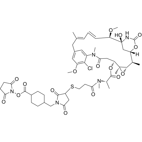
HY-101070
SMCC-DM1Drug-Linker Conjugate for ADC


Biological Activity
Description
Isotype
Human IgG1 kappa
Recommend Isotype Controls
Species Reactivity
Human
IC50 & Target
IL-11
In Vitro
In in vitro experiments using human fibroblasts, BI-765423 significantly inhibits fibroblast activation and reduced the progression of inflammation- and senescence-related pathological processes[1][2].
In in vitro cellular senescence transformation experiments using human cells, BI-765423 effectively counteractes cellular senescence transformation, demonstrating potential anti-aging activity[1].
MedChemExpress (MCE) has not independently confirmed the accuracy of these methods. They are for reference only.
In Vivo
MedChemExpress (MCE) has not independently confirmed the accuracy of these methods. They are for reference only.
Gene ID
Accession
Conjugated
Unconjugated
Reconsititution
The product can be reconstituted/diluted with sterile PBS or saline.
Format
-
Product Image

Application
ELISA, FACS, Functional assay


Chemical Information
-
Appearance Liquid
-
Molecular Weight 146.61 kDa
-
Color Colorless to light yellow
-
SMILES
[BI-765423]
-
Shipping
Shipping with dry ice.
-
Formulation
Please refer to the lot-specific COA for specific buffer information.
-
Storage
Please store the product under the recommended conditions in the Certificate of Analysis.


Purity & Documentation
-
Data Sheet (257 KB)
-
SDS (251 KB)
- English - EN (251 KB)
- Français - FR (251 KB)
- Deutsch - DE (251 KB)
- Norwegian - NO (251 KB)
- Español - ES (251 KB)
- Swedish - SV (251 KB)
- Italian - IT (251 KB)
- Korean - KR (251 KB)
- Portuguese - PT (251 KB)
-
Inhibitory Antibodies User Guide (603 KB)
References
[1]. Pudewell S, et al. Targeting the major pro-inflammatory interleukin-6-type cytokine receptor gp130 by antagonistic single domain antibodies. Front Immunol. 2025 Aug 15;16:1613004. [Content Brief]
[2]. Zhavoronkov A, et al. Age-related diseases as a testbed for anti-aging therapeutics: the case of idiopathic pulmonary fibrosis. Aging (Albany NY). 2025 Aug 16;17(8):1911-1928. [Content Brief]

Calculators
Concentration (start) × Volume (start) = Concentration (final) × Volume (final)


BI-765423 Related Classifications
HY-P991488 Related Classifications
 Powered by Bioz