



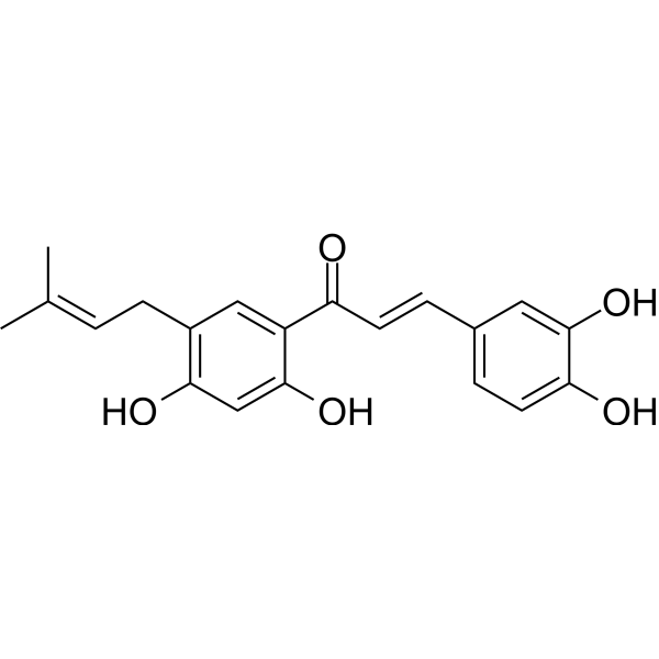
Broussochalcone A

Based on 1 Customer Validation
Broussochalcone A is an antioxidant and an inhibitor of Xanthine Oxidase (IC50=2.21 μM), with free radical scavenging activity. Broussochalcone A inhibits iron-induced lipid peroxidation and nitric oxide synthesis in lipopolysaccharide (LPS) -activated macrophages. Broussochalcone A also induces Apoptosis of human renal carcinoma cells by increasing ROS levels and activating FOXO3 signaling pathways.
商品は「研究用試薬」です。人や動物の医療用・臨床診断用・食品用の製品ではありません。
研究用途以外に使用した場合、当社は一切の責任を負いかねます。
- 純度 : 98.58%
- CAS 番号: 99217-68-2
- 分子式: C20H20O5
- 分子量:340.37
-
保管条件:Powder -20°C, 3 years , 4°C, 2 years ; In solvent -80°C, 6 months , -20°C, 1 month

To place orders, for customer services and technical support, please contact: MedChemExpress USA
Tel: 609-228-6898 E-mail: [email protected] [email protected]


-
 生物活性
生物活性
-
化学情報
-
溶剤 & 溶解度
- プロトコル
- 純度とドキュメンテーション
- 参考文献
-
 Help & FAQs




生物活性
製品説明
Cellular Effect
|
Cell Line

|
Type | Value | Description | References |
|---|---|---|---|---|
| Platelet | IC50 |
118.8 μM
Compound: 10
|
Antiplatelet activity against rabbit platelets assessed as platelet-activating facto-induced platelet aggregation by turbidimetric method
Antiplatelet activity against rabbit platelets assessed as platelet-activating facto-induced platelet aggregation by turbidimetric method
|
[PMID: 8864236] |
| Platelet | IC50 |
22.4 μM
Compound: 10
|
Antiplatelet activity against rabbit platelets assessed as collagen-induced platelet aggregation by turbidimetric method
Antiplatelet activity against rabbit platelets assessed as collagen-induced platelet aggregation by turbidimetric method
|
[PMID: 8864236] |
| Platelet | IC50 |
6.8 μM
Compound: 10
|
Antiplatelet activity against rabbit platelets assessed as arachidonic acid-induced platelet aggregation by turbidimetric method
Antiplatelet activity against rabbit platelets assessed as arachidonic acid-induced platelet aggregation by turbidimetric method
|
[PMID: 8864236] |
| RAW264.7 | IC50 |
11.3 μM
Compound: Table 4, R4C1
|
Inhibition of LPS-induced nitric oxide production in mouse RAW264.7 cells
Inhibition of LPS-induced nitric oxide production in mouse RAW264.7 cells
|
[PMID: 32208222] |
体外実験
Broussochalcone A (0.3, 1, and 3 μM; 10 min, Fe induction for another 30 min) inhibits Fe2+ (200 μM)-induced lipid peroxidation in rat brain homogenate[1].
Broussochalcone A (1-30 μM; 30 min) increases DPPH (100 μM)-scavenging activity dose-dependently[1].
Broussochalcone A (0.1-1 μM) inhibits cytochrome c reduction with an IC50 value of 0.5 μM, mostly due to its superoxide anion-scavenging activity and only partially to its inhibition of xanthine oxidase activity[1].
Broussochalcone A (1-20 μM; 24 h) inhibits nitrite production and iNOS protein expression[1].
MedChemExpress (MCE) has not independently confirmed the accuracy of these methods. They are for reference only.
-
Cell Line:LPS-activated RAW 264.7 macrophages
-
Concentration:1 μM, 3 μM, 10 μM, 20 μM
-
Incubation Time:30 min; then stimulated by LPS (1 μg/mL) for another 24 hr
-
Result:Caused inhibition of iNOS protein expression dose-dependently. Inhibited IkBα phosphorylation.


化学情報
-
CAS 番号 99217-68-2
-
性状 Solid
-
分子量 340.37
-
分子式 C20H20O5
-
Color Light yellow to brown
-
SMILES
O=C(C1=CC(C/C=C(C)\C)=C(O)C=C1O)/C=C/C2=CC=C(O)C(O)=C2
-
Structure Classification
-
輸送条件
Room temperature in continental US; may vary elsewhere.
-
保管条件
Powder -20°C 3 years 4°C 2 years In solvent -80°C 6 months -20°C 1 month


溶剤 & 溶解度
体外:
DMSO : 100 mg/mL (293.80 mM; Need ultrasonic; Hygroscopic DMSO has a significant impact on the solubility of product, please use newly opened DMSO)
Please refer to the solubility information to select the appropriate solvent. Once prepared, please aliquot and store the solution to prevent product inactivation from repeated freeze-thaw cycles.
Storage method and period of stock solution: -80°C, 6 months; -20°C, 1 month. When stored at -80°C, please use it within 6 months. When stored at -20°C, please use it within 1 month.
Please refer to the solubility information to select the appropriate solvent. Once prepared, please aliquot and store the solution to prevent product inactivation from repeated freeze-thaw cycles.
Storage method and period of stock solution: -80°C, 6 months; -20°C, 1 month. When stored at -80°C, please use it within 6 months. When stored at -20°C, please use it within 1 month.
濃度 (開始) × 体積 (開始) = 濃度 (終了) × 体積 (終了)
体内:
Select the appropriate dissolution method based on your experimental animal and administration route.
- For the following dissolution methods, please ensure to first prepare a clear stock solution using an In Vitro approach and then sequentially add co-solvents:
- To ensure reliable experimental results, the clarified stock solution can be appropriately stored based on storage conditions. As for the working solution for In Vivo experiments, it is recommended to prepare freshly and use it on the same day.
- The percentages shown for the solvents indicate their volumetric ratio in the final prepared solution. If precipitation or phase separation occurs during preparation, heat and/or sonication can be used to aid dissolution.
Add each solvent one by one: 10% DMSO 40% PEG300 5% Tween-80 45% Saline
Solubility: ≥ 2.5 mg/mL (7.34 mM); Clear solution
This protocol yields a clear solution of ≥ 2.5 mg/mL (saturation unknown).
Taking 1 mL working solution as an example, add 100 μL DMSO stock solution (25.0 mg/mL) to 400 μL PEG300, and mix evenly; then add 50 μL Tween-80 and mix evenly; then add 450 μL Saline to adjust the volume to 1 mL.
Preparation of Saline: Dissolve 0.9 g sodium chloride in ddH₂O and dilute to 100 mL to obtain a clear Saline solution.
Add each solvent one by one: 10% DMSO 90% (20% SBE-β-CD in Saline)
Solubility: ≥ 2.5 mg/mL (7.34 mM); Clear solution
This protocol yields a clear solution of ≥ 2.5 mg/mL (saturation unknown).
Taking 1 mL working solution as an example, add 100 μL DMSO stock solution (25.0 mg/mL) to 900 μL 20% SBE-β-CD in Saline, and mix evenly.
Preparation of 20% SBE-β-CD in Saline (4°C, storage for one week): 2 g SBE-β-CD powder is dissolved in 10 mL Saline, completely dissolve until clear.
In Vivo Dissolution Calculator
Please enter the basic information of animal experiments:
-
-
-
-
Recommended: Prepare an additional quantity of animals to account for potential losses during experiments.
Please enter your animal formula composition:
-
%DMSO +
Recommended: Keep the proportion of DMSO in working solution below 2% if your animal is weak.
-
%+
-
+%Tween-80 + +
-
%Saline +
The co-solvents required include: DMSO, . All of co-solvents are available by MedChemExpress (MCE). , Tween 80. All of co-solvents are available by MedChemExpress (MCE).
Working solution concentration: 0.22 mg/mL
Method for preparing stock solution: mg drug dissolved in μL DMSO. Stock solution concentration: mg/mL.
1. Take μL DMSO stock solution;
2. Add μL .
μL , mix evenly;
3. Then add μL Tween 80, mix evenly;
4. Then add μL
Please ensure that the stock solution in the first step is dissolved to a clear state, and add co-solvents in sequence. You can use ultrasonic heating (ultrasonic cleaner, recommended frequency 20-40 kHz), vortexing, etc. to assist dissolution.


純度とドキュメンテーション
-
データシート (272 KB)
-
SDS (254 KB)
- English - EN (254 KB)
- Français - FR (254 KB)
- Deutsch - DE (254 KB)
- Norwegian - NO (254 KB)
- Español - ES (254 KB)
- Swedish - SV (254 KB)
- Italian - IT (254 KB)
- Korean - KR (254 KB)
- Portuguese - PT (254 KB)
-
取扱説明書 (2659 KB)
参考文献
[1]. Cheng Z, et al. Broussochalcone A, a potent antioxidant and effective suppressor of inducible nitric oxide synthase in lipopolysaccharide-activated macrophages. Biochem Pharmacol. 2001 Apr 15;61(8):939-46. [Content Brief]
[2]. Lee HK, et al. Broussochalcone A Induces Apoptosis in Human Renal Cancer Cells via ROS Level Elevation and Activation of FOXO3 Signaling Pathway. Oxid Med Cell Longev. 2021 Oct 27;2021:2800706. [Content Brief]


Complete Stock Solution Preparation Table
Please refer to the solubility information to select the appropriate solvent. Once prepared, please aliquot and store the solution to prevent product inactivation from repeated freeze-thaw cycles.
Storage method and period of stock solution: -80°C, 6 months; -20°C, 1 month. When stored at -80°C, please use it within 6 months. When stored at -20°C, please use it within 1 month.
| Optional Solvent | Concentration Solvent Mass | 1 mg | 5 mg | 10 mg | 25 mg |
|---|---|---|---|---|---|
| DMSO | 1 mM | 2.9380 mL | 14.6899 mL | 29.3798 mL | 73.4495 mL |
| 5 mM | 0.5876 mL | 2.9380 mL | 5.8760 mL | 14.6899 mL | |
| 10 mM | 0.2938 mL | 1.4690 mL | 2.9380 mL | 7.3449 mL | |
| 15 mM | 0.1959 mL | 0.9793 mL | 1.9587 mL | 4.8966 mL | |
| 20 mM | 0.1469 mL | 0.7345 mL | 1.4690 mL | 3.6725 mL | |
| 25 mM | 0.1175 mL | 0.5876 mL | 1.1752 mL | 2.9380 mL | |
| 30 mM | 0.0979 mL | 0.4897 mL | 0.9793 mL | 2.4483 mL | |
| 40 mM | 0.0734 mL | 0.3672 mL | 0.7345 mL | 1.8362 mL | |
| 50 mM | 0.0588 mL | 0.2938 mL | 0.5876 mL | 1.4690 mL | |
| 60 mM | 0.0490 mL | 0.2448 mL | 0.4897 mL | 1.2242 mL | |
| 80 mM | 0.0367 mL | 0.1836 mL | 0.3672 mL | 0.9181 mL | |
| 100 mM | 0.0294 mL | 0.1469 mL | 0.2938 mL | 0.7345 mL |


Broussochalcone A 関連分類
HY-142125 関連分類
 Powered by Bioz