



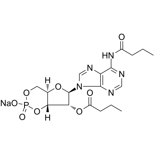
Bucladesine (Dibutyryl cAMP; DBcAMP) sodium (GMP) is a Bucladesine sodium (HY-B0764) produced by using GMP guidelines. Bucladesine (Dibutyryl cAMP; DBcAMP) is a membrane-permeable 3′, 5′-cyclic adenosine monophosphate (cAMP) analog. Bucladesine selectively activates cAMP dependent protein kinase (PKA) by increasing the intracellular level of cAMP. Bucladesine significantly attenuates MDMA-induced increases in hippocampal mitochondrial ROS formation, mitochondrial outer membrane damage, cytochrome c release, and hippocampal ADP/ATP ratio, thereby improving spatial learning and memory impairments. Bucladesine exhibit anti-nociceptive and anti-inflammation effect. Bucladesine can inhibit cancer cells proliferation, induce apoptosis. Bucladesine can be used for the researches of neurological disease, cancer, inflammation.
For research use only. We do not sell to patients.
- CAS No.: 16980-89-5
- Formula: C18H23N5NaO8P
- Molecular Weight:491.37
-
Storage:
Please store the product under the recommended conditions in the Certificate of Analysis.


To place orders, for customer services and technical support, please contact: MedChemExpress USA
Tel: 609-228-6898 E-mail: [email protected] [email protected]


-
 Biological Activity
-
Chemical Information
- Protocols
- Purity & Documentation
- References
-
 Help & FAQs


-
HY-B0764
Bucladesine sodiumcAMP Analog -
HY-12306
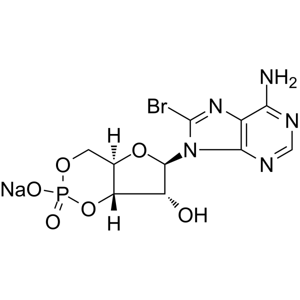
8-Bromo-cAMP sodium saltPKA Activator -

HY-B0764R
Bucladesine sodium (Standard)cAMP Analog -
HY-15979
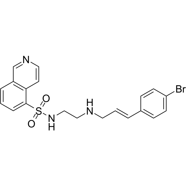
H-89PKA Inhibitor -
HY-B1511
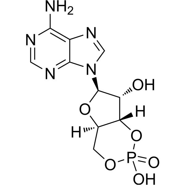
Cyclic AMPSecond Messenger -
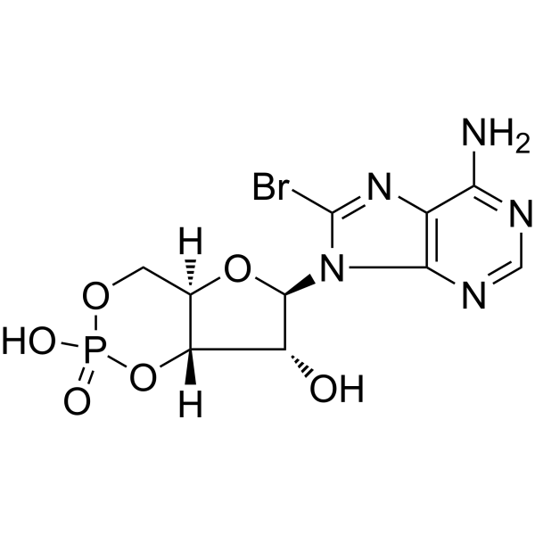
HY-12306A
8-Bromo-cAMPPKA Activator -
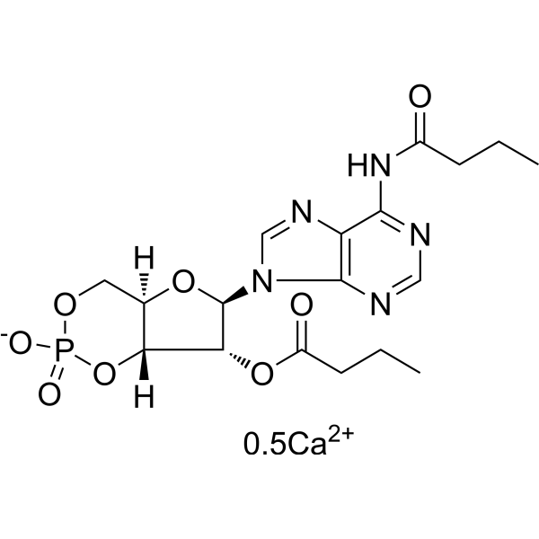
HY-B0764A
Bucladesine hemicalciumApoptosis Inducer -

HY-B1511R
Cyclic AMP (Standard)Second Messenger -
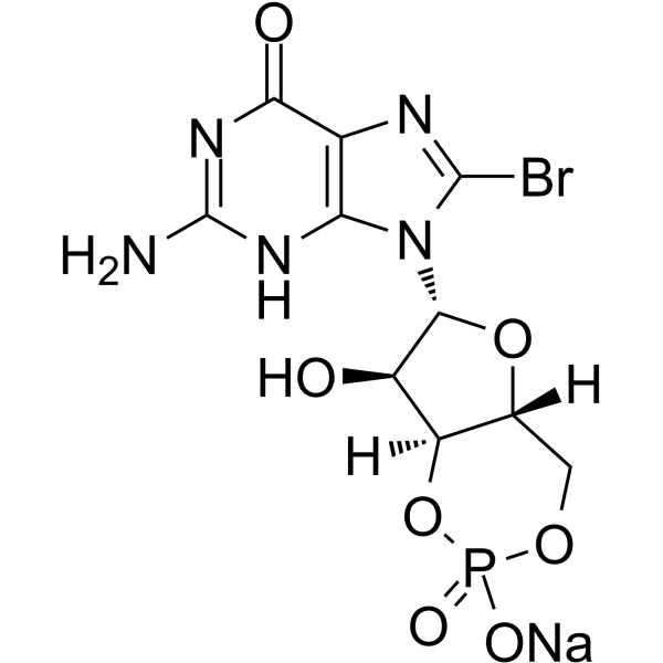
HY-101379A
8-Bromo-cGMP sodiumPKG Activator -
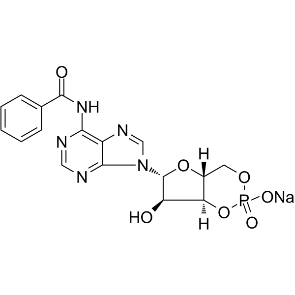
HY-103322
6-Bnz-cAMP sodium saltPKA Activator -
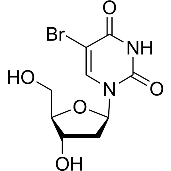
HY-15910
5-BrdUThymidine Analog -

HY-113469
Cyclic GMPSecond Messenger


Biological Activity
Description
Cellular Effect
|
Cell Line

|
Type | Value | Description | References |
|---|---|---|---|---|
| RAW264.7 | IC50 |
28.9 μM
Compound: dbcAMP
|
Antiinflammatory activity against mouse RAW264.7 cells assessed as inhibition of LPS-induced TNFalpha production after 4 hrs by ELISA
Antiinflammatory activity against mouse RAW264.7 cells assessed as inhibition of LPS-induced TNFalpha production after 4 hrs by ELISA
|
[PMID: 11000020] |
| Vero C1008 | IC50 |
>2.0 × 10⁵M
Compound: COVC-3360888908
|
Antiviral activity determined as inhibition of SARS-CoV-2 induced cytotoxicity of VERO-6 cells at 10 uM after 48 hours exposure to 0.01 MOI SARS CoV-2 virus by high content imaging
Antiviral activity determined as inhibition of SARS-CoV-2 induced cytotoxicity of VERO-6 cells at 10 uM after 48 hours exposure to 0.01 MOI SARS CoV-2 virus by high content imaging
|
10.6019/CHEMBL4651402 |
In Vitro
Bucladesine causes significant growth delay in multicellular tumor spheroids derived directly from human malignant brain tumors[1].
Bucladesine augments differentiation therapy (in combination with Sodium butyrate (HY-B0350A) and hyperthermia) in human and canine brain tumor cells in vitro[1].
Bucladesine suppresses the mitogenic effects of platelet-derived growth factor (PDGF), basic fibroblast growth factor (bFGF), and epidermal growth factor (EGF) on human astrocytoma cells; bucladesine alone neither potentiates nor inhibits human astrocytoma cell growth[1].
Bucladesine suppresses proliferation, enhances cytoplasmic process formation, increases cytoplasmic glial fibrillary acidic protein (GFAP) levels, and suppresses malignant glioma invasion by decreasing CD44 isoform expression in cultured malignant glioma cells, inducing biochemical and morphological changes[1].
Bucladesine decreases Bcl-2 levels and enhances SNAP-induced p53-sensitive cell death and MPP+-induced cell death, sensitizing human neuroblastoma cells to apoptosis[1].
Bucladesine inhibits proliferation and induces morphological differentiation of astrocytes in vitro via a cyclic adenosine monophosphate-dependent protein kinase pathway[1].
MedChemExpress (MCE) has not independently confirmed the accuracy of these methods. They are for reference only. Further protocols information, click here.
In Vivo
Bucladesine (50-300 nM/mouse; i.p.; single dose) exerts a biphasic anti-nociceptive effect on thermal-induced acute pain in male albino mice, with significant activity at 50 and 100 nM/mouse doses, no significant effect at 300 nM/mouse[3].
Bucladesine (0.5-1.5% cream, 5% solution; topical; single or two doses) produces a statistically significant reduction in Arachidonic acid (HY-109590)-induced acute skin inflammation in Mus musculus, with single administration of 1.5% cream yielding a 28% reduction in ear swelling and two administrations yielding a 24% reduction[4].
MedChemExpress (MCE) has not independently confirmed the accuracy of these methods. They are for reference only.


Chemical Information
-
CAS No. 16980-89-5
-
Molecular Weight 491.37
-
Formula C18H23N5NaO8P
-
SMILES
O=C(CCC)O[C@H]1[C@H](N2C(N=CN=C3NC(CCC)=O)=C3N=C2)O[C@@](CO4)([H])[C@@]1([H])OP4(O[Na])=O
-
Synonyms
Dibutyryl cAMP sodium (GMP); DBcAMP sodium (GMP)
-
Shipping
Room temperature in continental US; may vary elsewhere.
-
Storage
Please store the product under the recommended conditions in the Certificate of Analysis.

Protocols
-
Protocol for Shuttle Box Test (TDPA)
The Shuttle Box Test for TDPA, or temporally dissociated passive avoidance, measures hippocampus-dependent associative learning by testing whether a rodent avoids entering a dark compartment that was previously paired with foot shock after a temporal delay between dark-compartment entry and shock delivery. The main behavioral readout is crossover or step-through latency from the light chamber into the dark chamber; increased latency across training or retention trials reflects learned avoidance memory rather than motor performance alone.
-
Apoptosis
Apoptosis, also called programmed cell death, is generally characterized by distinct morphological characteristics.
-
TUNEL staining for apoptotic DNA fragmentation
TUNEL staining detects DNA strand breaks by using terminal deoxynucleotidyl transferase to add labeled nucleotides to exposed 3′-OH DNA termini, generating either microscopic staining in fixed cells or tissue sections, or fluorescence/cytometric signal in cell suspensions. TUNEL positivity reflects DNA fragmentation but should not be interpreted alone as definitive apoptosis, because TUNEL can also label necrotic, autolytic, mechanically damaged, or DNA-repair-associated DNA breaks.
-
Annexin V plus membrane-impermeant dye apoptosis staining
Annexin V-based apoptosis assays rely on the detection of phosphatidylserine (PS) externalization from the inner leaflet of the plasma membrane to the outer leaflet, an early biochemical hallmark of apoptosis. Fluorescently labeled Annexin V binds PS in a calcium-dependent manner, enabling identification of early apoptotic cells by flow cytometry or fluorescence microscopy. When combined with a membrane-impermeant DNA-binding dye (e. g. , propidium iodide), this approach allows discrimination between viable (Annexin V−/dye−), early apoptotic (Annexin V+/dye−), and late apoptotic or necrotic (Annexin V+/dye+) cell populations by assessing membrane integrity and PS exposure.
-
ROS/oxidative-stress fluorescent staining
ROS/oxidative-stress fluorescent staining uses cell-permeant fluorogenic probes that become fluorescent after oxidation inside cells or tissues; commonly used examples include DCFH-DA/DCFDA for broad cellular oxidant detection, DHE for superoxide-related signal detection, MitoSOX for mitochondrial superoxide-related signal detection, and CellROX probes for oxidative-stress-associated fluorescence readouts. The assay detects probe oxidation rather than a single ROS species unless the probe and analysis method have been chemically validated for that species. DCFH-DA enters cells, is deacetylated by intracellular esterases to DCFH, and produces fluorescent DCF after oxidation, so the readout is used as an operational measure of total cellular oxidative stress rather than a species-specific ROS measurement. DHE and MitoSOX can report superoxide-related oxidation, but red fluorescence alone can include non-specific ethidium-like oxidation products; HPLC or optimized spectral approaches are
-
Protocol for Water Maze
The Morris Water Maze is a rodent spatial learning and memory assay in which a mouse or rat swims in opaque water to find an escape platform; in the hidden-platform version, the animal cannot see the platform and must use distal extra-maze cues to learn its fixed spatial location. The assay primarily measures hippocampus-dependent spatial learning during acquisition trials and spatial reference memory during probe trials after platform removal; readouts include escape latency, swim path length, swim speed, quadrant occupancy, platform-site crossings, and proximity to the former platform location.
-
Apoptosis Solutions
Apoptosis is a regulated, generally non-lytic cell-death pathway that removes unwanted, damaged, infected, or abnormal cells through coordinated morphological changes, caspase activation, DNA fragmentation, and membrane remodeling. The intrinsic apoptosis pathway is controlled mainly by mitochondrial outer membrane permeabilization, BCL-2 family proteins, cytochrome c release, apoptosome formation, caspase-9 activation, and downstream executioner caspase-3/7 activation. The extrinsic apoptosis pathway is initiated by death receptors such as Fas, TNFR, and TRAIL receptors, which recruit adaptor proteins and activate caspase-8 before engaging executioner caspases or mitochondrial amplification through BID cleavage. Apoptosis is linked to many phenotypes, including cancer cell killing, tissue homeostasis, immune regulation, neurodegeneration, infection response, and treatment-induced cytotoxicity; unresolved questions include how apoptosis interacts with necroptosis, pyroptosis, ferroptos
-
Research Protocol for Inflammation-related Diseases
The NLRP3 inflammasome is a cytosolic innate immune signaling platform that integrates priming signals and danger-signal activation to promote caspase-1 activation, maturation of IL-1β and IL-18, and gasdermin D-mediated pyroptotic cell death. The core experimental logic is to determine whether inflammatory disease phenotypes are driven by increased NLRP3 expression, ASC-containing inflammasome assembly, caspase-1 cleavage, GSDMD cleavage, and extracellular release of IL-1β/IL-18 rather than by nonspecific cell injury alone. The pathway is strongly linked to inflammation-related disease phenotypes because monosodium urate crystals activate NALP3/NLRP3 inflammasome signaling in gout-like crystal inflammation, cholesterol crystals activate NLRP3 inflammasomes in atherogenesis models, and DSS-induced intestinal inflammation has been reported to involve NLRP3 inflammasome activity. However, experimental colitis studies also show context-dependent protective effects of NLRP3 inflammasome co
-
Research Protocol for Neurological Diseases
PINK1/Parkin-mediated mitophagy pathway is a mitochondrial quality-control signaling axis in which mitochondrial depolarization stabilizes PINK1 on damaged mitochondria, activates Parkin recruitment and E3 ubiquitin ligase activity, promotes ubiquitination of outer mitochondrial membrane proteins, recruits selective autophagy adaptors, and drives lysosomal degradation of damaged mitochondria. In neurological disease research, this pathway is experimentally important because neurons, especially dopaminergic neurons, are highly dependent on mitochondrial integrity, and defective mitochondrial turnover can lead to mitochondrial dysfunction, oxidative stress, impaired neuronal survival, α-synuclein accumulation, and neuroinflammatory damage-associated signals. The genetic disease link is strongest in Parkinson’s disease because mutations in PRKN/parkin cause autosomal recessive juvenile parkinsonism, mutations in PINK1 cause hereditary early-onset Parkinson’s disease, and Drosophila studie
-
Cell Viability Determination by MTT Colorimetric Assay
The following protocol uses the MTT colorimetric assay as a classic literature-established method for assessing cell viability/metabolic activity in cultured mammalian cells. MTT[3-(4,5-dimethylthiazol-2-yl)-2,5-diphenyltetrazolium bromide] is reduced by metabolically active cells to a colored formazan product; the amount of formazan is quantified spectrophotometrically and provides an indirect measure of metabolically active viable cells. Importantly, MTT reduction reflects cellular oxidoreductase/metabolic activity rather than an absolute direct count of living cells, so changes in cellular metabolism can alter the signal independently of cell number.


Purity & Documentation
References
[1]. Dalbasti T, et al. Local interstitial chemotherapy with sustained release bucladesine in de novo glioblastoma multiforme: a preliminary study. J Neurooncol. 2002;56(2):167-174. [Content Brief]
[2]. Taghizadeh G, et al. Bucladesine Attenuates Spatial Learning and Hippocampal Mitochondrial Impairments Induced by 3, 4-Methylenedioxymethamphetamine (MDMA). Neurotox Res. 2020;38(1):38-49. [Content Brief]
[3]. Salehi F, et al. Effect of bucladesine, pentoxifylline, and H-89 as cyclic adenosine monophosphate analog, phosphodiesterase, and protein kinase A inhibitor on acute pain. Fundam Clin Pharmacol. 2017;31(4):411-419. [Content Brief]
[4]. Rundfeldt C, et al. The stable cyclic adenosine monophosphate analogue, dibutyryl cyclo-adenosine monophosphate (bucladesine), is active in a model of acute skin inflammation. Arch Dermatol Res. 2012;304(4):313-317. [Content Brief]

Calculators
Concentration (start) × Volume (start) = Concentration (final) × Volume (final)


Bucladesine sodium (GMP) Related Classifications
HY-B0764G Related Classifications
 Powered by Bioz