



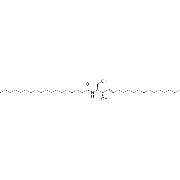
C18-Ceramide (d18:1/18:0)

Based on 1 Customer Validation
C18-Ceramide (d18:1/18:0) is a bioactive molecule with multiple functions in cells, not a traditional agonist or inhibitor targeting a single site. It can act on multiple cellular targets, such as proteins related to endoplasmic reticulum stress (e.g., ATF-4, XBP-1, CHOP), proteins in the PI3K/AKT signaling pathway, and SNARE complex proteins. It exerts activities like inducing cell death, promoting autophagy, and regulating exocytosis through mechanisms such as activating endoplasmic reticulum stress, inhibiting the PI3K/AKT signaling pathway, and affecting lipid raft - related functions. It can be used in research on the mechanism of neuronal injury in the field of neuroscience and in the treatment research of cancers such as glioma in the field of oncology.
Nos produits utilisent uniquement pour la recherche. Nous ne vendons pas aux patients.
- Pureté : 99.95%
- CAS No.: 2304-81-6
- Formule: C36H71NO3
- Masse moléculaire:565.95
-
Stockage:Powder -20°C, 3 years , 4°C, 2 years ; In solvent -80°C, 6 months , -20°C, 1 month

To place orders, for customer services and technical support, please contact: MedChemExpress USA
Tel: 609-228-6898 E-mail: [email protected] [email protected]


-
 Activité biologique
-
Chemical Information
-
Solvant et solubilité
- Protocole
- Pureté et documentation
- Références
-
 Help & FAQs


-
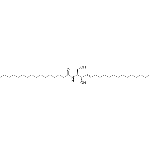
HY-100354
C16-Ceramide (d18:1/16:0)p53 Activator -
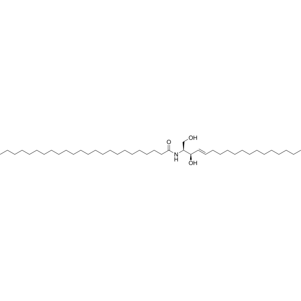
HY-134508
C24-CeramidePIP4K2C-mTOR Activator -
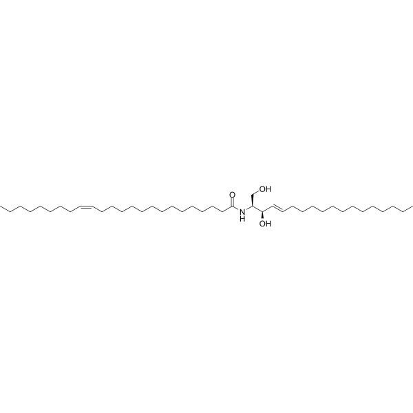
HY-134575
C24:1-CeramideCeramide -
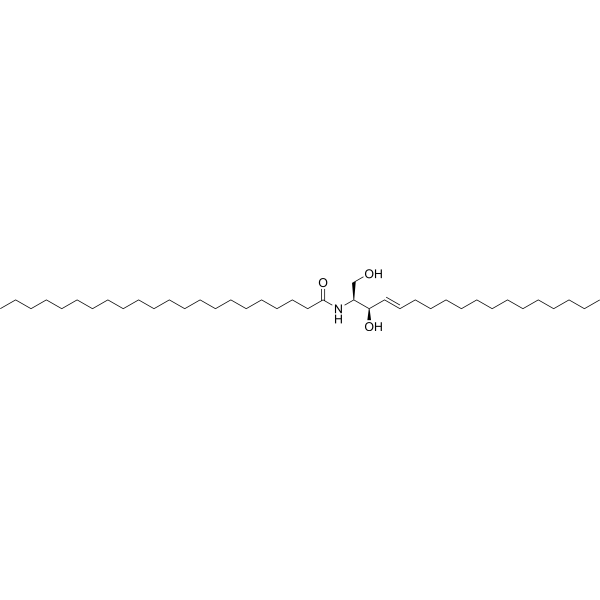
HY-154831
C22-Ceramide (d18:1/22:0)Ceramide -
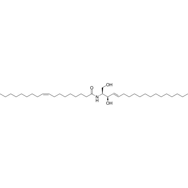
HY-141578
C18:1-CeramideCeramide -
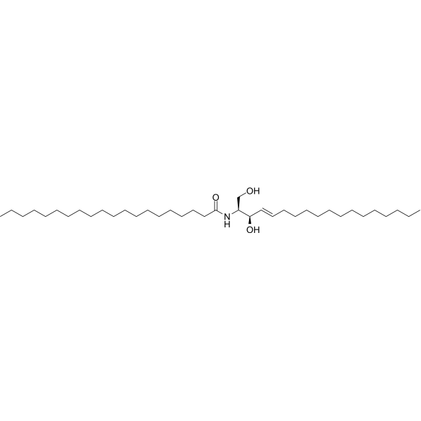
HY-112016
C20 Ceramide (d18:1/20:0)Ceramide -
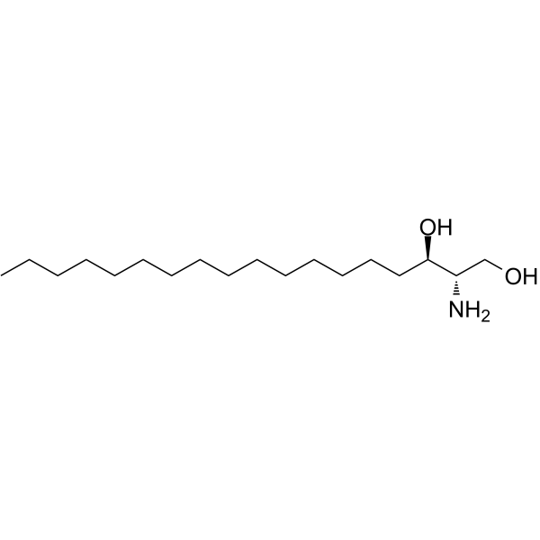
HY-W019838
D-Erythro-dihydrosphingosine -
HY-134508A
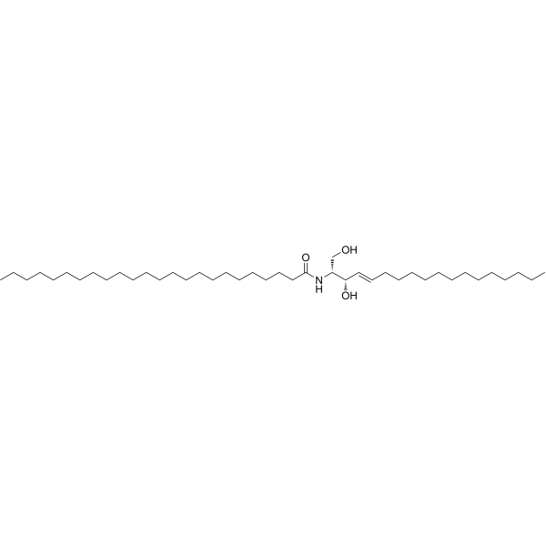
C24-Ceramide (d18:1/24:0)sphingolipid -
HY-108496
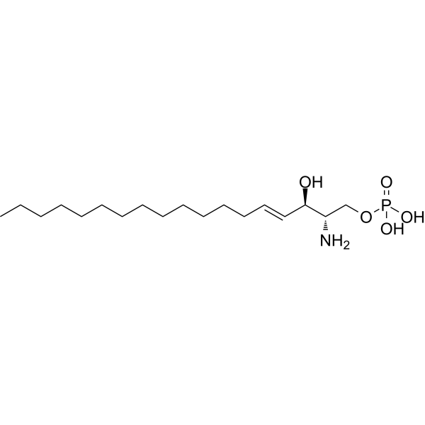
Sphingosine-1-phosphateS1PR Agonist -
HY-N6798
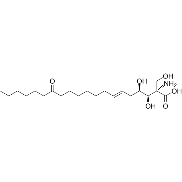
MyriocinSPT Inhibitor -
HY-101047
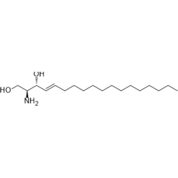
D-erythro-SphingosinePKC Inhibitor -

HY-100355R
C18-Ceramide (Standard)Ceramide
Voir tous les produits spécifiques à Isoform Endogenous Metabolite
More

Activité biologique
Description
In Vitro
In the PC12 cell capacitance measurement experiment, C18-Ceramide (d18:1/18:0) (2 μM; transient) can significantly increase the cell membrane capacitance, indicating that it can promote exocytosis[1].
In the PC12 cell total internal reflection fluorescence microscopy (TIRFM) experiment, C18-Ceramide (d18:1/18:0) (2 μM) can rapidly reduce the number of NPY-EGFP-labeled subcellular membrane vesicles, which undergo endocytosis by fusing with the cell membrane[1].
In U251 and A172 glioma cells, C18-Ceramide (d18:1/18:0) (20 μM; 48 h) inhibits glioma cell viability and induces cell death[2].
C18-Ceramide (d18:1/18:0) (20 μM; 48 h) activates endoplasmic reticulum stress and increases the mRNA and protein levels of ATF-4, XBP-1 (s) and CHOP[2].
C18-Ceramide (d18:1/18:0) induced lethal autophagy, increased the punctate distribution and density of GFP-LC3, promoted the conversion of LC3B-I to LC3B-II and decreased the level of p62[2].
C18-Ceramide (d18:1/18:0) inhibited the PI3K/AKT pathway and decreased the expression levels of PI3K and p-AKT (S473)[2].
C18-Ceramide (d18:1/18:0) (20μM; co-treatment with VM-26 for 48h) increased the chemosensitivity of U251 and A172 cells to VM-26 (HY-13761), synergistically inhibited cell growth, and induced more apoptosis[2].
MedChemExpress (MCE) has not independently confirmed the accuracy of these methods. They are for reference only.
-
Cell Line:U251, A172 glioma cells
-
Concentration:20 μM
-
Incubation Time:48 h
-
Result:Inhibited ell viability and induced cell death.
-
Cell Line:U251, A172 glioma cells
-
Concentration:20 μM
-
Incubation Time:48 h
-
Result:Increased the mRNA and protein levels of ATF-4, XBP-1(s), and CHOP.
-
Cell Line:U251, A172 glioma cells
-
Concentration:20 μM
-
Incubation Time:48 h
-
Result:Inhibited the expression levels of PI3K and p-AKT(S473).
-
Cell Line:U251, A172 glioma cells
-
Concentration:20μM (C18-Ceramide), with 0.25 μM VM-26 for U251 cells, or 10 μM VM-26 for A172 cells
-
Incubation Time:48 h
-
Result:Combined with VM-26, inhibited cell growth synergistically, and induced more apoptosis.
In Vivo
MedChemExpress (MCE) has not independently confirmed the accuracy of these methods. They are for reference only.


Chemical Information
-
CAS No. 2304-81-6
-
Appearance Solid
-
Masse moléculaire 565.95
-
Formule C36H71NO3
-
Color White to off-white
-
SMILES
CCCCCCCCCCCCCCCCCC(N[C@@H](CO)[C@H](O)/C=C/CCCCCCCCCCCCC)=O
-
Synonyms
C18-Ceramide
-
Structure Classification
-
Initial Source
-
Livraison
Room temperature in continental US; may vary elsewhere.
-
Stockage
Powder -20°C 3 years 4°C 2 years In solvent -80°C 6 months -20°C 1 month


Solvant et solubilité
In Vitro:
Ethanol : 5 mg/mL (8.83 mM; Need ultrasonic)
DMF : 1 mg/mL (1.77 mM; Need ultrasonic)
DMSO : < 1 mg/mL (insoluble or slightly soluble)
Please refer to the solubility information to select the appropriate solvent. Once prepared, please aliquot and store the solution to prevent product inactivation from repeated freeze-thaw cycles.
Storage method and period of stock solution: -80°C, 6 months; -20°C, 1 month. When stored at -80°C, please use it within 6 months. When stored at -20°C, please use it within 1 month.
Please refer to the solubility information to select the appropriate solvent. Once prepared, please aliquot and store the solution to prevent product inactivation from repeated freeze-thaw cycles.
Storage method and period of stock solution: -80°C, 6 months; -20°C, 1 month. When stored at -80°C, please use it within 6 months. When stored at -20°C, please use it within 1 month.
Concentration (start) × Volume (start) = Concentration (final) × Volume (final)


Pureté et documentation
-
Fiche technique (271 KB)
-
SDS (254 KB)
- English - EN (254 KB)
- Français - FR (254 KB)
- Deutsch - DE (254 KB)
- Norwegian - NO (254 KB)
- Español - ES (254 KB)
- Swedish - SV (254 KB)
- Italian - IT (254 KB)
- Korean - KR (254 KB)
- Portuguese - PT (254 KB)
-
Instruction de manipulation (2659 KB)
Références
[1]. Tang N, et al. Differential effects of ceramide species on exocytosis in rat PC12 cells. Exp Brain Res. 2007 Nov;183(2):241-7. [Content Brief]
[2]. Wang Z, et al. Overexpression of ceramide synthase 1 increases C18-ceramide and leads to lethal autophagy in human glioma. Oncotarget. 2017 Oct 23;8(61):104022-104036. [Content Brief]


Complete Stock Solution Preparation Table
Please refer to the solubility information to select the appropriate solvent. Once prepared, please aliquot and store the solution to prevent product inactivation from repeated freeze-thaw cycles.
Storage method and period of stock solution: -80°C, 6 months; -20°C, 1 month. When stored at -80°C, please use it within 6 months. When stored at -20°C, please use it within 1 month.
| Optional Solvent | Concentration Solvent Mass | 1 mg | 5 mg | 10 mg | 25 mg |
|---|---|---|---|---|---|
| DMF / Ethanol | 1 mM | 1.7669 mL | 8.8347 mL | 17.6694 mL | 44.1735 mL |
| Ethanol | 5 mM | 0.3534 mL | 1.7669 mL | 3.5339 mL | 8.8347 mL |


C18-Ceramide (d18:1/18:0) Related Classifications
HY-100355 Related Classifications
 Powered by Bioz