



Dimidium bromide

Based on 1 Customer Validation
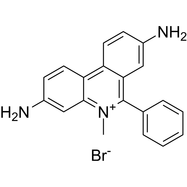
Dimidium bromide is a phenanthridine cationic fluorescent dye with trypanocidal activity and antibacterial activity. Dimidium bromide is a nucleic acid intercalating binding molecule that can bind both DNA and RNA, and its fluorescence signal is significantly enhanced upon binding; it can inhibit the biosynthesis of DNA and RNA. Dimidium bromide can stain the nucleoplasm and nucleoli of various living plant leaf cells, with a fluorescence emission peak at 600 nm under an excitation wavelength of 525 nm. Dimidium bromide can be used in fluorescence imaging-related research.
For research use only. We do not sell to patients.
- Purity : 98.64%
- CAS No.: 518-67-2
- Formula: C20H18BrN3
- Molecular Weight:380.28
-
Storage:
RT, sealed storage, away from moisture and light.
In solvent -80°C, 1 year , -20°C, 6 months

To place orders, for customer services and technical support, please contact: MedChemExpress USA
Tel: 609-228-6898 E-mail: [email protected] [email protected]


-
 Biological Activity
-
Chemical Information
-
Solvent & Solubility
- Protocols
- Purity & Documentation
- References
-
 Help & FAQs


-
HY-N0563
AlizarinNatural Dye -

HY-D0788R
Dimidium bromide (Standard)Reference Standard -
HY-D0711
Indocyanine greenFluorescent Agent -
HY-W110793
DithizoneBiochemical Assay Reagent -

HY-N0563R
Alizarin (Standard)natural dye -
HY-B1102
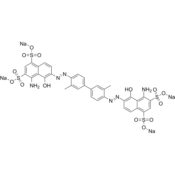
Evans BlueL-glutamate Uptake Inhibitor -
HY-D0908
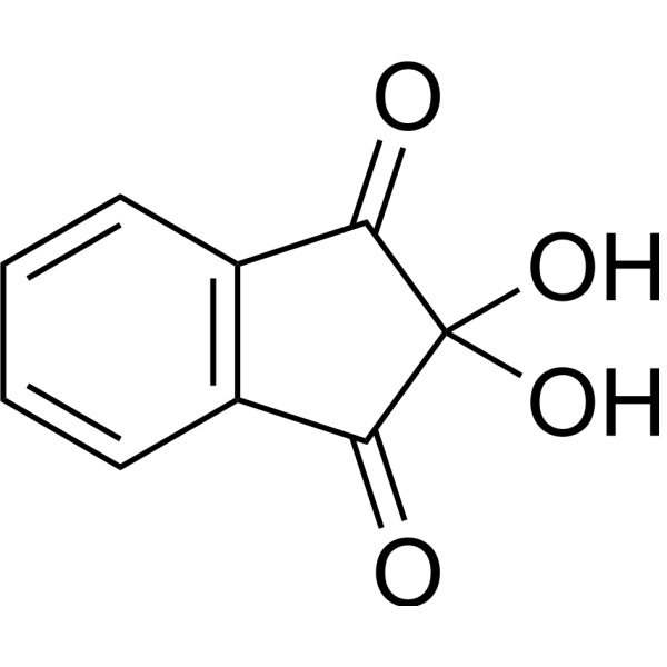
NinhydrinChromogenic Probe -
HY-D0169
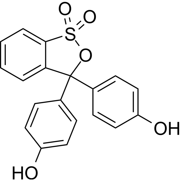
Phenol RedpH Indicator Dye -
HY-D0831
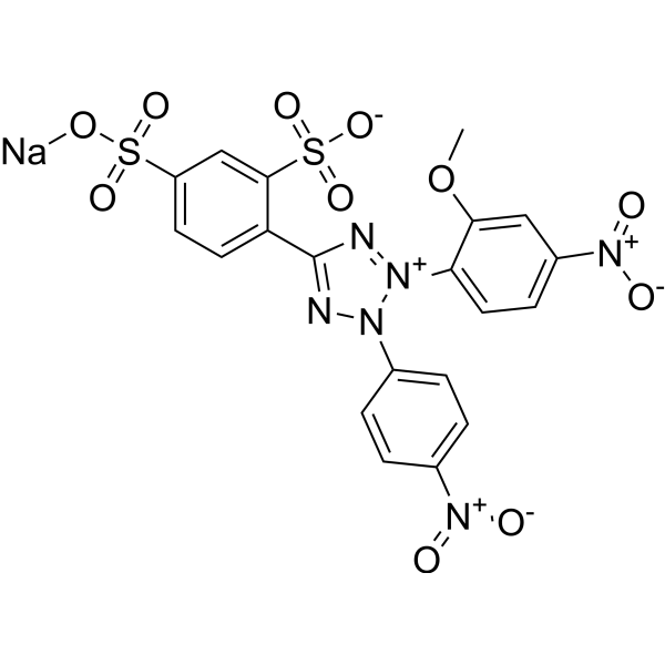
WST-8Tetrazolium Dye -
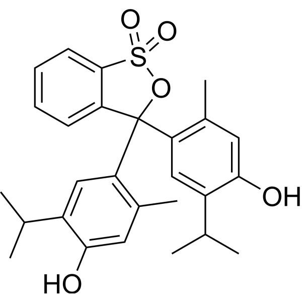
HY-D0219
Thymol blueAcid-base Indicator -
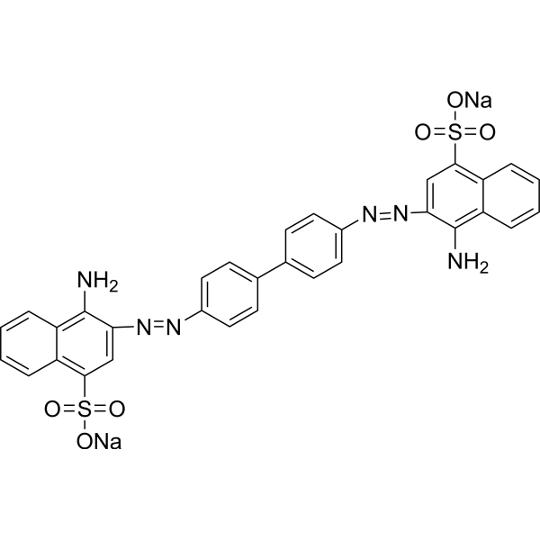
HY-D0236
Congo RedAzo Dye -

HY-W087928
Bromocresol purplepH Indicator
All Parasite Isoforms
More

Biological Activity
Description
In Vitro
Dimidium bromide (DimBr) (20 µM) colocalizes with both the nucleoplasmic marker H2B-sfGFP and the nucleolar marker Fibrillarin-sfGFP in N. benthamiana leaf epidermal cells, and can stain the nucleoplasm and nucleolus in living epidermal cells; it does not fully colocalize with the Cajal body marker Coilin-sfGFP, indicating that it does not stain all RNA-containing structures in the nucleus[3].
Dimidium bromide (2 µM) exhibits strong fluorescence when mixed with DNA or RNA in vitro, with comparable fluorescence intensity for both nucleic acids[3].
Dimidium bromide (20 µM; 10-20 min) can visualize nuclei in multiple cell types of N. benthamiana as well as in multiple plant species including Arabidopsis, lettuce, cucumber, and M. polymorpha[1].
Dimidium bromide (20 µM; 10 min) can stain nucleoli and cytoplasm but not nucleoplasm in CHO-K1 cells, indicating that the staining characteristics of nucleoplasm and cytoplasm differ among biological species; moreover, it enables time-lapse imaging of nuclear migration in living Arabidopsis leaf epidermal cells in response to blue light irradiation[1].
MedChemExpress (MCE) has not independently confirmed the accuracy of these methods. They are for reference only.
In Vivo
Dimidium bromide (0.2-0.24 mg/mouse; i.v.) has an intravenous LD50 of 11.0 mg/kg in mice, with toxic signs including convulsions and death from respiratory failure within 2 min[4].
MedChemExpress (MCE) has not independently confirmed the accuracy of these methods. They are for reference only.


Chemical Information
-
CAS No. 518-67-2
-
Appearance Solid
-
Molecular Weight 380.28
-
Formula C20H18BrN3
-
Color Pale purple to purple
-
SMILES
NC1=CC2=[N+](C(C3=CC=CC=C3)=C4C=C(N)C=CC4=C2C=C1)C.[Br-]
-
Shipping
Room temperature in continental US; may vary elsewhere.
-
Storage
RT, sealed storage, away from moisture and light
In solvent -80°C 1 year -20°C 6 months


Solvent & Solubility
In Vitro:
DMSO : 116.67 mg/mL (306.80 mM; Need ultrasonic; Hygroscopic DMSO has a significant impact on the solubility of product, please use newly opened DMSO)
Please refer to the solubility information to select the appropriate solvent. Once prepared, please aliquot and store the solution to prevent product inactivation from repeated freeze-thaw cycles.
Storage method and period of stock solution: -80°C, 1 year; -20°C, 6 months. When stored at -80°C, please use it within 1 year. When stored at -20°C, please use it within 6 months.
Please refer to the solubility information to select the appropriate solvent. Once prepared, please aliquot and store the solution to prevent product inactivation from repeated freeze-thaw cycles.
Storage method and period of stock solution: -80°C, 1 year; -20°C, 6 months. When stored at -80°C, please use it within 1 year. When stored at -20°C, please use it within 6 months.
Concentration (start) × Volume (start) = Concentration (final) × Volume (final)


Purity & Documentation
-
Data Sheet (287 KB)
-
SDS (394 KB)
- English - EN (394 KB)
- Français - FR (394 KB)
- Deutsch - DE (394 KB)
- Norwegian - NO (394 KB)
- Español - ES (394 KB)
- Swedish - SV (394 KB)
- Italian - IT (394 KB)
- Korean - KR (394 KB)
- Portuguese - PT (394 KB)
-
Handling Instructions (2659 KB)
References


Complete Stock Solution Preparation Table
Please refer to the solubility information to select the appropriate solvent. Once prepared, please aliquot and store the solution to prevent product inactivation from repeated freeze-thaw cycles.
Storage method and period of stock solution: -80°C, 1 year; -20°C, 6 months. When stored at -80°C, please use it within 1 year. When stored at -20°C, please use it within 6 months.
| Optional Solvent | Concentration Solvent Mass | 1 mg | 5 mg | 10 mg | 25 mg |
|---|---|---|---|---|---|
| DMSO | 1 mM | 2.6296 mL | 13.1482 mL | 26.2964 mL | 65.7410 mL |
| 5 mM | 0.5259 mL | 2.6296 mL | 5.2593 mL | 13.1482 mL | |
| 10 mM | 0.2630 mL | 1.3148 mL | 2.6296 mL | 6.5741 mL | |
| 15 mM | 0.1753 mL | 0.8765 mL | 1.7531 mL | 4.3827 mL | |
| 20 mM | 0.1315 mL | 0.6574 mL | 1.3148 mL | 3.2871 mL | |
| 25 mM | 0.1052 mL | 0.5259 mL | 1.0519 mL | 2.6296 mL | |
| 30 mM | 0.0877 mL | 0.4383 mL | 0.8765 mL | 2.1914 mL | |
| 40 mM | 0.0657 mL | 0.3287 mL | 0.6574 mL | 1.6435 mL | |
| 50 mM | 0.0526 mL | 0.2630 mL | 0.5259 mL | 1.3148 mL | |
| 60 mM | 0.0438 mL | 0.2191 mL | 0.4383 mL | 1.0957 mL | |
| 80 mM | 0.0329 mL | 0.1644 mL | 0.3287 mL | 0.8218 mL | |
| 100 mM | 0.0263 mL | 0.1315 mL | 0.2630 mL | 0.6574 mL |


Dimidium bromide Related Classifications
HY-D0788 Related Classifications
 Powered by Bioz