



Lipid X
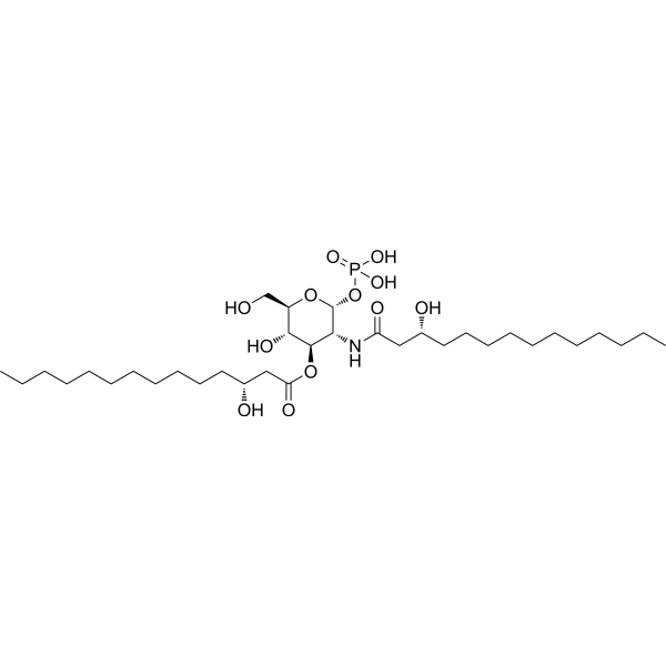
Lipid X is a 2,3-diacylglucosamine-1-phosphate that serves as the monosaccharide precursor of lipid A, possessing both LPS antagonist and weak agonist activities. Lipid X exerts protective effects by inhibiting tumor necrosis factor production, monocyte procoagulant activity, and neutrophil priming. Lipid X may induce transient pulmonary hypertension, neutropenia, and mild pyrogenic effects in laboratory animals. Lipid X has low toxicity and no in vitro antibacterial activity, but it significantly reduces mortality following Gram-negative bacterial infection and endotoxin exposure. Lipid X tends to accumulate in liver tissue, binds to circulating cellular components, and can be converted to lipid Y through transesterification. Lipid X can be used in research on Gram-negative bacterial sepsis, endotoxemia, and associated pulmonary hypertension.
For research use only. We do not sell to patients.
- CAS No.: 86559-73-1
- Formula: C34H66NO12P
- Molecular Weight:711.86
-
Storage:
Please store the product under the recommended conditions in the Certificate of Analysis.

To place orders, for customer services and technical support, please contact: MedChemExpress USA
Tel: 609-228-6898 E-mail: [email protected] [email protected]


-
 Biological Activity
-
Chemical Information
- Protocols
- Purity & Documentation
- References
-
 Help & FAQs




Biological Activity
Description
In Vitro
Lipid X (0-1.0 mM; 0-60 min) is converted to lipid Y by E. coli membrane-bound palmitoyltransferase, with formation linear with incubation time for up to 60 min and linear with membrane protein concentration up to 1.5 mg/mL, and exhibiting a concentration-dependent increase in product formation that approaches saturation at 1 mM lipid X[1].
Lipid X (10 μg/mL) exhibits no in vitro antimicrobial activity against E. coli (ATCC 25922) and does not enhance ticarcillin-mediated bacterial killing[2].
MedChemExpress (MCE) has not independently confirmed the accuracy of these methods. They are for reference only. Further protocols information, click here.
-
Cell Line:E. coli
-
Concentration:0-1.0 mM
-
Incubation Time:0-60 min
-
Result:Converted to lipid Y by E. coli membrane-bound palmitoyltransferase, with formation linear with incubation time for up to 60 min and linear with membrane protein concentration up to 1.5 mg/mL.
Exhibited a concentration-dependent increase in product formation that approaches saturation at 1 mM.
In Vivo
A single dose of Lipid X (750 μg) reduces the mortality rate of LPS-induced endotoxemia in C57BL/10 mice[3].
Lipid X protects sheep from LPS-induced death, alleviates LPS-induced pulmonary hypertension in both early and late stages, and protects neutropenic mice from fatal Gram-negative bacterial infection[3].
Pretreatment with Lipid X (100-200 μg/kg; intravenous injection; bolus; single administration 1 hour prior to endotoxin challenge) results in 100% survival of sheep subjected to lethal E. coli endotoxin challenge, and significantly alleviates endotoxin-induced pulmonary arterial hypertension and fever[4].
MedChemExpress (MCE) has not independently confirmed the accuracy of these methods. They are for reference only.
-
Animal Model:ICR mice (female)[2]
-
Dosage:1000 μg total (single dose); 500 μg (two doses, 6 hours before and after inoculation)
-
Administration:i.v.; single dose; two doses (6 hours before and after inoculation)
-
Result:Reduced cumulative mortality at 18 hours post-inoculation to 13-45% when administered alone.
Achieved 18% cumulative mortality at 18 hours post-inoculation with two 500-μg dose schedule (6 hours before and after inoculation).
Improved 24-hour survival to 67%, 48-hour survival to 23%, and 5-day cure rate to 18% when combined with ticarcillin 1200 mg/kg every 6 hours.
Increased survival 2- to 4-fold at 24 hours, 48 hours, and 5 days post-inoculation across a range of ticarcillin doses.
Reduced the ticarcillin dose required to protect 50% of mice by 4.5-fold at 1 day, 2-fold at 2 days, and 2.6-fold at 5 days.
Resulted in 94% survival during 48-hour antibiotic treatment period (6% cumulative mortality at 48 hours post-inoculation) when administered as single 1000-μg dose 6 hours post-inoculation alongside ticarcillin 150 mg/kg every 8 hours.


Chemical Information
-
CAS No. 86559-73-1
-
Molecular Weight 711.86
-
Formula C34H66NO12P
-
SMILES
O[C@H]1[C@H](OC(C[C@H](O)CCCCCCCCCCC)=O)[C@@H](NC(C[C@H](O)CCCCCCCCCCC)=O)[C@@H](OP(O)(O)=O)O[C@@H]1CO
-
Shipping
Room temperature in continental US; may vary elsewhere.
-
Storage
Please store the product under the recommended conditions in the Certificate of Analysis.

Protocols
-
Phagocytosis Functional Assay
A phagocytosis functional assay measures the ability of phagocytic cells, such as neutrophils, macrophages, monocytes, or microglia/macrophages, to bind and internalize particulate targets including bacteria, yeast particles, beads, or myelin particles. Fluorescent flow-cytometry assays detect target uptake as fluorescence associated with gated phagocytes, while pH-sensitive dyes such as pHrodo increase signal in acidic phagosomal compartments and therefore preferentially report internalized particles rather than particles remaining outside the cell. Microscopy or high-content imaging can be used to confirm intracellular localization and, in some protocols, to follow uptake kinetics.
-
Research Protocol for Infectious Diseases
Infectious-disease experiments test how pathogens interact with host barriers, innate immune receptors, inflammatory signaling, pathogen replication, and tissue injury; pattern-recognition receptors such as TLRs, RIG-I-like receptors, NOD-like receptors, and inflammasomes detect microbial molecules and activate NF-κB, interferon, and cytokine responses. The central hypothesis is that infection severity reflects the balance between pathogen burden and host response: protective inflammation restricts pathogen growth, whereas excessive or mislocalized inflammation contributes to tissue damage and disease phenotype. Unresolved questions include which host pathways are protective versus pathogenic, why some infection models fail to translate to human disease, and which combined readouts best predict clinically relevant infection outcomes.
-
LPS-Induced Endotoxemia/Systemic Inflammation
Lipopolysaccharide (LPS)-induced endotoxemia is a widely used in vivo model of acute systemic inflammation in which LPS, a Gram-negative bacterial endotoxin, activates innate immune signaling primarily through TLR4, leading to rapid and transient induction of pro-inflammatory cytokines such as TNF-α, IL-6, and IL-1β in circulation and tissues. This cytokine surge is commonly used as a measurable readout of systemic inflammatory activation and immune dysregulation, and is typically assessed within hours after intraperitoneal LPS administration in mouse models of endotoxemia. The model captures key features of systemic inflammatory response syndrome, including cytokine release, immune cell activation, and downstream tissue responses, and has been used to evaluate anti-inflammatory interventions such as cytokine modulation, lipid mediators, and immune cell-targeting therapies.
-
Research Protocol for Cardiovascular Diseases
Cardiovascular disease can be modeled as maladaptive cardiac remodeling, where ischemic injury or pressure overload activates inflammatory signaling, fibroblast activation, extracellular-matrix deposition, cardiomyocyte hypertrophy, vascular remodeling, and progressive ventricular dysfunction. The TGF-β/SMAD axis is a central profibrotic pathway after myocardial injury and pressure overload, while innate immune and cytokine pathways regulate leukocyte recruitment, scar formation, and adverse remodeling. Key unresolved questions include which inflammatory signals are reparative versus harmful, when fibrosis is protective versus maladaptive, and whether pathway inhibition improves function without weakening necessary infarct healing or compensatory remodeling.
-
Gram Staining of Tissue Sections
Gram staining of tissue sections is a histochemical technique used to differentiate Gram-positive and Gram-negative bacteria within histological specimens based on differences in bacterial cell wall structure and dye retention, adapted from classical bacteriological Gram staining into tissue-compatible “histological Gram stain” variants. In tissue applications, modifications of the Brown-Hopps and Brown-Brenn methods are commonly used to improve differentiation of microorganisms embedded within host connective tissue and to reduce overstaining or loss of Gram-negative signal, which are known limitations of earlier approaches. The principle relies on crystal violet-iodine complex retention in Gram-positive organisms and subsequent decolorization and counterstaining steps that allow contrast visualization of Gram-negative organisms against tissue background.
-
Bacterial live/dead nucleic-acid viability staining
The LIVE/DEAD bacterial viability staining method is based on differential permeability of nucleic-acid-binding fluorescent dyes, most commonly SYTO 9 and propidium iodide (PI), which enables discrimination of bacterial populations with intact versus compromised cytoplasmic membranes. SYTO 9 penetrates both intact and damaged bacterial membranes and binds nucleic acids to produce green fluorescence, whereas propidium iodide penetrates only cells with compromised membranes and fluoresces red while also reducing SYTO 9 signal through competitive binding and fluorescence interactions. The resulting fluorescence pattern is interpreted as a proxy for membrane integrity, which is widely used as an indicator of bacterial viability in microscopy, flow cytometry, and spectroscopic platforms. However, mechanistic studies show that SYTO 9 and PI interactions involve displacement and fluorescence resonance energy transfer effects, which can influence signal interpretation depending on dye ratios a


Purity & Documentation
References
[2]. Golenbock DT, et al. Lipid X protects mice against fatal Escherichia coli infection. Infect Immun. 1988;56(4):779-784. [Content Brief]
[3]. Golenbock DT, et al. Elimination and tissue distribution of the monosaccharide lipid A precursor, lipid X, in mice and sheep. Antimicrob Agents Chemother. 1988;32(1):37-41. [Content Brief]
[4]. Golenbock DT, et al. Lipid X ameliorates pulmonary hypertension and protects sheep from death due to endotoxin. Infect Immun. 1987;55(10):2471-2476. [Content Brief]

Calculators
Concentration (start) × Volume (start) = Concentration (final) × Volume (final)


Lipid X Related Classifications
HY-130581 Related Classifications
 Powered by Bioz