



BAI1
Based on 14 publication(s) in Google Scholar
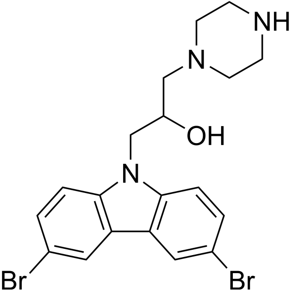
BAI1 is a selective and allosteric inhibitor of BAX, an apoptosis regulator. BAI1 directly binds to BAX and allosterically inhibits BAX activation. BAI1 has the potential for the research of diseases mediated by BAX-dependent cell death.
Nos produits utilisent uniquement pour la recherche. Nous ne vendons pas aux patients.
- Pureté : 99.68%
- CAS No.: 335165-68-9
- Formule: C19H21Br2N3O
- Masse moléculaire:467.20
-
Stockage:Powder -20°C, 3 years , 4°C, 2 years ; In solvent -80°C, 2 years , -20°C, 1 year

To place orders, for customer services and technical support, please contact: MedChemExpress USA
Tel: 609-228-6898 E-mail: [email protected] [email protected]


-
 Activité biologique
-
Chemical Information
-
Solvant et solubilité
- Protocole
- Pureté et documentation
- Références
-
 Help & FAQs


-
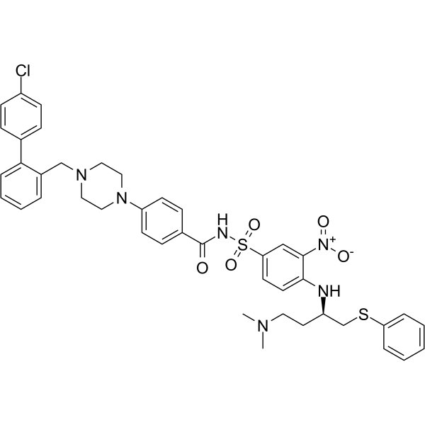
HY-50907
ABT-737Bcl-2 Family Inhibitor/BH3 Mimetic -
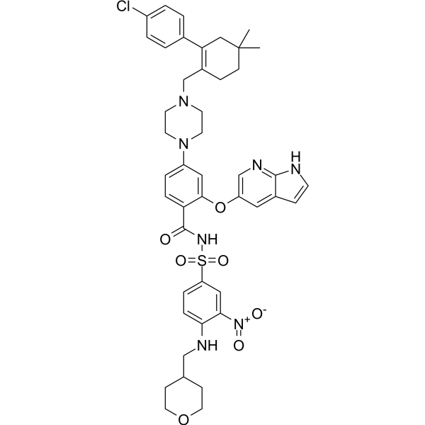
HY-15531
VenetoclaxBCL-2 Inhibitor -
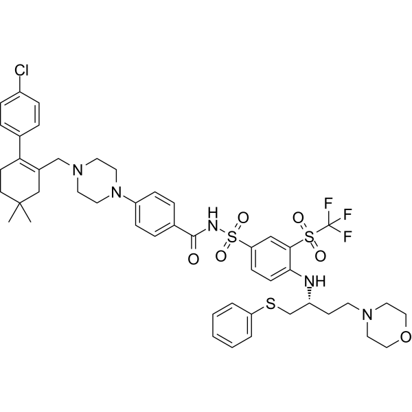
HY-10087
NavitoclaxBcl-2 Inhibitor -
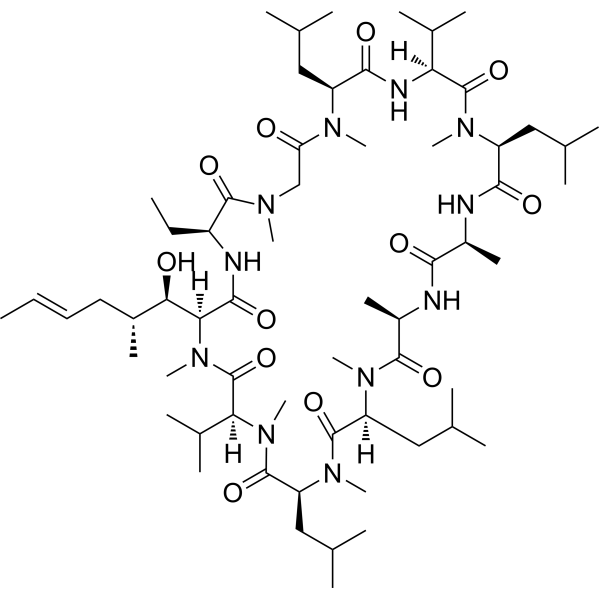
HY-B0579
Cyclosporin ACalcineurin Inhibitor -
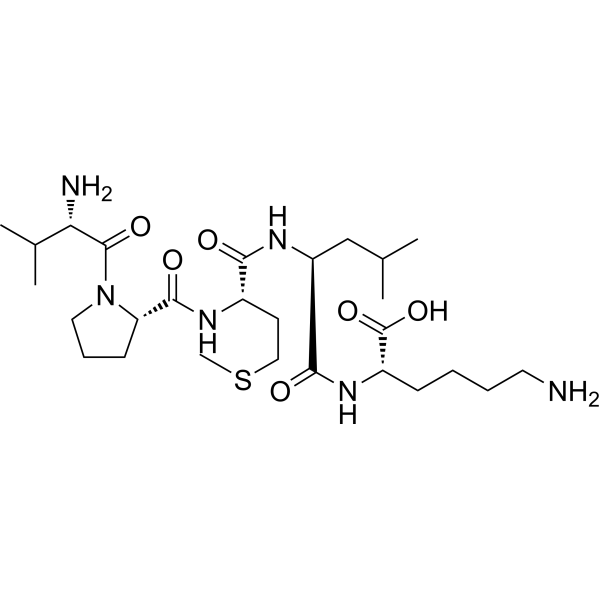
HY-P0081
Bax inhibitor peptide V5Bcl-2 Family Inhibitor -
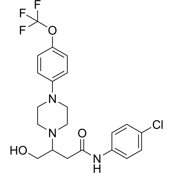
HY-129122
VBIT-4VDAC1 Inhibitor -
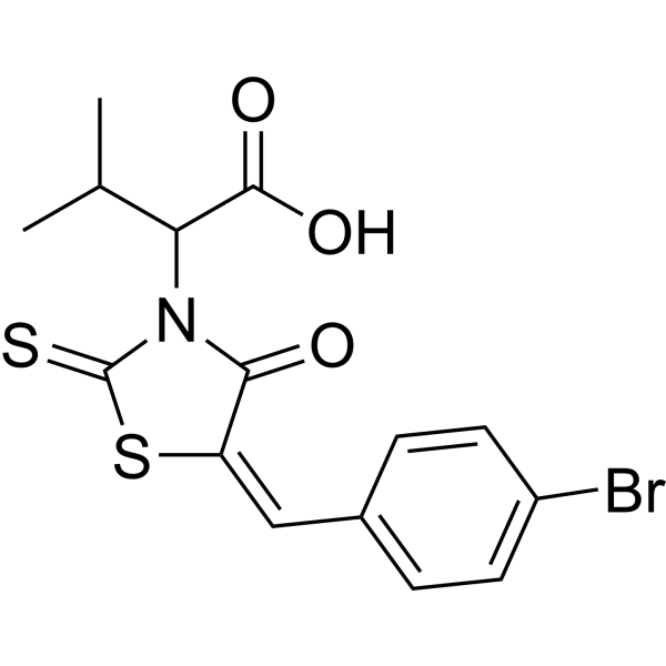
HY-100383
BH3I-1Bcl-2/Bcl-XL Inhibitor -
HY-16658B
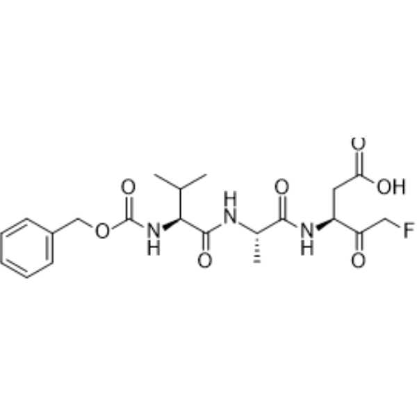
Z-VAD-FMKPan-Caspase Inhibitor -
HY-19741
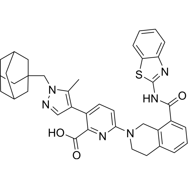
A-1331852Bcl-xL Inhibitor -
HY-118948
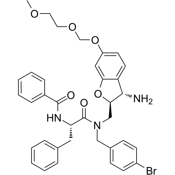
MSN-50Bax/Bak Inhibitor -
HY-120079
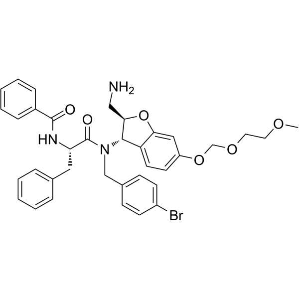
MSN-125Bax/Bak Oligomerization Inhibitor -

HY-15531R
Venetoclax (Standard)BCL-2 Inhibitor
Publications Citing Use of MedChemExpress (MCE) BAI1
More- Adv Funct Mater. 2023 Apr 28.
- Nat Commun. 2026 Jul 24;17(1):9041.
- Nat Commun. 2025 Aug 26;16(1):7936. [Abstract]
- Nat Commun. 2024 Dec 30;15(1):10806. [Abstract]
- Nat Commun. 2023 Nov 20;14(1):7547. [Abstract]
- Mater Today Bio. 2026 Apr 22:38:103132. [Abstract]
- Biochem Pharmacol. 2025 Jul 17:117177. [Abstract]
- Cell Mol Life Sci. 2025 Apr 7;82(1):142. [Abstract]
- Int Immunopharmacol. 2024 Oct 21;143(Pt 2):113424. [Abstract]
- Int J Mol Sci. 2023 May 11;24(10):8609. [Abstract]
- Biochim Biophys Acta Mol Basis Dis. 2026 Jul 28;1873(1):168388.
- Ren Fail. 2024 Dec;46(2):2415519. [Abstract]
- Nat Prod Commun. 2025 Apr 29.
- University of Rijeka. 2021 Jun.
 Customer Validation & Images

-
IF
-
Bio/Physico-chemical Assay
-
Histological Imaging/Staining
-
RT-PCR
-
Flow Cytometry


Activité biologique
Description
IC50 & Target
[1]|
Bax |
Cellular Effect
|
Cell Line

|
Type | Value | Description | References |
|---|---|---|---|---|
| HeLa | IC50 |
0.52 μM
Compound: 1
|
In vitro inhibition of Bax regulated release of cytochrome C was determined using liposome channel assay
In vitro inhibition of Bax regulated release of cytochrome C was determined using liposome channel assay
|
[PMID: 14521400] |
In Vitro
BAI1 selectively inhibits BAX-mediated apoptotic cell death[1].
?
BAI1 shows inhibition of tBID (HY-100464)-induced BAX-mediated membrane permeabilization in a dose dependent manner with an IC50 of 3.3 μΜ[1].
MedChemExpress (MCE) has not independently confirmed the accuracy of these methods. They are for reference only.
-
Cell Line:Mouse MEF
-
Concentration:0.3125 μM , 0.625 μM, 1 μM, 1.25 μM, 1.5 μM, 2.5 μM , 3 μM, 4 μM, 5 4 μM, 10 4 μM
-
Incubation Time:8 hours
-
Result:Inhibited cell death in BAX-dependent manner.


Chemical Information
-
CAS No. 335165-68-9
-
Appearance Solid
-
Masse moléculaire 467.20
-
Formule C19H21Br2N3O
-
Color White to off-white
-
SMILES
OC(CN1CCNCC1)CN2C3=C(C4=C2C=CC(Br)=C4)C=C(Br)C=C3
-
Livraison
Room temperature in continental US; may vary elsewhere.
-
Stockage
Powder -20°C 3 years 4°C 2 years In solvent -80°C 2 years -20°C 1 year


Publications (14)
-
Journal Impact Factor
-
Most Recent
-
-
-
Nat Commun
A multifaceted strategy for intra- and extracellular nucleic acid regulation to alleviate intervertebral disc degeneration. [Abstract]2025 Aug 26;16(1):7936. PMID: 40858573
BAI1 purchased from MedChemExpress. Usage Cited in: Nat Commun. 2025 Aug 26;16(1):7936. [Abstract]
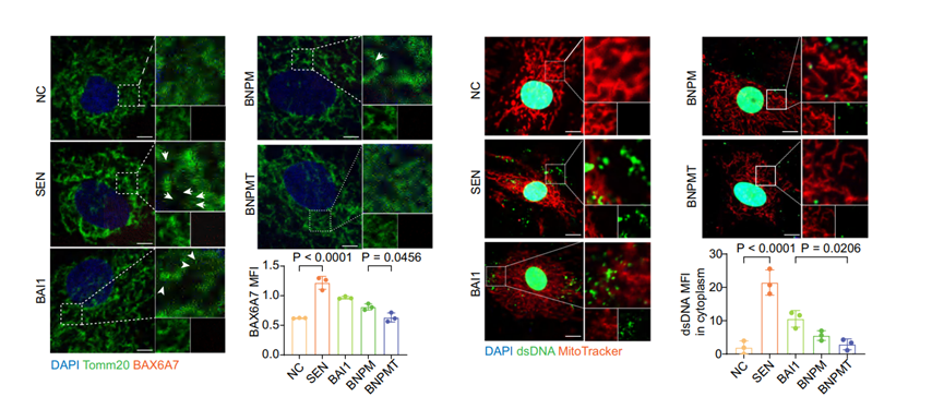
Immunofluorescence staining and quantification of BAX6A7 and dsDNA in aging NP cells treated with BAI1 (2.5 μM, 24 h), BNPM, or BNPMT.
-
Nat Commun
2024 Dec 30;15(1):10806. PMID: 39737939 -
Nat Commun
2023 Nov 20;14(1):7547. PMID: 37985759 -
Mater Today Bio
2026 Apr 22:38:103132. PMID: 42111771 -
Biochem Pharmacol
Targeting STING alleviates pyroptosis of bladder epithelial cells and ameliorates bladder fibrosis in neurogenic bladder. [Abstract]2025 Jul 17:117177. PMID: 40683470 -
Cell Mol Life Sci
Dietary advanced glycation end-products exacerbate sarcopenia onset by activating apoptosis through PRMT1-mediated CRTC3 arginine methylation. [Abstract]2025 Apr 7;82(1):142. PMID: 40192801
BAI1 purchased from MedChemExpress. Usage Cited in: Cell Mol Life Sci. 2025 Apr 7;82(1):142. [Abstract]
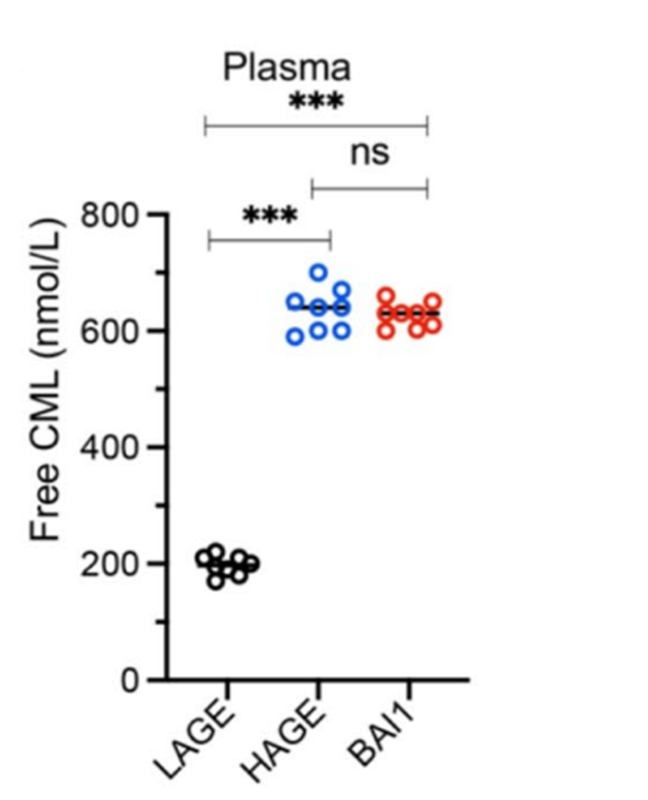
Levels of free and protein-bound CML were significantly increased in both the BAI1 (50 mg/kg, i.p.)-treated and HAGE groups compared to the LAGE group.
BAI1 purchased from MedChemExpress. Usage Cited in: Cell Mol Life Sci. 2025 Apr 7;82(1):142. [Abstract]
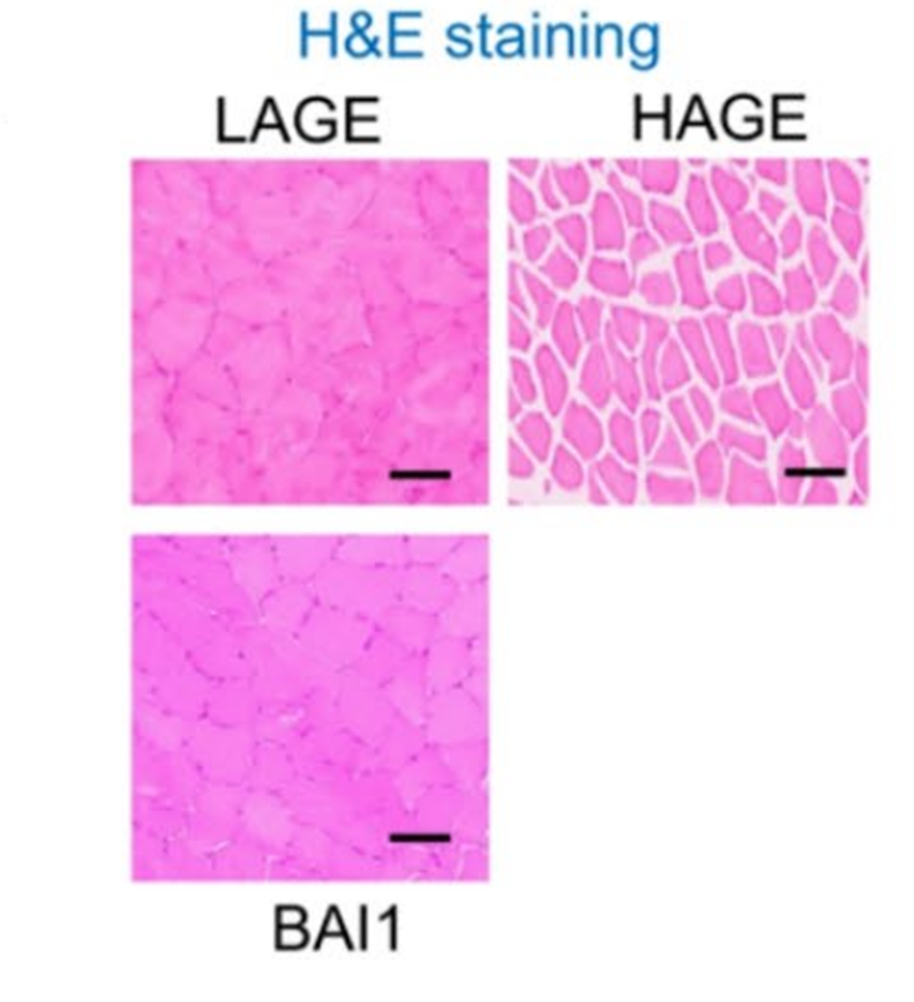
Histological analysis of skeletal muscle tissues demonstrated that BAI1 (50 mg/kg, i.p.) treatment significantly increased muscle fiber size and decreased connective tissue accumulation compared to the HAGE group.
-
Int Immunopharmacol
BAX pores facilitate mitochondrial DNA release in wasp sting-induced acute kidney injury. [Abstract]2024 Oct 21;143(Pt 2):113424. PMID: 39437488 -
Int J Mol Sci
Mitochondrial Trafficking of MLKL, Bak/Bax, and Drp1 Is Mediated by RIP1 and ROS which Leads to Decreased Mitochondrial Membrane Integrity during the Hyperglycemic Shift to Necroptosis. [Abstract]2023 May 11;24(10):8609. PMID: 37239951 -
-
Ren Fail
Piezo1 facilitates the initiation and progression of renal fibrosis by mediating cell apoptosis and mitochondrial dysfunction. [Abstract]2024 Dec;46(2):2415519. PMID: 39496543
BAI1 purchased from MedChemExpress. Usage Cited in: Ren Fail. 2024 Dec;46(2):2415519. [Abstract]
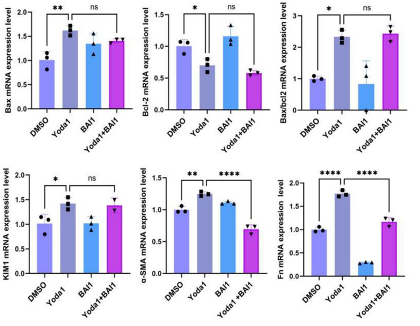
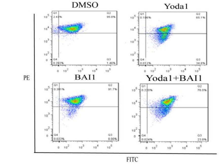
qPCR results indicated that Bax, Cytc, Caspase3, Cleaved-caspase3, KIM1, Fn and Collagen I expression were higher in the Yoda1 treatment group than in the DMSO group but lower in the Yoda1 + BAI1 (4 μM) group than in Yoda1 group.
BAI1 purchased from MedChemExpress. Usage Cited in: Ren Fail. 2024 Dec;46(2):2415519. [Abstract]
Flow cytometry scatter plot of JC1 staining after Yoda1 or/and BAI1 (4 μM) co-treatment, the FITC+ cells present the decreased of mitochondrial membrane potential.
-
-


Solvant et solubilité
In Vitro:
DMSO : 18.46 mg/mL (39.51 mM; ultrasonic and warming and heat to 60°C; Hygroscopic DMSO has a significant impact on the solubility of product, please use newly opened DMSO)
Please refer to the solubility information to select the appropriate solvent. Once prepared, please aliquot and store the solution to prevent product inactivation from repeated freeze-thaw cycles.
Storage method and period of stock solution: -80°C, 2 years; -20°C, 1 year. When stored at -80°C, please use it within 2 years. When stored at -20°C, please use it within 1 year.
Please refer to the solubility information to select the appropriate solvent. Once prepared, please aliquot and store the solution to prevent product inactivation from repeated freeze-thaw cycles.
Storage method and period of stock solution: -80°C, 2 years; -20°C, 1 year. When stored at -80°C, please use it within 2 years. When stored at -20°C, please use it within 1 year.
Concentration (start) × Volume (start) = Concentration (final) × Volume (final)
In Vivo:
Select the appropriate dissolution method based on your experimental animal and administration route.
- For the following dissolution methods, please ensure to first prepare a clear stock solution using an In Vitro approach and then sequentially add co-solvents:
- To ensure reliable experimental results, the clarified stock solution can be appropriately stored based on storage conditions. As for the working solution for In Vivo experiments, it is recommended to prepare freshly and use it on the same day.
- The percentages shown for the solvents indicate their volumetric ratio in the final prepared solution. If precipitation or phase separation occurs during preparation, heat and/or sonication can be used to aid dissolution.
Add each solvent one by one: 10% DMSO 40% PEG300 5% Tween-80 45% Saline
Solubility: ≥ 1.87 mg/mL (4.00 mM); Clear solution
This protocol yields a clear solution of ≥ 1.87 mg/mL (saturation unknown).
Taking 1 mL working solution as an example, add 100 μL DMSO stock solution (18.7 mg/mL) to 400 μL PEG300, and mix evenly; then add 50 μL Tween-80 and mix evenly; then add 450 μL Saline to adjust the volume to 1 mL.
Preparation of Saline: Dissolve 0.9 g sodium chloride in ddH₂O and dilute to 100 mL to obtain a clear Saline solution.
In Vivo Dissolution Calculator
Please enter the basic information of animal experiments:
-
-
-
-
Recommended: Prepare an additional quantity of animals to account for potential losses during experiments.
Please enter your animal formula composition:
-
%DMSO +
Recommended: Keep the proportion of DMSO in working solution below 2% if your animal is weak.
-
%+
-
+%Tween-80 + +
-
%Saline +
The co-solvents required include: DMSO, . All of co-solvents are available by MedChemExpress (MCE). , Tween 80. All of co-solvents are available by MedChemExpress (MCE).
Working solution concentration: 0.22 mg/mL
Method for preparing stock solution: mg drug dissolved in μL DMSO. Stock solution concentration: mg/mL.
1. Take μL DMSO stock solution;
2. Add μL .
μL , mix evenly;
3. Then add μL Tween 80, mix evenly;
4. Then add μL
Please ensure that the stock solution in the first step is dissolved to a clear state, and add co-solvents in sequence. You can use ultrasonic heating (ultrasonic cleaner, recommended frequency 20-40 kHz), vortexing, etc. to assist dissolution.


Pureté et documentation
-
Fiche technique (278 KB)
-
SDS (396 KB)
- English - EN (396 KB)
- Français - FR (396 KB)
- Deutsch - DE (396 KB)
- Norwegian - NO (396 KB)
- Español - ES (396 KB)
- Swedish - SV (396 KB)
- Italian - IT (396 KB)
- Korean - KR (396 KB)
- Portuguese - PT (396 KB)
-
Instruction de manipulation (2659 KB)
Références


Complete Stock Solution Preparation Table
Please refer to the solubility information to select the appropriate solvent. Once prepared, please aliquot and store the solution to prevent product inactivation from repeated freeze-thaw cycles.
Storage method and period of stock solution: -80°C, 2 years; -20°C, 1 year. When stored at -80°C, please use it within 2 years. When stored at -20°C, please use it within 1 year.
| Optional Solvent | Concentration Solvent Mass | 1 mg | 5 mg | 10 mg | 25 mg |
|---|---|---|---|---|---|
| DMSO | 1 mM | 2.1404 mL | 10.7021 mL | 21.4041 mL | 53.5103 mL |
| 5 mM | 0.4281 mL | 2.1404 mL | 4.2808 mL | 10.7021 mL | |
| 10 mM | 0.2140 mL | 1.0702 mL | 2.1404 mL | 5.3510 mL | |
| 15 mM | 0.1427 mL | 0.7135 mL | 1.4269 mL | 3.5674 mL | |
| 20 mM | 0.1070 mL | 0.5351 mL | 1.0702 mL | 2.6755 mL | |
| 25 mM | 0.0856 mL | 0.4281 mL | 0.8562 mL | 2.1404 mL | |
| 30 mM | 0.0713 mL | 0.3567 mL | 0.7135 mL | 1.7837 mL |


BAI1 Related Classifications
HY-103269 Related Classifications
 Powered by Bioz