



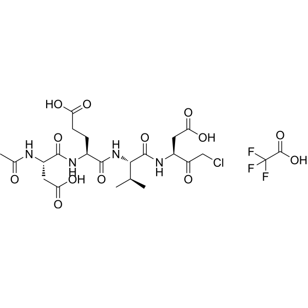
Ac-DEVD-CMK TFA
Based on 3 publication(s) in Google Scholar
Ac-DEVD-CMK (Caspase-3 Inhibitor III) TFA is a selective and irreversible caspase-3 inhibitor. Ac-DEVD-CMK TFA significantly inhibits apoptosis induced by high levels of glucose or 3,20-dibenzoate (IDB; HY-137295). Ac-DEVD-CMK TFA can be used in a variety of experimental approaches to inhibit apoptosis.
Nos produits utilisent uniquement pour la recherche. Nous ne vendons pas aux patients.
- Pureté : 98.09%
- Formule: C23H32ClF3N4O13
- Masse moléculaire:664.97
-
Stockage:
Sealed storage, away from moisture and light, under nitrogen.
Powder -80°C, 2 years , -20°C, 1 year* In solvent : -80°C, 6 months; -20°C, 1 month (sealed storage, away from moisture and light, under nitrogen)

To place orders, for customer services and technical support, please contact: MedChemExpress USA
Tel: 609-228-6898 E-mail: [email protected] [email protected]


-
 Activité biologique
-
Chemical Information
-
Solvant et solubilité
- Protocole
- Pureté et documentation
- Références
-
 Help & FAQs


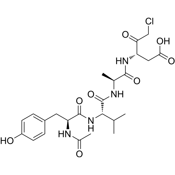
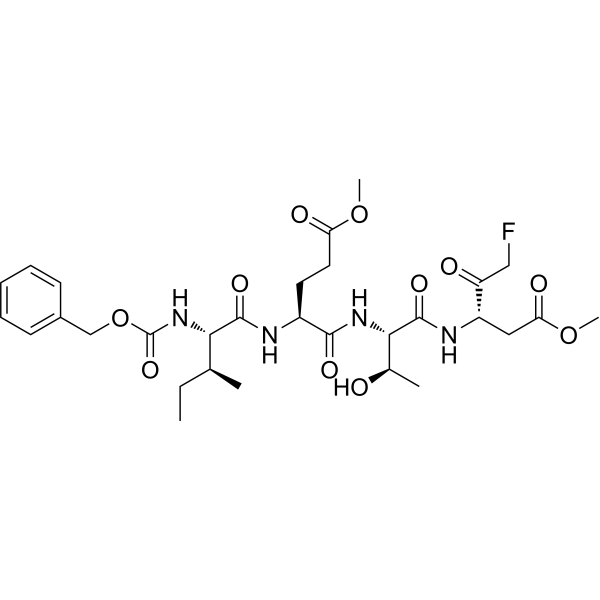
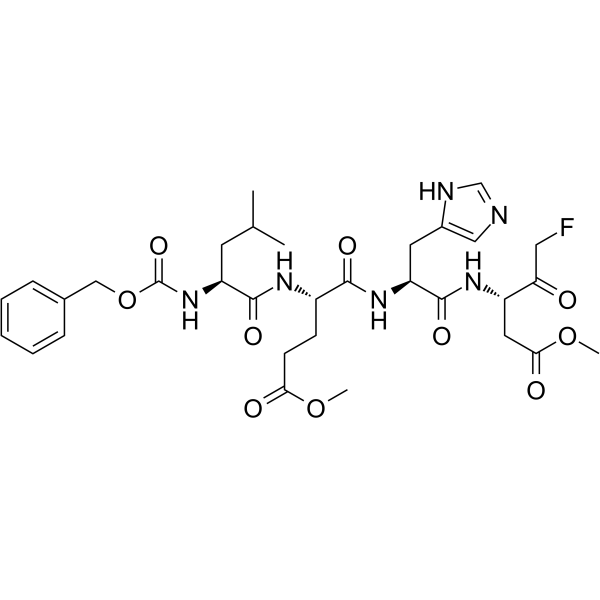
Publications Citing Use of MedChemExpress (MCE) Ac-DEVD-CMK TFA
MoreVoir tous les produits spécifiques à Isoform Caspase
More

Activité biologique
Description
In Vitro
Ac-DEVD-CMK TFA (100 μM; 24 h) inhibits IDB-induced apoptosis[3].
Ac-DEVD-CMK TFA inhibits (10 μM; 36 h) inhibits citrate (10 mM)-induced p21 cleavage and G2/M accumulation in human pharyngeal squamous carcinoma FaDu and Detroit 562 cell lines[4].
MedChemExpress (MCE) has not independently confirmed the accuracy of these methods. They are for reference only.
-
Cell Line:FaDu and Detroit 562 cell
-
Concentration:10 μM; with 10 mM citrate
-
Incubation Time:36 h
-
Result:Inhibited citrate to induce p21 cleavage.
In Vivo
Ac-DEVD-CMK TFA (25 mg/kg; ip; single dose) significantly attenuates APAP-induced liver injury (AILI) in susceptible Sdc1−/− mice[6].
MedChemExpress (MCE) has not independently confirmed the accuracy of these methods. They are for reference only.
-
Animal Model:Sdc1−/− mice induced by APAP[6]
-
Dosage:25 mg/kg
-
Administration:IP; single dose, 3 hours post-APAP
-
Result:Inhibited GSK‐3β or caspase‐3 activity to mitigate liver damage.


Chemical Information
-
Appearance Solid
-
Masse moléculaire 664.97
-
Formule C23H32ClF3N4O13
-
Color White to off-white
-
Synonyms
Caspase-3 Inhibitor III TFA
-
Livraison
Room temperature in continental US; may vary elsewhere.
-
Stockage
Sealed storage, away from moisture and light, under nitrogen
Powder -80°C 2 years -20°C 1 year * In solvent : -80°C, 6 months; -20°C, 1 month (sealed storage, away from moisture and light, under nitrogen)


Publications (3)
-
Journal Impact Factor
-
Most Recent
-
J Agric Food Chem
β-Hydroxybutyric Acid-Induced Bovine Endometrial Epithelial Cell Pyroptosis via Activating P2X7R. [Abstract]2025 Aug 27;73(34):21296-21308. PMID: 40742321 -
PLoS Pathog
2024 Jul 22;20(7):e1012408. PMID: 39038037 -
Exp Neurol
Muscone alleviates neuronal injury via increasing stress granules formation and reducing apoptosis in acute ischemic stroke. [Abstract]2024 Mar:373:114678. PMID: 38185313


Solvant et solubilité
In Vitro:
DMSO : 100 mg/mL (150.38 mM; Need ultrasonic; Hygroscopic DMSO has a significant impact on the solubility of product, please use newly opened DMSO)
H2O : 50 mg/mL (75.19 mM; ultrasonic and warming and heat to 60°C)
Please refer to the solubility information to select the appropriate solvent. Once prepared, please aliquot and store the solution to prevent product inactivation from repeated freeze-thaw cycles.
Storage method and period of stock solution: -80°C, 6 months; -20°C, 1 month (sealed storage, away from moisture and light, under nitrogen). When stored at -80°C, please use it within 6 months. When stored at -20°C, please use it within 1 month.
* Note: If you choose water as the stock solution, please dilute it to the working solution, then filter and sterilize it with a 0.22 μm filter before use.
Please refer to the solubility information to select the appropriate solvent. Once prepared, please aliquot and store the solution to prevent product inactivation from repeated freeze-thaw cycles.
Storage method and period of stock solution: -80°C, 6 months; -20°C, 1 month (sealed storage, away from moisture and light, under nitrogen). When stored at -80°C, please use it within 6 months. When stored at -20°C, please use it within 1 month.
* Note: If you choose water as the stock solution, please dilute it to the working solution, then filter and sterilize it with a 0.22 μm filter before use.
Concentration (start) × Volume (start) = Concentration (final) × Volume (final)


Pureté et documentation
-
Fiche technique (277 KB)
-
SDS (252 KB)
- English - EN (252 KB)
- Français - FR (252 KB)
- Deutsch - DE (252 KB)
- Norwegian - NO (252 KB)
- Español - ES (252 KB)
- Swedish - SV (252 KB)
- Italian - IT (252 KB)
- Korean - KR (252 KB)
- Portuguese - PT (252 KB)
-
Instruction de manipulation (2659 KB)
Références
[1]. Eon Jeong Nam, et al. Syndecan-1 limits the progression of liver injury and promotes liver repair in acetaminophen-induced liver injury in mice. Hepatology. 2017 Nov;66(5):1601-1615. [Content Brief]
[2]. Hung KC, et al. Citrate-Induced p85α⁻PTEN Complex Formation Causes G2/M Phase Arrest in Human Pharyngeal Squamous Carcinoma Cell Lines. Int J Mol Sci. 2019 Apr 29;20(9):2105. [Content Brief]
[3]. Nam EJ, et al. Syndecan-1 limits the progression of liver injury and promotes liver repair in acetaminophen-induced liver injury in mice. Hepatology. 2017 Nov;66(5):1601-1615. [Content Brief]
[4]. Lu Cai, et al. Hyperglycemia-induced apoptosis in mouse myocardium: mitochondrial cytochrome C-mediated caspase-3 activation pathway. Diabetes. 2002 Jun;51(6):1938-48. [Content Brief]
[5]. M M Mocanu, et al. Caspase inhibition and limitation of myocardial infarct size: protection against lethal reperfusion injury. Br J Pharmacol. 2000 May;130(2):197-200. [Content Brief]
[6]. M Blanco-Molina, et al. Ingenol esters induce apoptosis in Jurkat cells through an AP-1 and NF-kappaB independent pathway. Chem Biol. 2001 Aug;8(8):767-78. [Content Brief]


Complete Stock Solution Preparation Table
Please refer to the solubility information to select the appropriate solvent. Once prepared, please aliquot and store the solution to prevent product inactivation from repeated freeze-thaw cycles.
Storage method and period of stock solution: -80°C, 6 months; -20°C, 1 month (sealed storage, away from moisture and light, under nitrogen). When stored at -80°C, please use it within 6 months. When stored at -20°C, please use it within 1 month.
| Optional Solvent | Concentration Solvent Mass | 1 mg | 5 mg | 10 mg | 25 mg |
|---|---|---|---|---|---|
| H2O / DMSO | 1 mM | 1.5038 mL | 7.5191 mL | 15.0383 mL | 37.5957 mL |
| 5 mM | 0.3008 mL | 1.5038 mL | 3.0077 mL | 7.5191 mL | |
| 10 mM | 0.1504 mL | 0.7519 mL | 1.5038 mL | 3.7596 mL | |
| 15 mM | 0.1003 mL | 0.5013 mL | 1.0026 mL | 2.5064 mL | |
| 20 mM | 0.0752 mL | 0.3760 mL | 0.7519 mL | 1.8798 mL | |
| 25 mM | 0.0602 mL | 0.3008 mL | 0.6015 mL | 1.5038 mL | |
| 30 mM | 0.0501 mL | 0.2506 mL | 0.5013 mL | 1.2532 mL | |
| 40 mM | 0.0376 mL | 0.1880 mL | 0.3760 mL | 0.9399 mL | |
| 50 mM | 0.0301 mL | 0.1504 mL | 0.3008 mL | 0.7519 mL | |
| 60 mM | 0.0251 mL | 0.1253 mL | 0.2506 mL | 0.6266 mL | |
| DMSO | 80 mM | 0.0188 mL | 0.0940 mL | 0.1880 mL | 0.4699 mL |
| 100 mM | 0.0150 mL | 0.0752 mL | 0.1504 mL | 0.3760 mL |
* Note: If you choose water as the stock solution, please dilute it to the working solution, then filter and sterilize it with a 0.22 μm filter before use.


Ac-DEVD-CMK TFA Related Classifications
HY-P0034A Related Classifications
 Powered by Bioz