



Belantamab
Based on 3 publication(s) in Google Scholar
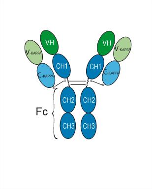
Belantamab (GSK2857916) is a humanized IgG1 anti-BCMA/TNFRSF17 monoclonal antibody. Belantamab is linked to MMAF (HY-15579) through a non-cleavable ADC linker to synthesize the antibody-active molecule conjugate (ADC) Belantamab mafodotin (HY-P3239). After binding to BCMA on the surface of tumor cells, Belantamab mafodotin enters the cell through receptor-mediated endocytosis. After entering the cell, Belantamab mafodotin releases MMAF, blocks cell division by inhibiting tubulin polymerization, arrests the cell cycle and induces cell apoptosis. Belantamab can be used for the study of multiple myeloma, especially relapsed/refractory multiple myeloma.
Nos produits utilisent uniquement pour la recherche. Nous ne vendons pas aux patients.
- Pureté : 99.90%
- CAS No.: 2061894-48-0
- Masse moléculaire:146.06 kDa
-
Stockage:
Please store the product under the recommended conditions in the Certificate of Analysis.


To place orders, for customer services and technical support, please contact: MedChemExpress USA
Tel: 609-228-6898 E-mail: [email protected] [email protected]


-
 Activité biologique
-
Chemical Information
- Protocole
- Pureté et documentation
- Références
-
 Help & FAQs


-

HY-P99151
BrentuximabAnti-CD30 Monoclonal Antibody -

HY-P99042
PolatuzumabAnti-CD79b mAb -

HY-P99045
SacituzumabAnti-TROP-2 Antibody -

HY-P99016
EnfortumabAnti-Nectin-4 Monoclonal Antibody -

HY-P99843
DatopotamabAnti-TROP2 Antibody -

HY-P99854
DisitamabAnti-HER2 Antibody -

HY-P3239
Belantamab mafodotinAnti-BCMA ADC -

HY-P99271
TisotumabAnti-TF Antibody -

HY-P9907
TrastuzumabHER2 Blocking Antibody -

HY-P99001
Human IgG1 kappa, Isotype ControlIgG1κ Isotype Control -

HY-P9908
AdalimumabTNF-α Inhibitor -

HY-P990007
TulisokibartAnti-TNFSf15/TL1A Antibody
Publications Citing Use of MedChemExpress (MCE) Belantamab
MoreVoir tous les produits spécifiques à Isoform TNF Receptor
More

Activité biologique
Description
Isotype
Human IgG1 kappa
Recommend Isotype Controls
Species Reactivity
Human
IC50 & Target
TNFRSF17/BCMA/CD269
In Vitro
Belantamab (0.000001-10000 μg/mL; 3 days) does not inhibit growth of BCMA-expressing NCI-H929 human multiple myeloma cells or EL4-hBCMA murine lymphoma cells[2].
Belantamab (0.1-10 μg/mL; 24-72 h) does not induce immunogenic cell death in NCI-H929 human multiple myeloma cells, as measured by ER stress activation, DAMP exposure or release, or reduced cell viability[2].
Belantamab (10-100 μg/mL; 24-96 h) does not induce immunogenic cell death in EL4-hBCMA murine lymphoma cells[2].
MedChemExpress (MCE) has not independently confirmed the accuracy of these methods. They are for reference only.
In Vivo
Belantamab (15 mg/kg; i.p.; twice weekly; 3 weeks) does not enhance intratumoral CD8+ T-cell infiltration in C57BL/6 mice bearing EL4-hBCMA tumors[2].
MedChemExpress (MCE) has not independently confirmed the accuracy of these methods. They are for reference only.
-
Animal Model:C57BL/6 bearing EL4-hBCMA tumors (female, 8-10 weeks old)[2]
-
Dosage:30 mg/kg
-
Administration:I.p.; twice weekly; 3 weeks
-
Result:Increased Fc-dependent (ADCC-mediated) antitumor activity.
Did not induce complete regressions or produce tumor-free animals.
- Detection of Apoptosis
- TUNEL staining for apoptotic DNA fragmentation
- Flow cytometric DNA-content cell-cycle staining
- Annexin V plus membrane-impermeant dye apoptosis staining
- BrdU Incorporation Assay
- CFSE Dye Dilution Proliferation Assay
- Dye-dilution cell tracking and proliferation staining
- Apoptosis Solutions
- Protocol for Cell Cycle
Essai clinique
| NCT Number | Sponsor | Condition | Start Date |
Phase

|
|---|---|---|---|---|
| NCT01329991 | Plexxikon| | 2011-05 | PHASE1 |
Gene ID
Accession
Conjugated
Unconjugated
Reconsititution
The product can be reconstituted/diluted with sterile PBS or saline.
Format
-
Human IgG1 kappa
Application
ELISA, FACS, Functional assay
Verified Bioactivity
-
 Immobilized BCMA/TNFRSF17 Protein, Human can bind Belantamab. The EC50 for this effect is 8.0 ng/mL.
 -
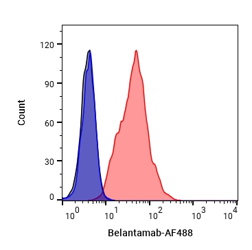 Flow Cytometry analysis of K562 cells labelling CD269 (red) with Belantamab (HY-P9980). Cells were fixed with 4% paraformaldehyde and permeabilised with 90% methanol. Then cells were stained with the primary antibody at 1/200 dilution for an hour at 4℃. Goat Anti-Human IgG H&L (AF488) (HY-P83776) was used as the secondary antibody at 1/1,000 dilution for 30 minutes at 4℃. Human IgG1 kappa (HY-P99001, blue) was used as the isotype control, cells without incubation with primary antibody were used as the unlabeled control (black).
 -
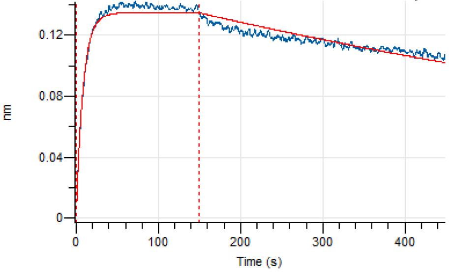 Loaded Belantamab on AHC2 biosensor, can bind BCMA/TNFRSF17 Protein, Human (HEK293, His, HY-P72847) with an affinity constant of 3.934E-10 M as determined in BLI assay.


Chemical Information
-
CAS No. 2061894-48-0
-
Appearance Liquid
-
Masse moléculaire 146.06 kDa
-
Color Colorless to light yellow
-
SMILES
[Belantamab]
-
Synonyms
GSK2857914
-
Livraison
Shipping with dry ice.
-
Formulation
Please refer to the lot-specific COA for specific buffer information.
-
Stockage
Please store the product under the recommended conditions in the Certificate of Analysis.


Publications (3)
-
Journal Impact Factor
-
Most Recent
-
ACS Nano
Post-Conjugation Process for Antibody-Conjugated Lipid Nanoparticles Enabling Tunable Antibody Surface Density for Targeted RNA Delivery. [Abstract]2026 Jun 2;20(21):15092-15102. PMID: 42160160 -
-


Pureté et documentation
-
Fiche technique (262 KB)
-
SDS (251 KB)
- English - EN (251 KB)
- Français - FR (251 KB)
- Deutsch - DE (251 KB)
- Norwegian - NO (251 KB)
- Español - ES (251 KB)
- Swedish - SV (251 KB)
- Italian - IT (251 KB)
- Korean - KR (251 KB)
- Portuguese - PT (251 KB)
-
Inhibitory Antibodies User Guide (603 KB)
Références
[1]. Lucijanic M, et al. Belantamab Mafodotin, Bortezomib, and Dexamethasone for Multiple Myeloma. N Engl J Med. 2024 Oct 10;391(14):1364. [Content Brief]
[2]. Montes de Oca R, et al. Belantamab Mafodotin (GSK2857916) Drives Immunogenic Cell Death and Immune-mediated Antitumor Responses In Vivo. Mol Cancer Ther. 2021;20(10):1941-1955. [Content Brief]

Calculators
Concentration (start) × Volume (start) = Concentration (final) × Volume (final)


Belantamab Related Classifications
HY-P9980 Related Classifications
 Powered by Bioz