



HSV-TK substrate
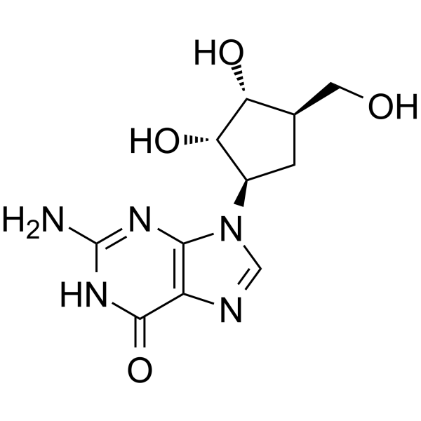
HSV-TK substrate is a substrate for HSV-TK, and induces multi-log cytotoxicity in HSV-TK-expressing and bystander cells. HSV-TK substrate shows antitumor activity.
Nos produits utilisent uniquement pour la recherche. Nous ne vendons pas aux patients.
- CAS No.: 111687-37-7
- Formule: C11H15N5O4
- Masse moléculaire:281.27
-
Stockage:
Please store the product under the recommended conditions in the Certificate of Analysis.

To place orders, for customer services and technical support, please contact: MedChemExpress USA
Tel: 609-228-6898 E-mail: [email protected] [email protected]


-
 Activité biologique
-
Chemical Information
- Protocole
- Pureté et documentation
- Références
-
 Help & FAQs


-
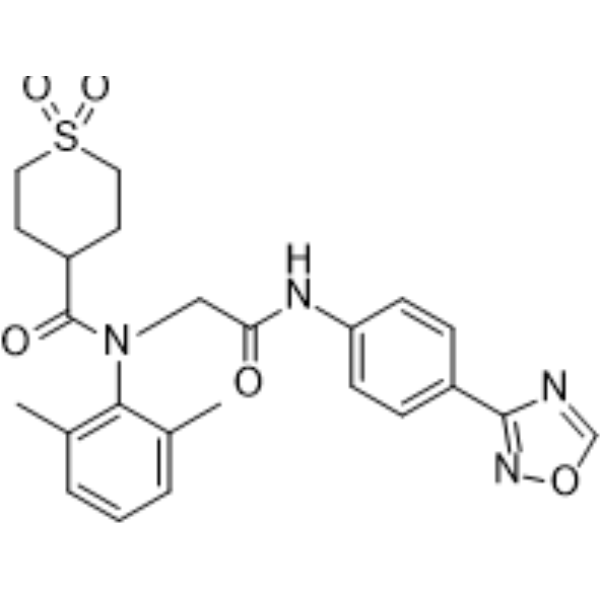
HY-14809
AmenamevirHSV Inhibitor -
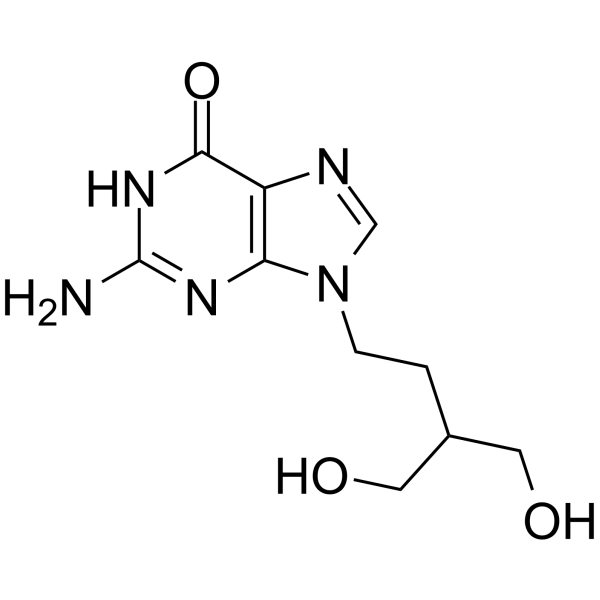
HY-17424
PenciclovirAnti-herpesvirus Agent -
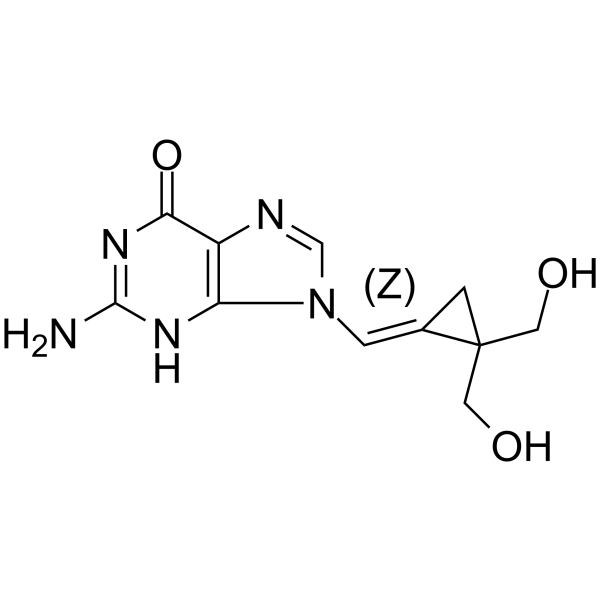
HY-16721
CyclopropavirAnti-herpesvirus Agent -

HY-14809R
Amenamevir (Standard)HSV Inhibitor -

HY-17424R
Penciclovir (Standard)Anti-herpesvirus Agent -
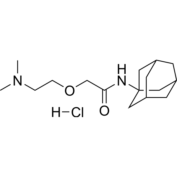
HY-U00124B
Tromantadine hydrochlorideHSV Replication Inhibitor -
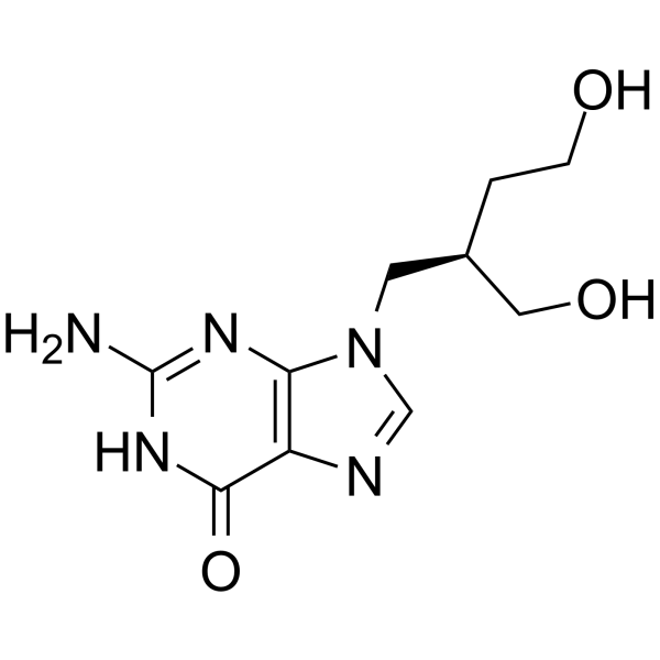
HY-116174
OmaciclovirHerpesvirus Inhibitor -
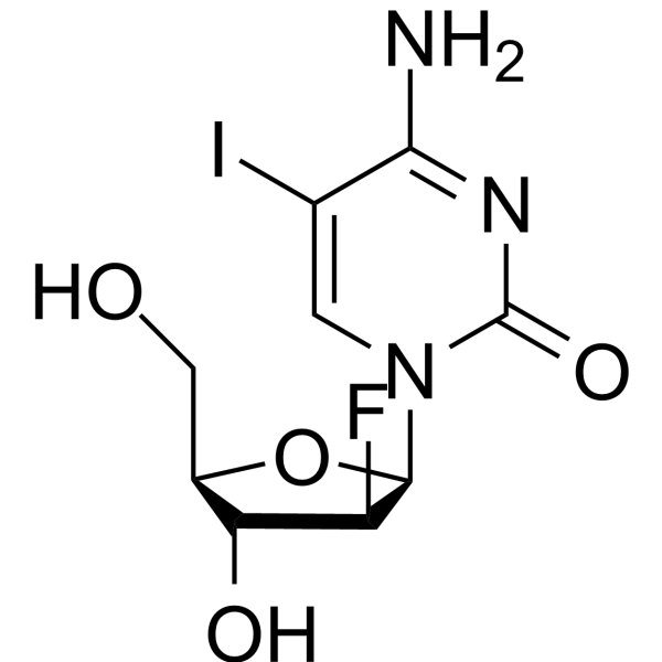
HY-50735
FiacitabineDNA Polymerase Inhibitor -
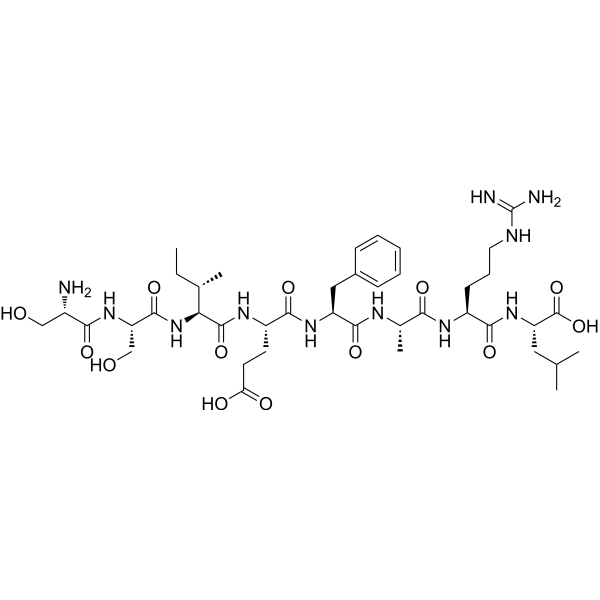
HY-P1862
HSV-gB2 (498-505)HSV Glycoprotein Epitope -
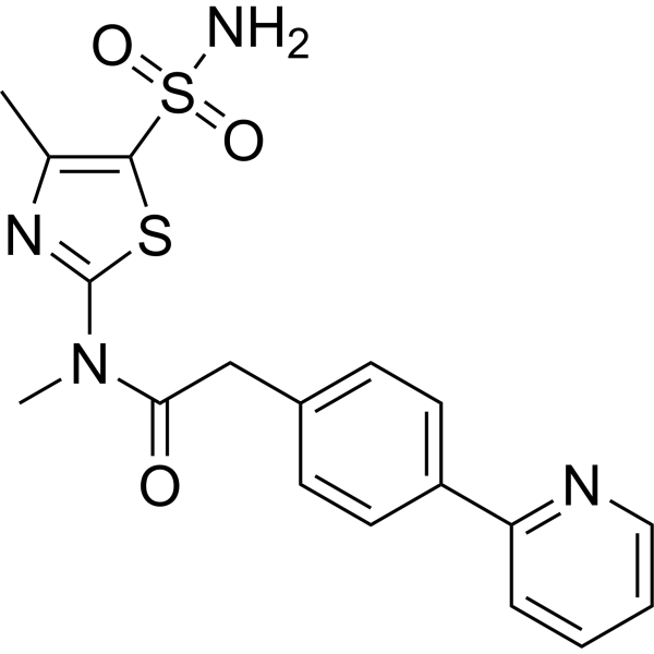
HY-15303
PritelivirHSV Inhibitor -
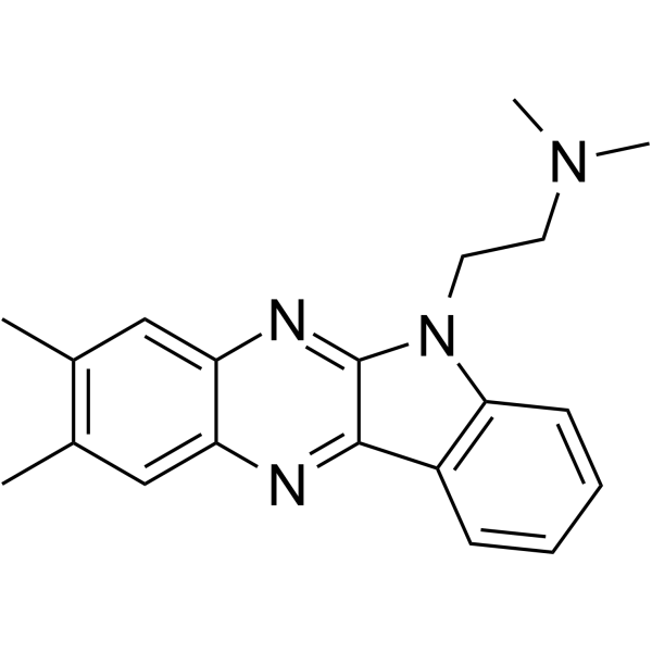
HY-100272
B220Antiviral Agent -
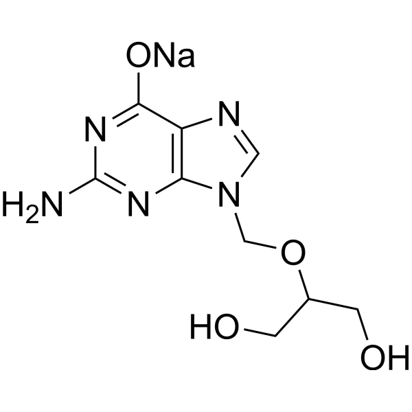
HY-13637A
Ganciclovir sodiumAntiviral Agent


Activité biologique
Description


Chemical Information
-
CAS No. 111687-37-7
-
Masse moléculaire 281.27
-
Formule C11H15N5O4
-
SMILES
O=C1NC(N)=NC2=C1N=CN2[C@H]3[C@H](O)[C@H](O)[C@@H](CO)C3
-
Livraison
Room temperature in continental US; may vary elsewhere.
-
Stockage
Please store the product under the recommended conditions in the Certificate of Analysis.


Pureté et documentation
Références

Calculators
Concentration (start) × Volume (start) = Concentration (final) × Volume (final)


HSV-TK substrate Related Classifications
HY-126218 Related Classifications
 Powered by Bioz