



CCT128930 hydrochloride
Based on 7 publication(s) in Google Scholar
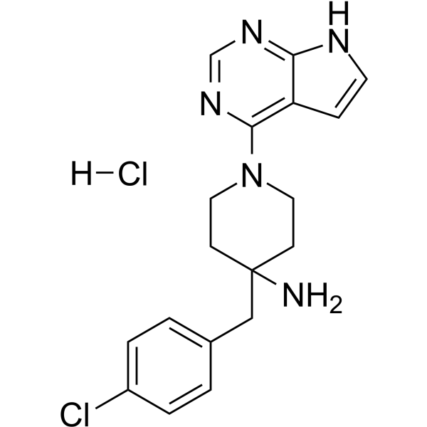
CCT128930 hydrochloride is a potent and selective inhibitor of AKT (IC50=6 nM). CCT128930 hydrochloride has 28-fold selectivity over the closely related PKA kinase (IC50=168 nM) through the targeting of Met282 of AKT (Met173 of PKA-AKT chimera), as well as 20-fold selectivity over p70S6K (IC50=120 nM). CCT128930 hydrochloride induces cell cycle arrest, DNA damage, and autophagy. Antitumor activity.
For research use only. We do not sell to patients.
- Purity : 98.97%
- CAS No.: 2453324-32-6
- Formula: C18H21Cl2N5
- Molecular Weight:378.30
-
Storage:
4°C, sealed storage, away from moisture
* In solvent : -80°C, 6 months; -20°C, 1 month (sealed storage, away from moisture)

To place orders, for customer services and technical support, please contact: MedChemExpress USA
Tel: 609-228-6898 E-mail: [email protected] [email protected]


-
 Biological Activity
-
Chemical Information
-
Solvent & Solubility
- Protocols
- Purity & Documentation
- References
-
 Help & FAQs


-
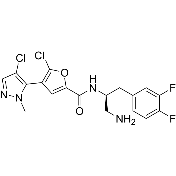
HY-15965
UprosertibAkt Inhibitor -
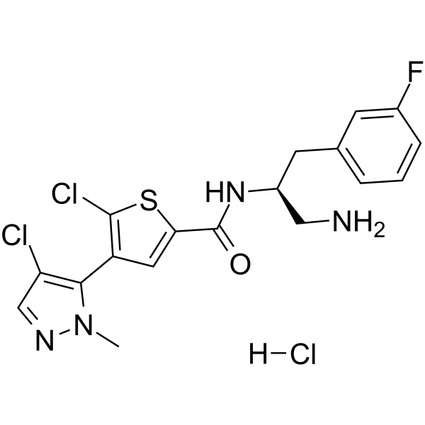
HY-15727A
Afuresertib hydrochlorideAkt Inhibitor -
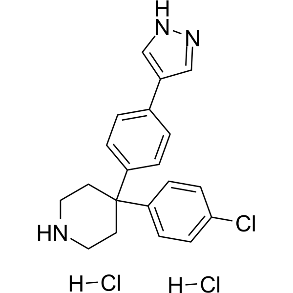
HY-12059A
AT7867 dihydrochloride -
HY-18749
SC79Akt Activator -
HY-15431
CapivasertibAkt Inhibitor -
HY-13254
A-674563Akt1 Inhibitor -
HY-10355
AKT inhibitor VIIIAKT Inhibitor -
HY-15186
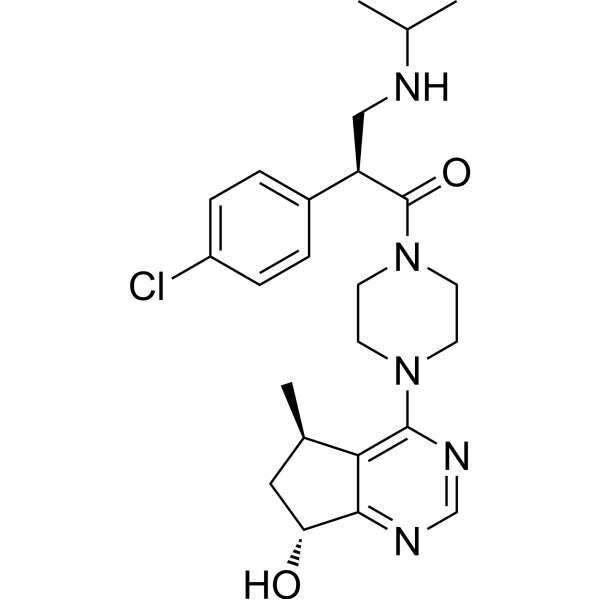
IpatasertibPan-Akt Inhibitor -
HY-10425
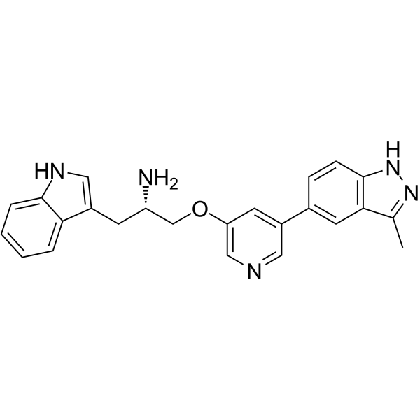
Rizavasertibpan-Akt Inhibitor -
HY-13254A
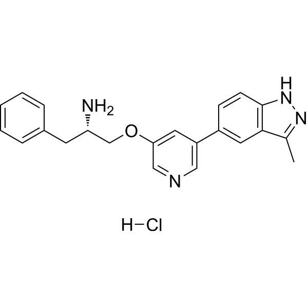
A-674563 hydrochlorideAkt1 Inhibitor -
HY-16666
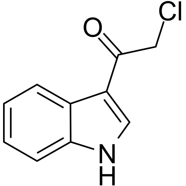
3CAIAkt Inhibitor -
HY-N0004
OridoninBacterial Inhibitor
Publications Citing Use of MedChemExpress (MCE) CCT128930 hydrochloride
More- Cell Death Dis. 2026 Jun 3. [Abstract]
- J Transl Med. 2025 Dec 23;23(1):1412. [Abstract]
- Mol Cancer Ther. 2024 Oct 1;23(10):1404-1417. [Abstract]
- Biochem Biophys Res Commun. 2021 Jun 30:560:132-138. [Abstract]
- Patent. US20240066113A1.
- J Healthc Eng. 2022 Jan 5:2022:1473597. [Abstract]
- Oncotarget. 2016 May 17;7(20):29131-42. [Abstract]
 Customer Validation & Images

-
Bio/Physico-chemical Assay
-
Cell Proliferation/Viability Assay
-
WB
-
ELISA
-
IF


Biological Activity
Description
IC50 & Target
[1]|
Akt2 6 nM (IC50) |
PKA 168 nM (IC50) |
p70S6K 120 nM (IC50) |
Autophagy |
Apoptosis |
In Vitro
The GI50 values of CCT128930 hydrochloride for growth inhibition are 6.3 μM for U87MG human glioblastoma cells, 0.35 μM for LNCaP human prostate cancer cells, and 1.9 μM for PC3 human prostate cancer cells, all of which are PTEN-deficient human tumor cell lines[1].
CCT128930 (0.1-60 μM; 1 hour; U87MG human glioblastoma cells) hydrochloride shows an initial induction of AKT phosphorylation at serine 473 up to 20 μM, followed by a decreased in phosphorylation at higher concentrations[1].
CCT128930 hydrochloride inhibits direct substrates of AKT (Ser9 GSK3β, pThr246 PRAS40 and pT24 FOXO1/p32 FOXO3a) at ≥5 μM, and the downstream target, pSer235/236 S6RP at ≥10 μM, with generally constant levels of the respective total proteins and GAPDH[1].
CCT128930 (18.9 μM; U87MG human glioblastoma cells) hydrochloride causes an increase in phosphorylation of pSer473 AKT after 30 minutes, which is sustained for 48 hours. Total AKT protein signal decreases gradually from 8 hours to 48 hours of treatment[1].
CCT128930 (PTEN-null U87MG human glioblastoma cells; over a 24-hour time period) hydrochloride results in an increase in G0/G1 phase cells from 43.6% to 64.8% after 24 hours of treatment[1].
CCT128930 (0-10 μM; 24 hours) hydrochloride increases, but not inhibites, the phosphorylation of Akt in HepG2 and A549 cells. CCT128930 (0-20 μM; 24 hours) hydrochloride inhibits cell proliferation by inducing cell cycle arrest in G1 phase through downregulation of cyclinD1 and Cdc25A, and upregulation of p21, p27 and p53. CCT128930 (20 μM) hydrochloride triggers cell apoptosis with activation of caspase-3, caspase-9, and PARP. CCT128930 (0-20 μM; 24 hours) hydrochloride increases phosphorylation of ERK and JNK in HepG2 cells. CCT128930 (0-20 μM; 24 hours) hydrochloride activates DNA damage response of HepG2 cell characterized by phosphorylation of H2AX, ATM (ataxia-telangiectasia mutated), Chk1 and Chk2[2].
MedChemExpress (MCE) has not independently confirmed the accuracy of these methods. They are for reference only. Further protocols information, click here.
In Vivo
| Tissue | Route | T1/2 (h) |
Tmax (h) |
Cmax (μM) |
Vss (L) |
Cl (L/h) |
AUC0-∞ (µMh) |
Bioavailability (%) |
| Plasma | i.v. | 0.95 | 0.083 | 6.36 | 0.25 | 0.325 | 4.62 | 100 |
| Plasma | i.p. | 2.33 | 0.5 | 1.28 | N/A | 0.372 | 1.33 | 28.8 |
| Tumor | i.p. | 3.89 | 1 | 8.02 | N/A | 0.06* | 25.8 | N/A |
| Plasma | p.o. | 0.57 | 0.5 | 0.432 | N/A | 0.317 | 0.392 | 8.5 |
*Apparent clearance.
MedChemExpress (MCE) has not independently confirmed the accuracy of these methods. They are for reference only.
-
Animal Model:6-8 weeks old female CrTacNCr-Fox1nu mice[1]
-
Dosage:25 mg/kg (U87MG human glioblastoma xenografts) or 40 mg/kg (BT474 human breast cancer xenografts)
-
Administration:i.p. daily for 5 days (U87MG human glioblastoma xenografts); i.p. twice daily for 5 days (BT474 human breast cancer xenografts)
-
Result:Giving a treated:control (T/C) ratio on day 12 of 48%. There was no weight loss associated with this regime in U87MG human glioblastoma xenografts.
Had a profound antitumor effect with complete growth arrest and a T/C ratio of 29% on day 22. This regimen was associated with minimal weight loss, with a nadir of only 94.8% of the initial body weight on day 15 of treatment in BT474 human breast cancer xenografts.


Chemical Information
-
CAS No. 2453324-32-6
-
Appearance Solid
-
Molecular Weight 378.30
-
Formula C18H21Cl2N5
-
Color White to yellow
-
SMILES
NC1(CC2=CC=C(Cl)C=C2)CCN(C3=C4C(NC=C4)=NC=N3)CC1.[H]Cl
-
Shipping
Room temperature in continental US; may vary elsewhere.
-
Storage
4°C, sealed storage, away from moisture
* In solvent : -80°C, 6 months; -20°C, 1 month (sealed storage, away from moisture)


Publications (7)
-
Journal Impact Factor
-
Most Recent
-
Cell Death Dis
PEBP4 alleviates muscle wasting in lung cancer cachexia via KEAP1-NRF2-mediated redox homeostasis. [Abstract]2026 Jun 3. PMID: 42236665 -
J Transl Med
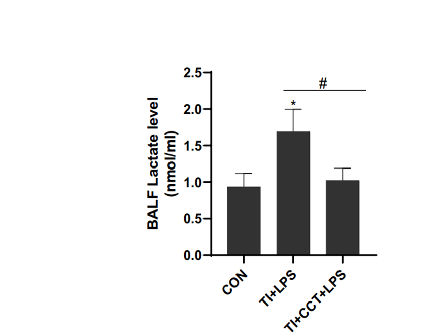
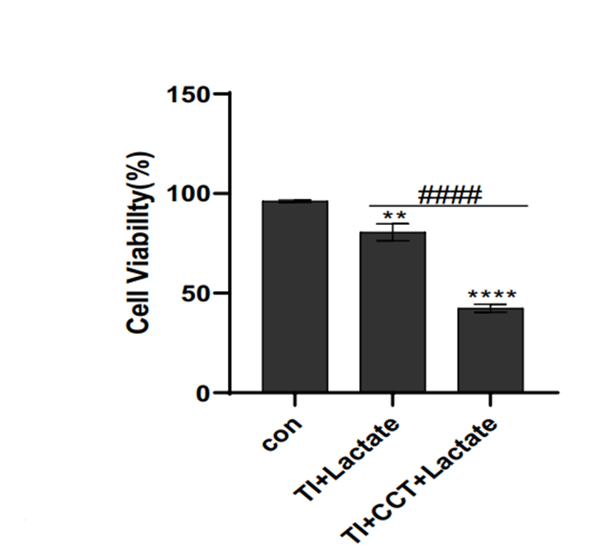
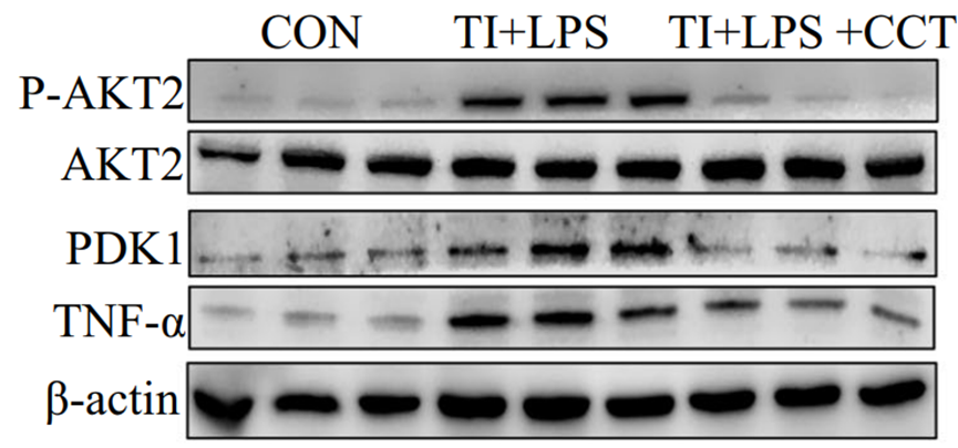
Trained immunity attenuated acute lung injury by activating alveolar macrophages via AKT2-PDK1 axis-mediated metabolic reprogramming. [Abstract]2025 Dec 23;23(1):1412. PMID: 41437043
CCT128930 hydrochloride purchased from MedChemExpress. Usage Cited in: J Transl Med. 2025 Dec 23;23(1):1412. [Abstract]
Lactate secretion levels were measured following CCT128930 (CCT) treatment and compared with the TI group.
CCT128930 hydrochloride purchased from MedChemExpress. Usage Cited in: J Transl Med. 2025 Dec 23;23(1):1412. [Abstract]
CCK-8 assay demonstrated reduced cellular lactate tolerance after CCT128930 (CCT) treatment.
CCT128930 hydrochloride purchased from MedChemExpress. Usage Cited in: J Transl Med. 2025 Dec 23;23(1):1412. [Abstract]
Western blot and QPCR analyses revealed decreased PDK1 and inflammatory marker expression after CCT128930 (CCT) treatment.
CCT128930 hydrochloride purchased from MedChemExpress. Usage Cited in: J Transl Med. 2025 Dec 23;23(1):1412. [Abstract]
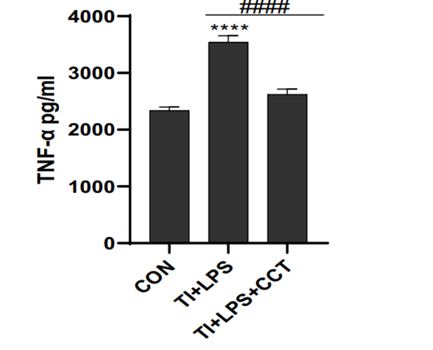
TNF-α ELISA showed significant reduction after CCT128930 (CCT) treatment.
CCT128930 hydrochloride purchased from MedChemExpress. Usage Cited in: J Transl Med. 2025 Dec 23;23(1):1412. [Abstract]
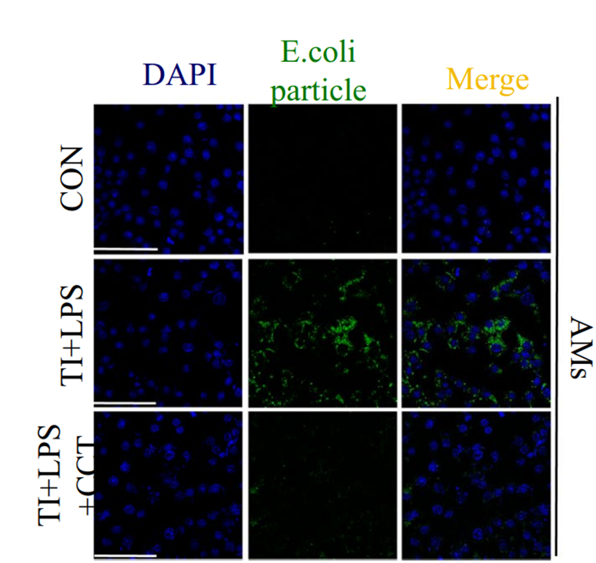
CCT128930 (CCT) treatment impaired phagocytic capacity.
-
Mol Cancer Ther
AKT Inhibition Sensitizes to Polo-Like Kinase 1 Inhibitor Onvansertib in Prostate Cancer. [Abstract]2024 Oct 1;23(10):1404-1417. PMID: 38894678 -
Biochem Biophys Res Commun
2021 Jun 30:560:132-138. PMID: 33989904 -
-
J Healthc Eng
Study on Toll-Like Receptor 2-Mediated Inflammation-Induced Familial Hypertension Combined with Hyperlipemia and Its Mechanism. [Abstract]2022 Jan 5:2022:1473597. PMID: 35035808 -
Oncotarget
Dual inhibition of AKT/FLT3-ITD by A674563 overcomes FLT3 ligand-induced drug resistance in FLT3-ITD positive AML. [Abstract]2016 May 17;7(20):29131-42. PMID: 27074558


Solvent & Solubility
In Vitro:
DMSO : 20 mg/mL (52.87 mM; ultrasonic and warming and heat to 60°C; Hygroscopic DMSO has a significant impact on the solubility of product, please use newly opened DMSO)
Please refer to the solubility information to select the appropriate solvent. Once prepared, please aliquot and store the solution to prevent product inactivation from repeated freeze-thaw cycles.
Storage method and period of stock solution: -80°C, 6 months; -20°C, 1 month (sealed storage, away from moisture). When stored at -80°C, please use it within 6 months. When stored at -20°C, please use it within 1 month.
Please refer to the solubility information to select the appropriate solvent. Once prepared, please aliquot and store the solution to prevent product inactivation from repeated freeze-thaw cycles.
Storage method and period of stock solution: -80°C, 6 months; -20°C, 1 month (sealed storage, away from moisture). When stored at -80°C, please use it within 6 months. When stored at -20°C, please use it within 1 month.
Concentration (start) × Volume (start) = Concentration (final) × Volume (final)
In Vivo:
Select the appropriate dissolution method based on your experimental animal and administration route.
- For the following dissolution methods, please ensure to first prepare a clear stock solution using an In Vitro approach and then sequentially add co-solvents:
- To ensure reliable experimental results, the clarified stock solution can be appropriately stored based on storage conditions. As for the working solution for In Vivo experiments, it is recommended to prepare freshly and use it on the same day.
- The percentages shown for the solvents indicate their volumetric ratio in the final prepared solution. If precipitation or phase separation occurs during preparation, heat and/or sonication can be used to aid dissolution.
Add each solvent one by one: 10% DMSO 40% PEG300 5% Tween-80 45% Saline
Solubility: ≥ 2 mg/mL (5.29 mM); Clear solution
This protocol yields a clear solution of ≥ 2 mg/mL (saturation unknown).
Taking 1 mL working solution as an example, add 100 μL DMSO stock solution (20.0 mg/mL) to 400 μL PEG300, and mix evenly; then add 50 μL Tween-80 and mix evenly; then add 450 μL Saline to adjust the volume to 1 mL.
Preparation of Saline: Dissolve 0.9 g sodium chloride in ddH₂O and dilute to 100 mL to obtain a clear Saline solution.
Add each solvent one by one: 10% DMSO 90% (20% SBE-β-CD in Saline)
Solubility: ≥ 2 mg/mL (5.29 mM); Clear solution
This protocol yields a clear solution of ≥ 2 mg/mL (saturation unknown).
Taking 1 mL working solution as an example, add 100 μL DMSO stock solution (20.0 mg/mL) to 900 μL 20% SBE-β-CD in Saline, and mix evenly.
Preparation of 20% SBE-β-CD in Saline (4°C, storage for one week): 2 g SBE-β-CD powder is dissolved in 10 mL Saline, completely dissolve until clear.
In Vivo Dissolution Calculator
Please enter the basic information of animal experiments:
-
-
-
-
Recommended: Prepare an additional quantity of animals to account for potential losses during experiments.
Please enter your animal formula composition:
-
%DMSO +
Recommended: Keep the proportion of DMSO in working solution below 2% if your animal is weak.
-
%+
-
+%Tween-80 + +
-
%Saline +
The co-solvents required include: DMSO, . All of co-solvents are available by MedChemExpress (MCE). , Tween 80. All of co-solvents are available by MedChemExpress (MCE).
Working solution concentration: 0.22 mg/mL
Method for preparing stock solution: mg drug dissolved in μL DMSO. Stock solution concentration: mg/mL. * In solvent : -80°C, 6 months; -20°C, 1 month (sealed storage, away from moisture)
1. Take μL DMSO stock solution;
2. Add μL .
μL , mix evenly;
3. Then add μL Tween 80, mix evenly;
4. Then add μL
Please ensure that the stock solution in the first step is dissolved to a clear state, and add co-solvents in sequence. You can use ultrasonic heating (ultrasonic cleaner, recommended frequency 20-40 kHz), vortexing, etc. to assist dissolution.

Protocols
-
Cell Cytotoxicity Assay
Cytotoxicity assays are usually based on the assessment of cell membrane damage, which can also be indirectly detected by measuring cell viability. Detection methods include MTT assay, CKK-8 assay, LDH assay and ATP assay, etc.
-
Flow cytometric DNA-content cell-cycle staining
Flow cytometric DNA-content cell-cycle staining measures the fluorescence intensity of DNA-bound fluorochromes in single cells or nuclei to estimate DNA content distributions, allowing assignment of populations to G0/G1, S, and G2/M phases by DNA histogram deconvolution. Propidium iodide (PI) intercalates into DNA, and PI fluorescence is proportional to cellular DNA content when staining is performed under conditions that make DNA accessible and minimize non-DNA signal. Cells with G2/M DNA content are expected to show approximately twice the fluorescence intensity of G0/G1 cells, while S-phase cells occupy intermediate fluorescence values. PI-based DNA-content analysis can also detect cells with fractional DNA content, often reported as sub-G1, when DNA fragmentation and extraction during staining reduce retained DNA signal in apoptotic cells. DAPI is an alternative DNA fluorochrome for univariate DNA-content analysis, while bivariate approaches combining DNA content with proliferation
-
BrdU Incorporation Assay
Bromodeoxyuridine (BrdU) incorporation assay is based on the principle that BrdU, a thymidine analog, is incorporated into newly synthesized DNA during the S phase of the cell cycle, thereby serving as a marker of DNA replication and cellular proliferation. Incorporated BrdU can be detected using anti-BrdU antibodies following DNA denaturation, enabling visualization or quantification of proliferating cells through immunochemical detection methods such as immunofluorescence or immunohistochemistry.
-
Autophagy
Autophagy is a process in which eukaryotic cells use lysosomes to degrade their own cytoplasmic proteins and damaged organelles under the regulation of autophagy related gene (Atg). Microtubule-associated proteins light chain 3 (LC3) is recognized as autophagy marker, which transfers from cytoplasmic LC3 (LC3-I) to membrane type (LC3-II). LC3-II/I ratio could be detected by Western Blot and fluorescence microscopy.
-
Protocol for Cell Cycle
Cell-cycle analysis by flow cytometry measures DNA content in single cells to estimate the fraction of cells in G0/G1, S, and G2/M phases. Propidium iodide intercalates into DNA, and after RNA removal with RNase, fluorescence intensity reflects cellular DNA content: 2N cells are assigned to G0/G1, cells between 2N and 4N to S phase, and 4N cells to G2/M. DNA-content analysis alone cannot reliably separate G0 from G1 or G2 from M. Ki-67 can distinguish quiescent G0 cells from cycling cells, EdU or BrdU incorporation marks active DNA synthesis in S phase, and phospho-histone H3 staining identifies mitotic cells within the 4N population.
-
Lysosome and acidic-vesicle live-cell staining
Lysosome and acidic-vesicle live-cell staining detects acidic intracellular compartments by using membrane-permeant acidotropic probes that accumulate in low-pH vesicles, including lysosomes, late endosomes, autolysosomes, and acidic phagosomes. LysoTracker staining is commonly used as an intensity-based readout of acidic lysosomal compartment abundance or enlargement, while acridine orange produces green fluorescence in less concentrated compartments and red fluorescence after concentration-dependent accumulation in acidic vesicular organelles. Loss or reduction of acridine-orange red signal can be used as a readout of lysosomal membrane permeabilization or reduced acidic-vesicle integrity. This protocol is designed for live cultured cells and can be adapted for fluorescence microscopy, high-content imaging, plate-reader readout, or flow cytometry when the selected literature supports the readout. Because these dyes report acidotropic accumulation rather than lysosome identity alone,
-
Subcutaneous Cell-Line-Derived Xenograft
Subcutaneous cell-line-derived xenograft (CDX) models are established by implanting cultured human cancer cell lines into immunodeficient mice, where the injected cells form localized tumors that can be monitored in vivo as a measure of tumorigenic potential, growth kinetics, and treatment response. These models are widely used in oncology research because they allow reproducible tumor formation and enable comparative assessment of tumor growth between different cell lines or genetic manipulations in a controlled in vivo microenvironment. Subcutaneous implantation of cancer cells in immunodeficient mice is a standard approach for evaluating tumor growth behavior and therapeutic response across multiple cancer types, including prostate, esophageal, pancreatic, and colon cancer models.
-
Macroautophagy Solutions
Macroautophagy is a conserved lysosome-dependent degradation pathway in which cytoplasmic material is sequestered into double-membrane autophagosomes and delivered to lysosomes for degradation and recycling. The pathway supports cellular homeostasis during nutrient limitation, organelle stress, protein-aggregate accumulation, infection, differentiation, and tissue remodeling by coupling cargo sequestration, autophagosome maturation, lysosomal fusion, and degradation of cargo-derived macromolecules. The core molecular sequence includes initiation by nutrient- and stress-regulated autophagy machinery, autophagosome nucleation, LC3/ATG8-family conjugation to autophagosomal membranes, cargo selection through receptors such as SQSTM1/p62, autophagosome-lysosome fusion, and lysosomal degradation. LC3 was identified as a mammalian homolog of yeast Atg8 that localizes to autophagosomal membranes after processing, and p62/SQSTM1 was shown to connect ubiquitinated cargo with autophagic degradati
-
Genotoxicity/Mutagenicity Study
The bacterial reverse mutation assay detects point mutations that restore amino-acid prototrophy in auxotrophic Salmonella typhimurium or Escherichia coli tester strains; after exposure to a test article, mutagenic activity is read out as an increased number of revertant colonies on minimal agar compared with the vehicle control. The assay uses tester strains with different mutation targets so that base-substitution and frameshift mutagens can be detected, and testing is performed with and without exogenous mammalian metabolic activation because some chemicals require biotransformation to become mutagenic.


Purity & Documentation
-
Data Sheet (283 KB)
-
SDS (393 KB)
- English - EN (393 KB)
- Français - FR (393 KB)
- Deutsch - DE (393 KB)
- Norwegian - NO (393 KB)
- Español - ES (393 KB)
- Swedish - SV (393 KB)
- Italian - IT (393 KB)
- Korean - KR (393 KB)
- Portuguese - PT (393 KB)
-
Handling Instructions (2659 KB)
References
[1]. Yap TA et al. Preclinical pharmacology, antitumor activity, and development of pharmacodynamic markers for the novel, potent AKT inhibitor CCT128930. Mol Cancer Ther. 2011 Feb;10(2):360-71. [Content Brief]
[2]. Wang FZ, et al. CCT128930 induces cell cycle arrest, DNA damage, and autophagy independent of Akt inhibition. Biochimie. 2014;103:118-125. [Content Brief]


Complete Stock Solution Preparation Table
Please refer to the solubility information to select the appropriate solvent. Once prepared, please aliquot and store the solution to prevent product inactivation from repeated freeze-thaw cycles.
Storage method and period of stock solution: -80°C, 6 months; -20°C, 1 month (sealed storage, away from moisture). When stored at -80°C, please use it within 6 months. When stored at -20°C, please use it within 1 month.
| Optional Solvent | Concentration Solvent Mass | 1 mg | 5 mg | 10 mg | 25 mg |
|---|---|---|---|---|---|
| DMSO | 1 mM | 2.6434 mL | 13.2170 mL | 26.4340 mL | 66.0851 mL |
| 5 mM | 0.5287 mL | 2.6434 mL | 5.2868 mL | 13.2170 mL | |
| 10 mM | 0.2643 mL | 1.3217 mL | 2.6434 mL | 6.6085 mL | |
| 15 mM | 0.1762 mL | 0.8811 mL | 1.7623 mL | 4.4057 mL | |
| 20 mM | 0.1322 mL | 0.6609 mL | 1.3217 mL | 3.3043 mL | |
| 25 mM | 0.1057 mL | 0.5287 mL | 1.0574 mL | 2.6434 mL | |
| 30 mM | 0.0881 mL | 0.4406 mL | 0.8811 mL | 2.2028 mL | |
| 40 mM | 0.0661 mL | 0.3304 mL | 0.6609 mL | 1.6521 mL | |
| 50 mM | 0.0529 mL | 0.2643 mL | 0.5287 mL | 1.3217 mL |


CCT128930 hydrochloride Related Classifications
HY-13260A Related Classifications
 Powered by Bioz