



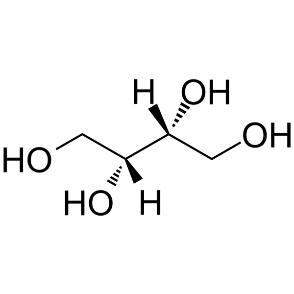
D-Threitol

Based on 1 Customer Validation
D-Threitol is a naturally occurring four-carbon sugar alcohol and an α-glucosidase (α‑glucosidase) inhibitor with an IC50 of 49 mM against yeast α-glucosidase, and it is orally active. D-Threitol modulates gut microbiota composition, increases microbial diversity, enriches beneficial bacterial genera, and restores fecal short-chain fatty acid levels. D-Threitol reduces body weight gain and fat accumulation, improves glucose tolerance and insulin sensitivity, and alleviates liver, kidney, and pancreatic tissue damage. D-Threitol inhibits hepatic accumulation of ceramides and diacylglycerols. D-Threitol preserves glomerular morphology and islet structure in diabetes models. D-Threitol is also a low-calorie sweetener. D-Threitol serves as a cryoprotective polyol that stabilizes protein structure at low temperatures. D-Threitol is used in research on type 2 diabetes and as an antifreeze agent for the Alaskan beetle.
Nos produits utilisent uniquement pour la recherche. Nous ne vendons pas aux patients.
- Pureté : 99.95%
- CAS No.: 2418-52-2
- Formule: C4H10O4
- Masse moléculaire:122.12
-
Stockage:Powder -20°C, 3 years ; In solvent -80°C, 6 months , -20°C, 1 month

To place orders, for customer services and technical support, please contact: MedChemExpress USA
Tel: 609-228-6898 E-mail: [email protected] [email protected]


-
 Activité biologique
-
Chemical Information
-
Solvant et solubilité
- Protocole
- Pureté et documentation
- Références
-
 Help & FAQs


Voir tous les produits spécifiques à Isoform Endogenous Metabolite
More

Activité biologique
Description
IC50 & Target
[1]|
α‑glucosidase |
In Vivo
D-threitol (500 mg/kg; p.o.; daily; 8 weeks) improves insulin sensitivity, reduces hepatic steatosis and levels of lipotoxic ceramides (with reductions of 24%-62% in major species) and diacylglycerols (53.1% reduction), and ameliorates systemic metabolic dysfunction in db/db type 2 diabetic mice[2].
MedChemExpress (MCE) has not independently confirmed the accuracy of these methods. They are for reference only.
-
Animal Model:C57BL/6J (male; 6 weeks old; initial body weight 20 g; high-fat diet feeding for 6 weeks followed by intraperitoneal injection of streptozotocin at 40 mg/kg for three consecutive days to induce type 2 diabetes mellitus)[1]
-
Dosage:500 mg/kg/day
-
Administration:oral gavage; daily; 8 weeks
-
Result:Exhibited slower weight gain beginning in the fifth week of treatment, with a significant reduction by the ninth week relative to the untreated diabetic model group.
Showed significantly lower postprandial blood glucose compared with the model group.
Had significantly reduced oral glucose tolerance test area under the curve, indicating improved glucose tolerance.
Had significantly lower serum insulin levels and HOMA-IR values than the model group, demonstrating ameliorated insulin resistance.
Reduced abdominal white adipose tissue weight by 17.45% compared with the model group.
Reduced serum total cholesterol, triglycerides, and high-density lipoprotein cholesterol by 8.97%, 14.86%, and 8.68%, respectively, relative to the model group.
Notably decreased serum alanine aminotransferase and aspartate aminotransferase levels, indicating protection against liver injury.
Reduced hepatocellular vacuolation and improved hepatic architecture, reduced renal tubular epithelial cell vacuolation, and preserved pancreatic islet structure with clear borders and prevented islet atrophy.
Increased the number of observed OTUs to 600, increased Shannon and Pielou indices, and decreased the Simpson index, indicating restored α-diversity.
Reduced the Firmicutes/Bacteroidetes ratio to 4.10 from 6.08 in the model group.
Enriched beneficial genera including Allobaculum, Lactobacillus, and Akkermansia, while decreasing Oscillospira, Ruminococcus, and Coprococcus.
Restored fecal levels of acetate, propionate, and butyrate, and reversed elevated levels of isobutyrate and isovalerate.
-
Animal Model:C57BLKs/J db/db (male; 8 weeks old; homozygous leptin receptor deficiency)[2]
-
Dosage:500 mg/kg
-
Administration:p.o.; daily; 8 weeks
-
Result:Reduced water intake by 20.9% relative to vehicle control.
Reduced visceral white adipose tissue mass by 24.4% (from 1.94 g to 1.47 g) relative to vehicle control.
Reduced HOMA-IR index by 21.0% (from 20.6 to 16.3) relative to vehicle control.
Resulted in mean fasting blood glucose of 23.0 mmol/L versus 25.7 mmol/L in vehicle control group.
Significantly reduced ITT area under the curve relative to vehicle control.
Reduced serum alanine aminotransferase by 28.1% (75.4 U/L vs. 104.8 U/L) relative to vehicle control.
Reduced serum triglycerides by 22.3% (1.75 mmol/L vs. 2.25 mmol/L) relative to vehicle control.
Reduced serum total cholesterol by 23.2% (2.41 mmol/L vs. 3.14 mmol/L) relative to vehicle control.
Reduced serum LDL-cholesterol by 40.2% (0.19 mmol/L vs. 0.32 mmol/L) relative to vehicle control.
Reduced hepatic diacylglycerol content by 53.1% relative to vehicle control.
Reduced 32 hepatic ceramide species, with C16:0, C18:0, C20:0, C22:0, C24:0, and C24:1 ceramides showing reductions ranging from 24% to 62% (log2 fold change ranging from −0.64 to −1.49) relative to vehicle control.
Significantly downregulated hepatic Sptlc2 and Cers6 mRNA (de novo ceramide synthesis genes) and upregulated Asah1 and Cerk mRNA (ceramide degradation and conversion genes) relative to vehicle control.
Showed nearly absent hepatocellular lipid vacuoles, attenuated inflammatory infiltration in the liver, more intact glomerular morphology with reduced mesangial expansion in the kidney, and better preserved pancreatic islet structure relative to vehicle control.
Reduced body weight by 8.38% (38.5 g vs. 42.0 g at week 8) relative to sucrose-treated group at the same dose.
Reduced visceral white adipose tissue mass by 27.8% relative to sucrose-treated group at the same dose.
Reduced fasting blood glucose by 16.5% relative to sucrose-treated group at the same dose.
Reduced HOMA-IR by 31.2% relative to sucrose-treated group at the same dose.
Reduced serum ALT by 42.7% relative to sucrose-treated group at the same dose.
Reduced serum AST by 43.5% relative to sucrose-treated group at the same dose.
Resulted in substantially lower serum triglycerides, total cholesterol, and LDL-cholesterol relative to sucrose-treated group at the same dose.


Chemical Information
-
CAS No. 2418-52-2
-
Appearance Solid
-
Masse moléculaire 122.12
-
Formule C4H10O4
-
Color White to light yellow
-
SMILES
OC[C@H]([C@@H](CO)O)O
-
Structure Classification
-
Initial Source
-
Livraison
Room temperature in continental US; may vary elsewhere.
-
Stockage
Powder -20°C 3 years In solvent -80°C 6 months -20°C 1 month


Solvant et solubilité
In Vitro:
DMSO : 250 mg/mL (2047.17 mM; Need ultrasonic; Hygroscopic DMSO has a significant impact on the solubility of product, please use newly opened DMSO)
Please refer to the solubility information to select the appropriate solvent. Once prepared, please aliquot and store the solution to prevent product inactivation from repeated freeze-thaw cycles.
Storage method and period of stock solution: -80°C, 6 months; -20°C, 1 month. When stored at -80°C, please use it within 6 months. When stored at -20°C, please use it within 1 month.
Please refer to the solubility information to select the appropriate solvent. Once prepared, please aliquot and store the solution to prevent product inactivation from repeated freeze-thaw cycles.
Storage method and period of stock solution: -80°C, 6 months; -20°C, 1 month. When stored at -80°C, please use it within 6 months. When stored at -20°C, please use it within 1 month.
Concentration (start) × Volume (start) = Concentration (final) × Volume (final)
In Vivo:
Select the appropriate dissolution method based on your experimental animal and administration route.
- For the following dissolution methods, please ensure to first prepare a clear stock solution using an In Vitro approach and then sequentially add co-solvents:
- To ensure reliable experimental results, the clarified stock solution can be appropriately stored based on storage conditions. As for the working solution for In Vivo experiments, it is recommended to prepare freshly and use it on the same day.
- The percentages shown for the solvents indicate their volumetric ratio in the final prepared solution. If precipitation or phase separation occurs during preparation, heat and/or sonication can be used to aid dissolution.
Add each solvent one by one: 10% DMSO 40% PEG300 5% Tween-80 45% Saline
Solubility: ≥ 2.08 mg/mL (17.03 mM); Clear solution
This protocol yields a clear solution of ≥ 2.08 mg/mL (saturation unknown).
Taking 1 mL working solution as an example, add 100 μL DMSO stock solution (20.8 mg/mL) to 400 μL PEG300, and mix evenly; then add 50 μL Tween-80 and mix evenly; then add 450 μL Saline to adjust the volume to 1 mL.
Preparation of Saline: Dissolve 0.9 g sodium chloride in ddH₂O and dilute to 100 mL to obtain a clear Saline solution.
Add each solvent one by one: 10% DMSO 90% (20% SBE-β-CD in Saline)
Solubility: ≥ 2.08 mg/mL (17.03 mM); Clear solution
This protocol yields a clear solution of ≥ 2.08 mg/mL (saturation unknown).
Taking 1 mL working solution as an example, add 100 μL DMSO stock solution (20.8 mg/mL) to 900 μL 20% SBE-β-CD in Saline, and mix evenly.
Preparation of 20% SBE-β-CD in Saline (4°C, storage for one week): 2 g SBE-β-CD powder is dissolved in 10 mL Saline, completely dissolve until clear.
In Vivo Dissolution Calculator
Please enter the basic information of animal experiments:
-
-
-
-
Recommended: Prepare an additional quantity of animals to account for potential losses during experiments.
Please enter your animal formula composition:
-
%DMSO +
Recommended: Keep the proportion of DMSO in working solution below 2% if your animal is weak.
-
%+
-
+%Tween-80 + +
-
%Saline +
The co-solvents required include: DMSO, . All of co-solvents are available by MedChemExpress (MCE). , Tween 80. All of co-solvents are available by MedChemExpress (MCE).
Working solution concentration: 0.22 mg/mL
Method for preparing stock solution: mg drug dissolved in μL DMSO. Stock solution concentration: mg/mL.
1. Take μL DMSO stock solution;
2. Add μL .
μL , mix evenly;
3. Then add μL Tween 80, mix evenly;
4. Then add μL
Please ensure that the stock solution in the first step is dissolved to a clear state, and add co-solvents in sequence. You can use ultrasonic heating (ultrasonic cleaner, recommended frequency 20-40 kHz), vortexing, etc. to assist dissolution.

Protocole
-
Cell Cytotoxicity Assay
Cytotoxicity assays are usually based on the assessment of cell membrane damage, which can also be indirectly detected by measuring cell viability. Detection methods include MTT assay, CKK-8 assay, LDH assay and ATP assay, etc.
-
Research Protocol for Endocrine Diseases
Endocrine diseases often arise from disrupted hormone production, hormone signaling, or target-tissue responsiveness; for diabetes-focused endocrine disease models, insulin signaling regulates glucose uptake, hepatic glucose output, lipid metabolism, and β-cell compensation. Type 2 diabetes develops through interacting defects in insulin resistance, β-cell dysfunction, adipose inflammation, hepatic glucose overproduction, altered incretin signaling, and ectopic lipid metabolism. A major unresolved question is whether endocrine dysfunction is driven primarily by target-tissue insulin resistance, intrinsic β-cell failure, immune/inflammatory stress, or combined multi-organ failure that differs by disease stage.
-
Research Protocol for Microbiome Analysis
Microbiome analysis characterizes microbial communities in biological or environmental samples by measuring community composition, diversity, taxonomic structure, functional potential, and associations with host or environmental phenotypes. 16S rRNA gene amplicon sequencing is commonly used for bacterial and archaeal taxonomic profiling, while shotgun metagenomics provides higher taxonomic resolution and direct functional information, including microbial genes, pathways, viruses, fungi, and antimicrobial-resistance genes when sequencing depth and host-DNA contamination are adequately controlled. Microbiome results are strongly affected by sample collection, storage, DNA extraction, contamination, sequencing method, reference database, and bioinformatic pipeline; therefore, standardized protocols, negative controls, mock communities, and transparent analysis workflows are required. Unresolved issues include low-biomass contamination, compositional-data bias, inconsistent species-level c
-
Research Protocol for Metabolic Diseases
AMP-activated protein kinase, AMPK, is a conserved cellular energy sensor that responds to reduced cellular energy status and coordinates metabolism by increasing ATP-generating catabolic pathways while suppressing ATP-consuming anabolic processes. In metabolic disease research, the AMPK pathway is experimentally relevant because it regulates hepatic lipid synthesis, fatty acid oxidation, glucose production, skeletal-muscle glucose disposal, mTORC1-linked biosynthesis, autophagy, mitochondrial homeostasis, and whole-body energy balance. The central pathway logic is that energy stress, metformin, exercise-like stimulation, or direct AMPK activators increase AMPKα Thr172 phosphorylation and downstream substrate phosphorylation, including ACC and RAPTOR. Phosphorylation of ACC suppresses lipogenesis and supports fatty acid oxidation, whereas phosphorylation of RAPTOR suppresses mTORC1 signaling and links cellular energy status to growth and protein synthesis control. The pathway is linked


Pureté et documentation
-
Fiche technique (289 KB)
-
SDS (394 KB)
- English - EN (394 KB)
- Français - FR (394 KB)
- Deutsch - DE (394 KB)
- Norwegian - NO (394 KB)
- Español - ES (394 KB)
- Swedish - SV (394 KB)
- Italian - IT (394 KB)
- Korean - KR (394 KB)
- Portuguese - PT (394 KB)
-
Instruction de manipulation (2659 KB)
Références


Complete Stock Solution Preparation Table
Please refer to the solubility information to select the appropriate solvent. Once prepared, please aliquot and store the solution to prevent product inactivation from repeated freeze-thaw cycles.
Storage method and period of stock solution: -80°C, 6 months; -20°C, 1 month. When stored at -80°C, please use it within 6 months. When stored at -20°C, please use it within 1 month.
| Optional Solvent | Concentration Solvent Mass | 1 mg | 5 mg | 10 mg | 25 mg |
|---|---|---|---|---|---|
| DMSO | 1 mM | 8.1887 mL | 40.9433 mL | 81.8867 mL | 204.7167 mL |
| 5 mM | 1.6377 mL | 8.1887 mL | 16.3773 mL | 40.9433 mL | |
| 10 mM | 0.8189 mL | 4.0943 mL | 8.1887 mL | 20.4717 mL | |
| 15 mM | 0.5459 mL | 2.7296 mL | 5.4591 mL | 13.6478 mL | |
| 20 mM | 0.4094 mL | 2.0472 mL | 4.0943 mL | 10.2358 mL | |
| 25 mM | 0.3275 mL | 1.6377 mL | 3.2755 mL | 8.1887 mL | |
| 30 mM | 0.2730 mL | 1.3648 mL | 2.7296 mL | 6.8239 mL | |
| 40 mM | 0.2047 mL | 1.0236 mL | 2.0472 mL | 5.1179 mL | |
| 50 mM | 0.1638 mL | 0.8189 mL | 1.6377 mL | 4.0943 mL | |
| 60 mM | 0.1365 mL | 0.6824 mL | 1.3648 mL | 3.4119 mL | |
| 80 mM | 0.1024 mL | 0.5118 mL | 1.0236 mL | 2.5590 mL | |
| 100 mM | 0.0819 mL | 0.4094 mL | 0.8189 mL | 2.0472 mL |


D-Threitol Related Classifications
HY-W012846 Related Classifications
 Powered by Bioz