



BT424

Based on 1 Customer Validation
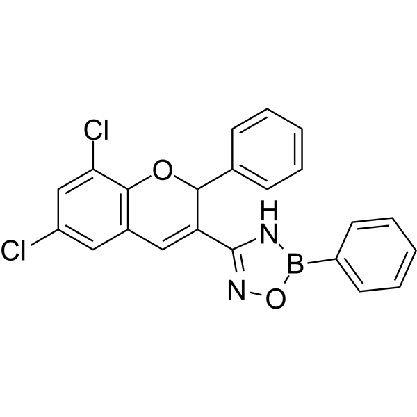
BT424 is a specific HCK inhibitor. BT424 can regulate macrophage activation and autophagy in vitro. BT424 ameliorates inflammation and kidney fibrosis in UUO model.
Nos produits utilisent uniquement pour la recherche. Nous ne vendons pas aux patients.
- Pureté : 98.89%
- CAS No.: 2755180-37-9
- Formule: C22H15BCl2N2O2
- Masse moléculaire:421.08
-
Stockage:
4°C, stored under nitrogen
* In solvent : -80°C, 6 months; -20°C, 1 month (stored under nitrogen)

To place orders, for customer services and technical support, please contact: MedChemExpress USA
Tel: 609-228-6898 E-mail: [email protected] [email protected]


-
 Activité biologique
-
Chemical Information
-
Solvant et solubilité
- Protocole
- Pureté et documentation
- Références
-
 Help & FAQs


-
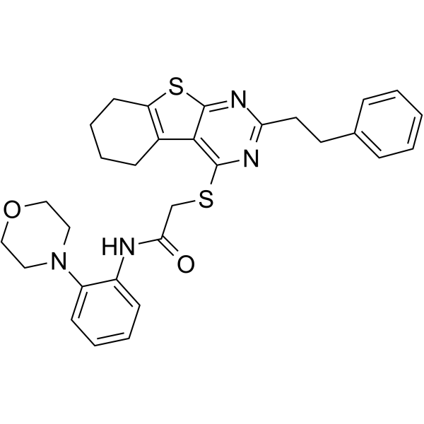
HY-139147
iHCK-37Hck Inhibitor -
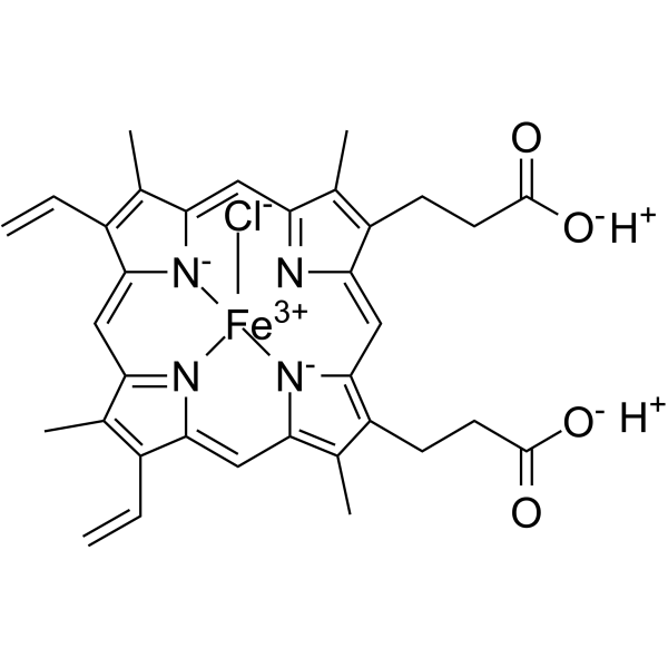
HY-19424
HeminHeme Oxygenase-1 Inducer -

HY-P9926
DupilumabIL-4Rα Inhibitor -
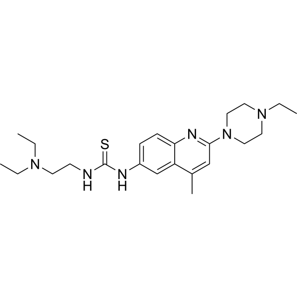
HY-124691
D-I03RAD52 Inhibitor -
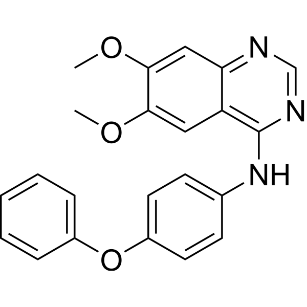
HY-101053
Src Inhibitor 1Src Inhibitor -
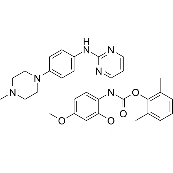
HY-12299
WH-4-023Src Inhibitor -
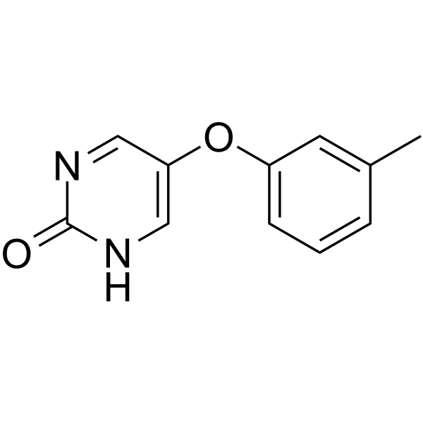
HY-59047
TolimidoneSrc Activator -
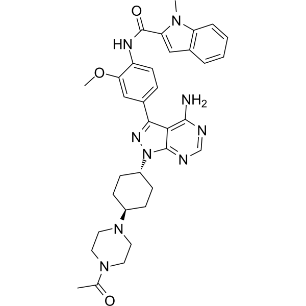
HY-11011
A-770041Lck Inhibitor -
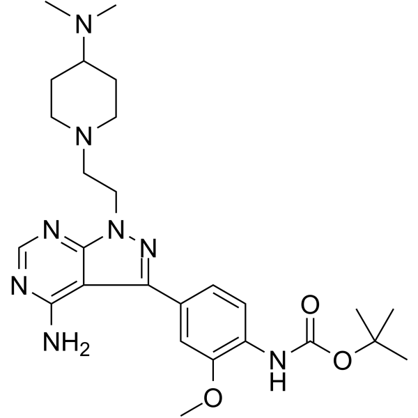
HY-112096
eCF506YES1/SRC inhibitor -
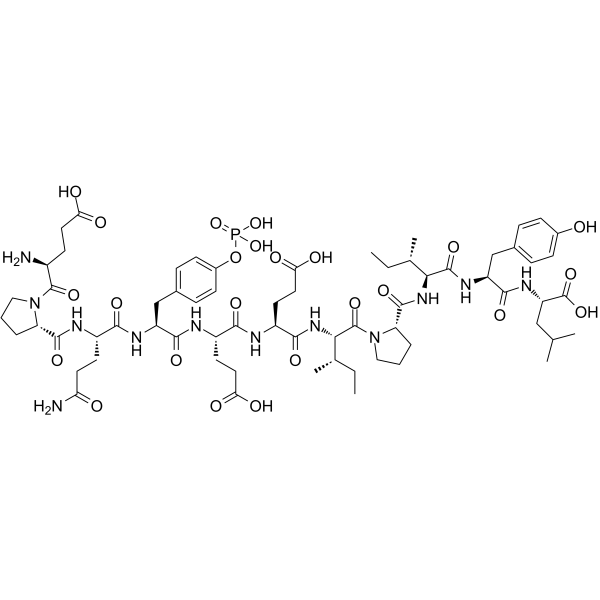
HY-P3279
EPQpYEEIPIYLSrc Family Activator -
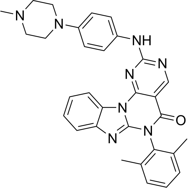
HY-12072
Lck InhibitorLck Inhibitor -
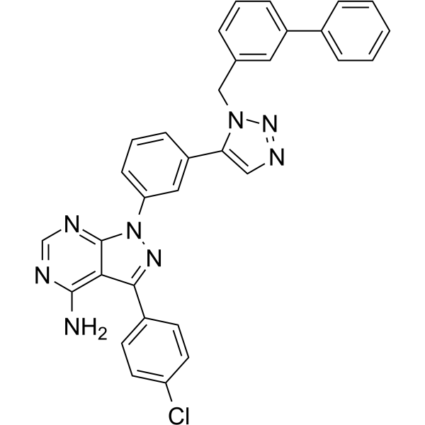
HY-108488
KB SRC 4c-Src Inhibitor


Activité biologique
Description


Chemical Information
-
CAS No. 2755180-37-9
-
Appearance Solid
-
Masse moléculaire 421.08
-
Formule C22H15BCl2N2O2
-
Color White to off-white
-
SMILES
ClC1=CC2=C(OC(C(C3=NOB(C4=CC=CC=C4)N3)=C2)C5=CC=CC=C5)C(Cl)=C1
-
Livraison
Room temperature in continental US; may vary elsewhere.
-
Stockage
4°C, stored under nitrogen
* In solvent : -80°C, 6 months; -20°C, 1 month (stored under nitrogen)


Solvant et solubilité
In Vitro:
DMSO : 100 mg/mL (237.48 mM; Need ultrasonic; Hygroscopic DMSO has a significant impact on the solubility of product, please use newly opened DMSO)
Please refer to the solubility information to select the appropriate solvent. Once prepared, please aliquot and store the solution to prevent product inactivation from repeated freeze-thaw cycles.
Storage method and period of stock solution: -80°C, 6 months; -20°C, 1 month (stored under nitrogen). When stored at -80°C, please use it within 6 months. When stored at -20°C, please use it within 1 month.
Please refer to the solubility information to select the appropriate solvent. Once prepared, please aliquot and store the solution to prevent product inactivation from repeated freeze-thaw cycles.
Storage method and period of stock solution: -80°C, 6 months; -20°C, 1 month (stored under nitrogen). When stored at -80°C, please use it within 6 months. When stored at -20°C, please use it within 1 month.
Concentration (start) × Volume (start) = Concentration (final) × Volume (final)
In Vivo:
Select the appropriate dissolution method based on your experimental animal and administration route.
- For the following dissolution methods, please ensure to first prepare a clear stock solution using an In Vitro approach and then sequentially add co-solvents:
- To ensure reliable experimental results, the clarified stock solution can be appropriately stored based on storage conditions. As for the working solution for In Vivo experiments, it is recommended to prepare freshly and use it on the same day.
- The percentages shown for the solvents indicate their volumetric ratio in the final prepared solution. If precipitation or phase separation occurs during preparation, heat and/or sonication can be used to aid dissolution.
In Vivo Dissolution Calculator
Please enter the basic information of animal experiments:
-
-
-
-
Recommended: Prepare an additional quantity of animals to account for potential losses during experiments.
Please enter your animal formula composition:
-
%DMSO +
Recommended: Keep the proportion of DMSO in working solution below 2% if your animal is weak.
-
%+
-
+%Tween-80 + +
-
%Saline +
The co-solvents required include: DMSO, . All of co-solvents are available by MedChemExpress (MCE). , Tween 80. All of co-solvents are available by MedChemExpress (MCE).
Working solution concentration: 0.22 mg/mL
Method for preparing stock solution: mg drug dissolved in μL DMSO. Stock solution concentration: mg/mL. * In solvent : -80°C, 6 months; -20°C, 1 month (stored under nitrogen)
1. Take μL DMSO stock solution;
2. Add μL .
μL , mix evenly;
3. Then add μL Tween 80, mix evenly;
4. Then add μL
Please ensure that the stock solution in the first step is dissolved to a clear state, and add co-solvents in sequence. You can use ultrasonic heating (ultrasonic cleaner, recommended frequency 20-40 kHz), vortexing, etc. to assist dissolution.


Pureté et documentation
-
Fiche technique (273 KB)
-
SDS (252 KB)
- English - EN (252 KB)
- Français - FR (252 KB)
- Deutsch - DE (252 KB)
- Norwegian - NO (252 KB)
- Español - ES (252 KB)
- Swedish - SV (252 KB)
- Italian - IT (252 KB)
- Korean - KR (252 KB)
- Portuguese - PT (252 KB)
-
Instruction de manipulation (2659 KB)
Références


Complete Stock Solution Preparation Table
Please refer to the solubility information to select the appropriate solvent. Once prepared, please aliquot and store the solution to prevent product inactivation from repeated freeze-thaw cycles.
Storage method and period of stock solution: -80°C, 6 months; -20°C, 1 month (stored under nitrogen). When stored at -80°C, please use it within 6 months. When stored at -20°C, please use it within 1 month.
| Optional Solvent | Concentration Solvent Mass | 1 mg | 5 mg | 10 mg | 25 mg |
|---|---|---|---|---|---|
| DMSO | 1 mM | 2.3748 mL | 11.8742 mL | 23.7485 mL | 59.3711 mL |
| 5 mM | 0.4750 mL | 2.3748 mL | 4.7497 mL | 11.8742 mL | |
| 10 mM | 0.2375 mL | 1.1874 mL | 2.3748 mL | 5.9371 mL | |
| 15 mM | 0.1583 mL | 0.7916 mL | 1.5832 mL | 3.9581 mL | |
| 20 mM | 0.1187 mL | 0.5937 mL | 1.1874 mL | 2.9686 mL | |
| 25 mM | 0.0950 mL | 0.4750 mL | 0.9499 mL | 2.3748 mL | |
| 30 mM | 0.0792 mL | 0.3958 mL | 0.7916 mL | 1.9790 mL | |
| 40 mM | 0.0594 mL | 0.2969 mL | 0.5937 mL | 1.4843 mL | |
| 50 mM | 0.0475 mL | 0.2375 mL | 0.4750 mL | 1.1874 mL | |
| 60 mM | 0.0396 mL | 0.1979 mL | 0.3958 mL | 0.9895 mL | |
| 80 mM | 0.0297 mL | 0.1484 mL | 0.2969 mL | 0.7421 mL | |
| 100 mM | 0.0237 mL | 0.1187 mL | 0.2375 mL | 0.5937 mL |


BT424 Related Classifications
HY-155657 Related Classifications
 Powered by Bioz