



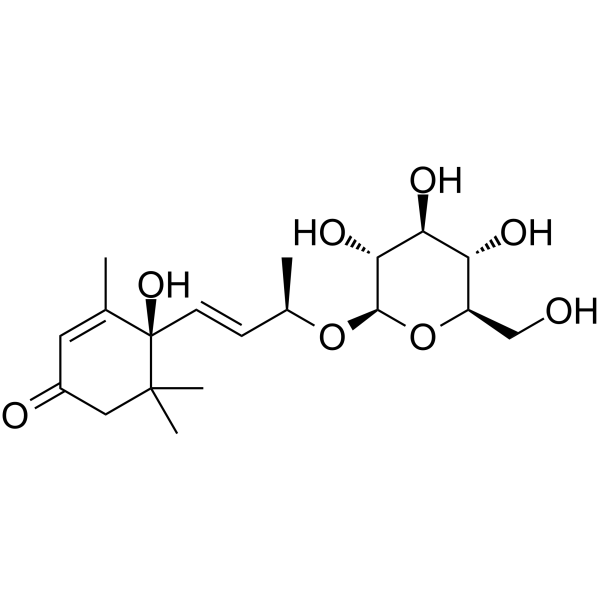
Roseoside

Based on 1 Customer Validation
Roseoside is an inhibitor of DNA gyrase and HAV 3C protease, and also inhibits HCV NS5A/B replicase in human systems with an IC50 of 20 μM. Roseoside binds to the active site of enzymes and stabilizes the interaction by forming hydrogen bonds with key amino acid residues. Roseoside inhibits the growth of Gram-positive bacteria, Gram-negative bacteria, and Candida albicans, and interferes with HCV RNA replication in vitro by inhibiting HCV NS5A/B replicase (IC50=20 μM). Roseoside shows no cytotoxicity and serves as a research tool for studies related to bacterial infections, candidiasis, HAV and HCV.
Nos produits utilisent uniquement pour la recherche. Nous ne vendons pas aux patients.
- Pureté : 96.99%
- CAS No.: 54835-70-0
- Formule: C19H30O8
- Masse moléculaire:386.44
-
Stockage:
4°C, protect from light
* In solvent : -80°C, 6 months; -20°C, 1 month (protect from light)

To place orders, for customer services and technical support, please contact: MedChemExpress USA
Tel: 609-228-6898 E-mail: [email protected] [email protected]


-
 Activité biologique
-
Chemical Information
- Protocole
- Pureté et documentation
- Références
-
 Help & FAQs


Voir tous les produits spécifiques à Isoform DNA/RNA Synthesis
More

Activité biologique
Description
IC50 & Target
|
DNA Polymerase |
Cellular Effect
|
Cell Line

|
Type | Value | Description | References |
|---|---|---|---|---|
| HEK-293T | IC50 |
55.7 μM
Compound: 7
|
Cytoprotective activity against CdCl2-induced cell death in human HEK293T cells assessed as increase in cell viability preincubated for 2 hrs followed by CdCl2 treatment and measured after 48 hrs by MTT assay
Cytoprotective activity against CdCl2-induced cell death in human HEK293T cells assessed as increase in cell viability preincubated for 2 hrs followed by CdCl2 treatment and measured after 48 hrs by MTT assay
|
[PMID: 31593459] |
In Vitro
Roseoside potently inhibits the growth of Escherichia coli, Klebsiella pneumoniae, Staphylococcus aureus, Bacillus subtilis, and Candida albicans with MIC values of 4.53 µM, 9.05 µM, 3.23 µM, 5.56 µM, and 2.59 µM, respectively[1].
Roseoside (31.25-1000 µg/mL) exhibits low cytotoxicity toward Vero cells, with an IC50 of 591.50 µg/mL and a maximum non-toxic concentration of 250 µg/mL[1].
Roseoside exhibits favorable in silico ADME properties, including moderate gastrointestinal absorption, no blood-brain barrier permeability, and a bioavailability score of 0.55, supporting its potential as an orally bioactive agent[1].
Roseoside (40 μM; 48 h) significantly inhibits HCV infection in naïve Huh7.5 cells, reducing relative HCV RNA levels to 48% of the control[2].
Roseoside (5-40 μM; 48 h) inhibits HCV infection in naïve Huh7.5 cells in vitro in a concentration-dependent manner (EC50 = 38.92 μM over 48 h) with no marked cytotoxicity at concentrations ≤40 μM[2].
Roseoside (40 μM; maintained from 4 h post-inoculation) inhibits HCV replication in naïve Huh7.5 cells in a time-dependent manner, reducing intracellular HCV RNA to 62% and 49% of the control at 36 and 48 h post-inoculation, respectively[2].
Roseoside (20-40 μM; 15 min pre-incubation at room temperature, followed by 1 h reaction) inhibits HCV NS5A/B replicase activity in vitro, reducing activity to 50% and 45% of the control at concentrations of 20 μM and 40 μM, respectively[2].
MedChemExpress (MCE) has not independently confirmed the accuracy of these methods. They are for reference only. Further protocols information, click here.
-
Cell Line:Naïve Huh7.5 human hepatocellular carcinoma cells
-
Concentration:40 μM
-
Incubation Time:24 h, 36 h, 48 h (maintained from 4 h post-inoculation)
-
Result:Reduced intracellular HCV RNA levels to 67%, 62%, and 49% at 24, 36, and 48 h post-inoculation, respectively, relative to the DMSO control.
Significantly decreased extracellular viral RNA levels at later time points.
Showed a time-dependent inhibitory effect on viral replication, with significant reduction in HCV RNA at 36 and 48 h post-inoculation.


Chemical Information
-
CAS No. 54835-70-0
-
Appearance Solid
-
Masse moléculaire 386.44
-
Formule C19H30O8
-
Color White to off-white
-
SMILES
C[C@@H](O[C@@H]([C@@H]([C@H]1O)O)O[C@@H]([C@H]1O)CO)/C=C/[C@](C(C)(C2)C)(C(C)=CC2=O)O
-
Livraison
Room temperature in continental US; may vary elsewhere.
-
Stockage
4°C, protect from light
* In solvent : -80°C, 6 months; -20°C, 1 month (protect from light)

Protocole
-
Cell Cytotoxicity Assay
Cytotoxicity assays are usually based on the assessment of cell membrane damage, which can also be indirectly detected by measuring cell viability. Detection methods include MTT assay, CKK-8 assay, LDH assay and ATP assay, etc.
-
Mammalian live/dead viability and cytotoxicity staining
Live/dead viability and cytotoxicity staining assays are based on the simultaneous detection of intracellular esterase activity in metabolically active (viable) cells and membrane integrity loss in non-viable cells. In commonly used dual-staining approaches, membrane-permeant fluorogenic substrates are converted by intracellular esterases into fluorescent products in live cells, while impermeant DNA-binding dyes selectively enter cells with compromised plasma membranes and label nucleic acids in dead or dying cells, enabling discrimination between viable and non-viable populations by fluorescence microscopy or flow cytometry.
-
Gram Staining of Tissue Sections
Gram staining of tissue sections is a histochemical technique used to differentiate Gram-positive and Gram-negative bacteria within histological specimens based on differences in bacterial cell wall structure and dye retention, adapted from classical bacteriological Gram staining into tissue-compatible “histological Gram stain” variants. In tissue applications, modifications of the Brown-Hopps and Brown-Brenn methods are commonly used to improve differentiation of microorganisms embedded within host connective tissue and to reduce overstaining or loss of Gram-negative signal, which are known limitations of earlier approaches. The principle relies on crystal violet-iodine complex retention in Gram-positive organisms and subsequent decolorization and counterstaining steps that allow contrast visualization of Gram-negative organisms against tissue background.
-
Cell Viability Determination by MTT Colorimetric Assay
The following protocol uses the MTT colorimetric assay as a classic literature-established method for assessing cell viability/metabolic activity in cultured mammalian cells. MTT[3-(4,5-dimethylthiazol-2-yl)-2,5-diphenyltetrazolium bromide] is reduced by metabolically active cells to a colored formazan product; the amount of formazan is quantified spectrophotometrically and provides an indirect measure of metabolically active viable cells. Importantly, MTT reduction reflects cellular oxidoreductase/metabolic activity rather than an absolute direct count of living cells, so changes in cellular metabolism can alter the signal independently of cell number.


Pureté et documentation
-
Fiche technique (273 KB)
-
SDS (252 KB)
- English - EN (252 KB)
- Français - FR (252 KB)
- Deutsch - DE (252 KB)
- Norwegian - NO (252 KB)
- Español - ES (252 KB)
- Swedish - SV (252 KB)
- Italian - IT (252 KB)
- Korean - KR (252 KB)
- Portuguese - PT (252 KB)
-
Instruction de manipulation (2659 KB)
Références

Calculators
Concentration (start) × Volume (start) = Concentration (final) × Volume (final)


Roseoside Related Classifications
HY-N1341 Related Classifications
 Powered by Bioz