



Chromeceptin
Based on 2 publication(s) in Google Scholar
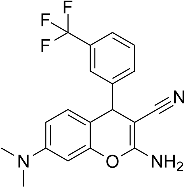
Chromeceptin (94G6) is an IGF signaling pathway inhibitor. Chromeceptin suppresses IGF2 expression at mRNA and protein levels in hepatocyte and HCC cells. Chromeceptin inhibits the phosphorylation levels of AKT and mTOR.
For research use only. We do not sell to patients.
- Purity : 99.80%
- CAS No.: 331859-86-0
- Formula: C19H16F3N3O
- Molecular Weight:359.35
-
Storage:Powder -20°C, 3 years , 4°C, 2 years ; In solvent -80°C, 6 months , -20°C, 1 month

To place orders, for customer services and technical support, please contact: MedChemExpress USA
Tel: 609-228-6898 E-mail: [email protected] [email protected]


-
 Biological Activity
-
Chemical Information
-
Solvent & Solubility
- Protocols
- Purity & Documentation
- References
-
 Help & FAQs


-
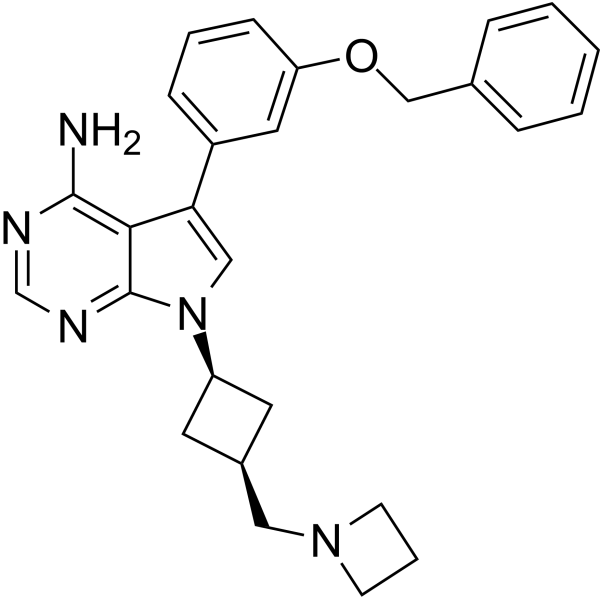
HY-50866
NVP-AEW541IGF-1R/IR Inhibitor -
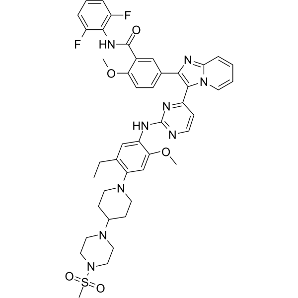
HY-10524
GSK1904529AIGF-1R Inhibitor -

HY-P7019
IGF2 Protein, Human -

HY-P99274
XentuzumabIGF1/IGF2 Antagonist -
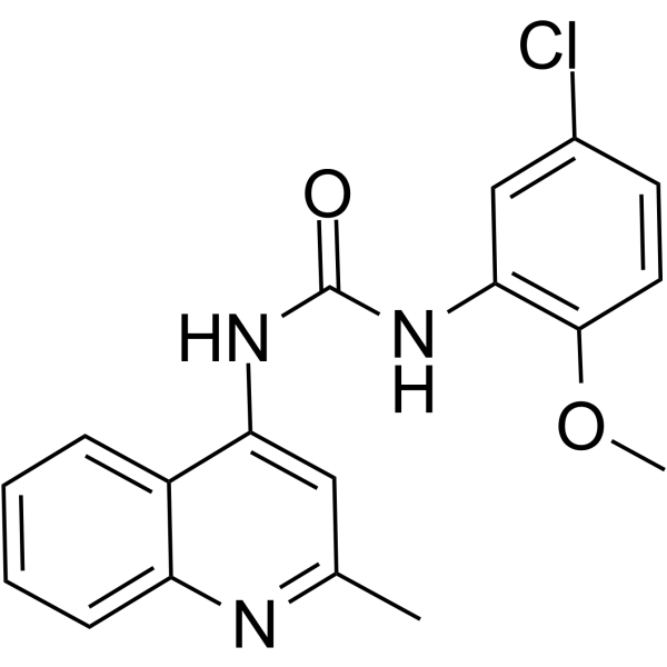
HY-13686
PQ401IGF-1R Inhibitor -
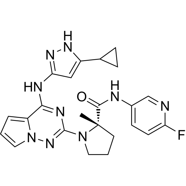
HY-10200
BMS-754807IGF-1R/IR Inhibitor -
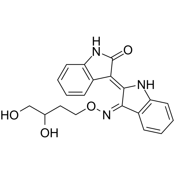
HY-18785
Indirubin Derivative E804IGF-1R Inhibitor -
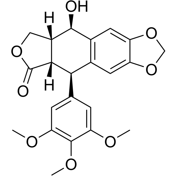
HY-15494
PicropodophyllinApoptosis Inducer -
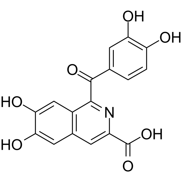
HY-110135
NBI-31772IGFBP Inhibitor -
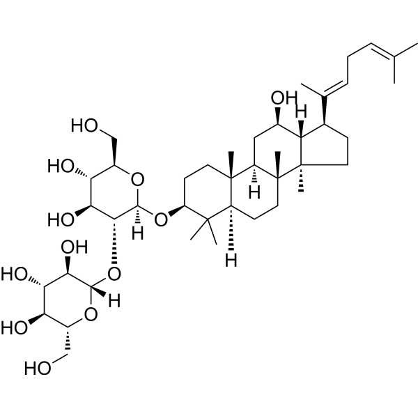
HY-N0908
Ginsenoside Rg5IGF-1R Agonist -
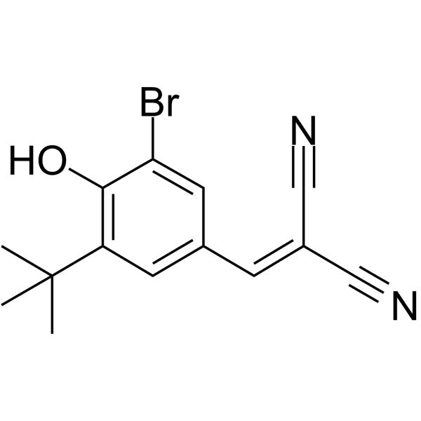
HY-10253
AG1024IGF-1R Inhibitor -
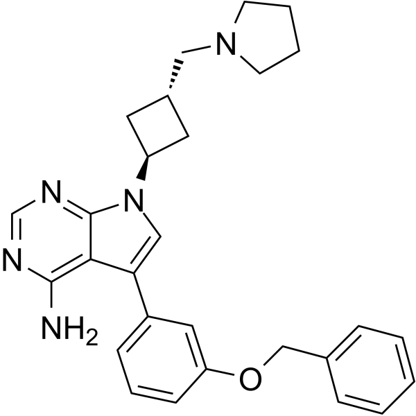
HY-10252
NVP-ADW742IGF-1R Inhibitor
Publications Citing Use of MedChemExpress (MCE) Chromeceptin
More

Biological Activity
Description
In Vitro
Chromeceptin (94G6) (5 μM) inhibits IGF2 expression in Hep3B-derived TS cells in a time-dependent manner[1].
Chromeceptin decreases the Phosphorylation levels of protein kinase B (AKT, Ser463) and rapamycin (mTOR, Ser 2481 and Ser 2448) in TS cells[1].
Chromeceptin represses the phosphorylation at ser371 in mTOR effectors ribosomal protein S6 kinase (p70S6K)[1].
MedChemExpress (MCE) has not independently confirmed the accuracy of these methods. They are for reference only. Further protocols information, click here.


Chemical Information
-
CAS No. 331859-86-0
-
Appearance Solid
-
Molecular Weight 359.35
-
Formula C19H16F3N3O
-
Color White to light yellow
-
SMILES
N#CC1=C(N)OC2=CC(N(C)C)=CC=C2C1C3=CC=CC(C(F)(F)F)=C3
-
Synonyms
94G6
-
Shipping
Room temperature in continental US; may vary elsewhere.
-
Storage
Powder -20°C 3 years 4°C 2 years In solvent -80°C 6 months -20°C 1 month


Publications (2)
-
Journal Impact Factor
-
Most Recent
-
Int J Biol Sci
Mechanical stretch-induced IGF2 overexpression in epidermal keratinocytes promotes hypertrophic scar formation through the IGF1R/p-c-Jun axis. [Abstract]2025 Jan 1;21(1):109-127. PMID: 39744430 -


Solvent & Solubility
In Vitro:
DMSO : 100 mg/mL (278.28 mM; Need ultrasonic; Hygroscopic DMSO has a significant impact on the solubility of product, please use newly opened DMSO)
H2O : < 0.1 mg/mL (insoluble)
Please refer to the solubility information to select the appropriate solvent. Once prepared, please aliquot and store the solution to prevent product inactivation from repeated freeze-thaw cycles.
Storage method and period of stock solution: -80°C, 6 months; -20°C, 1 month. When stored at -80°C, please use it within 6 months. When stored at -20°C, please use it within 1 month.
Please refer to the solubility information to select the appropriate solvent. Once prepared, please aliquot and store the solution to prevent product inactivation from repeated freeze-thaw cycles.
Storage method and period of stock solution: -80°C, 6 months; -20°C, 1 month. When stored at -80°C, please use it within 6 months. When stored at -20°C, please use it within 1 month.
Concentration (start) × Volume (start) = Concentration (final) × Volume (final)
In Vivo:
For the following dissolution methods, please prepare the working solution directly:
It is recommended to prepare fresh solutions and use them promptly within a short period of time.
The percentages shown for the solvents indicate their volumetric ratio in the final prepared solution. If precipitation or phase separation occurs during preparation, heat and/or sonication can be used to aid dissolution.
Add each solvent one by one: 50% PEG300 50% Saline
Solubility: 10 mg/mL (27.83 mM); Suspended solution; Need ultrasonic

Protocols
-
Kinase activity and phosphorylation assays
Kinase activity assays measure the ability of kinases to transfer phosphate groups from ATP to specific substrates, while phosphorylation assays detect the presence and levels of phosphorylated proteins. Common methods include radiolabeled ATP incorporation (e. g. ,), ADP release detection via bioluminescence (e. g. ,[3]), enzyme-linked immunosorbent assays (ELISA) for phospho-specific epitopes (e. g. ,[6]), and microtiter-based formats for high-throughput screening (e. g. ,[8]). The ADP-Glo assay quantifies kinase activity by measuring ADP produced during phosphorylation using a luciferase-based system. Radiometric assays involve autoradiography or scintillation counting after incorporation of 32P-labeled ATP into substrate proteins. ELISA-based approaches rely on phospho-specific antibodies to detect activated kinases in cell lysates or purified samples.
-
RNA extraction experimental
By lysing cells, releasing RNA, and removing impurities such as proteins and DNA, high-purity RNA products are finally obtained. The commonly used traditional method is the guanidine isothiocyanate/phenol/chloroform method (Trizol), which is suitable for a variety of animal materials including animal tissues, microorganisms, cultured cells, etc., and most plant materials.
-
Western Blot
Western blotting (WB) is a commonly used experimental method in molecular biology, biochemistry, and immunogenetics for identifying and quantifying target proteins. It combines gel electrophoresis with immunoassay, enabling researchers to analyze protein expression, post-translational modifications, and molecular weight.
-
Liver Cancer Modeling
Liver cancer can be classified into primary liver cancer and secondary liver cancer. Secondary liver cancer is the metastatic liver cancer. Primary liver cancer includes hepatocellular carcinoma (HCC), intrahepatic cholangiocarcinoma (ICC) and fibrolamellar HCC, of which HCC is the most common form, accounting for approximately 90% of primary liver cancers[1]. HCC mouse models include chemical agent-induced models, transplanted tumor models, and genetic engineered models.
-
Protocol for Kinase activity and phosphorylation assays
Kinase activity assays measure transfer of phosphate from ATP to a protein or peptide substrate, generating phosphorylated substrate, ADP, or incorporated radiolabeled phosphate as the readout; phosphorylation assays measure site-specific phosphorylation in cells or tissues as a proxy for kinase-pathway activation, inhibition, or substrate regulation. Phosphorylation can be detected by phospho-specific Western blot, immunoprecipitation kinase assay, phospho-immunofluorescence, phospho-flow cytometry, luminescent ADP detection, radiolabeled ATP incorporation, or reporter-based pathway assays, and these readouts can be applied to cancer cells, primary neurons, mouse tumors, organoids, inflammatory macrophages, ferroptosis studies, and mitophagy studies when the kinase target is biologically relevant.


Purity & Documentation
-
Data Sheet (276 KB)
-
SDS (393 KB)
- English - EN (393 KB)
- Français - FR (393 KB)
- Deutsch - DE (393 KB)
- Norwegian - NO (393 KB)
- Español - ES (393 KB)
- Swedish - SV (393 KB)
- Italian - IT (393 KB)
- Korean - KR (393 KB)
- Portuguese - PT (393 KB)
-
Handling Instructions (2659 KB)
References


Complete Stock Solution Preparation Table
Please refer to the solubility information to select the appropriate solvent. Once prepared, please aliquot and store the solution to prevent product inactivation from repeated freeze-thaw cycles.
Storage method and period of stock solution: -80°C, 6 months; -20°C, 1 month. When stored at -80°C, please use it within 6 months. When stored at -20°C, please use it within 1 month.
| Optional Solvent | Concentration Solvent Mass | 1 mg | 5 mg | 10 mg | 25 mg |
|---|---|---|---|---|---|
| DMSO | 1 mM | 2.7828 mL | 13.9140 mL | 27.8280 mL | 69.5701 mL |
| 5 mM | 0.5566 mL | 2.7828 mL | 5.5656 mL | 13.9140 mL | |
| 10 mM | 0.2783 mL | 1.3914 mL | 2.7828 mL | 6.9570 mL | |
| 15 mM | 0.1855 mL | 0.9276 mL | 1.8552 mL | 4.6380 mL | |
| 20 mM | 0.1391 mL | 0.6957 mL | 1.3914 mL | 3.4785 mL | |
| 25 mM | 0.1113 mL | 0.5566 mL | 1.1131 mL | 2.7828 mL | |
| 30 mM | 0.0928 mL | 0.4638 mL | 0.9276 mL | 2.3190 mL | |
| 40 mM | 0.0696 mL | 0.3479 mL | 0.6957 mL | 1.7393 mL | |
| 50 mM | 0.0557 mL | 0.2783 mL | 0.5566 mL | 1.3914 mL | |
| 60 mM | 0.0464 mL | 0.2319 mL | 0.4638 mL | 1.1595 mL | |
| 80 mM | 0.0348 mL | 0.1739 mL | 0.3479 mL | 0.8696 mL | |
| 100 mM | 0.0278 mL | 0.1391 mL | 0.2783 mL | 0.6957 mL |


Chromeceptin Related Classifications
HY-115449 Related Classifications
 Powered by Bioz