



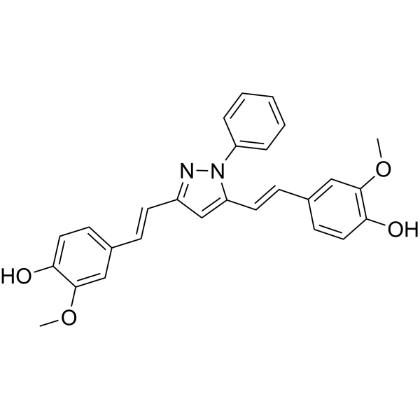
CNB-001
CNB-001 is a potent and orally active 5-lipoxygenase (5-LOX) inhibitor. CNB-001 can decreases 5-LOX expression and increase proteasome activity. CNB-001 can inhibit accumulation of soluble Amyloid-β and ubiquitinated aggregated proteins. CNB-001 can inhibit apoptosis, ROS production and stabilize mitochondrial membrane potential. CNB-001 can reduce insulin resistance and increase glucose uptake. CNB-001 also exhibits anti-ischemic, anti-inflammatory effects. CNB-001 can be used for the researches of inflammation, neurological and metabolic disease, such as Alzheimer's disease, stroke and diabetes.
For research use only. We do not sell to patients.
- CAS No.: 1019110-87-2
- Formula: C27H24N2O4
- Molecular Weight:440.49
-
Storage:
4°C, sealed storage, away from moisture
* In solvent : -80°C, 6 months; -20°C, 1 month (sealed storage, away from moisture)

To place orders, for customer services and technical support, please contact: MedChemExpress USA
Tel: 609-228-6898 E-mail: [email protected] [email protected]


-
 Biological Activity
-
Chemical Information
- Protocols
- Purity & Documentation
- References
-
 Help & FAQs




Biological Activity
Description
|
5-LOX 70 nM (IC50) |
15-LOX |
In Vitro
CNB-001 (1 μM, 24-96 h) inhibits and promotes the clearance of Aβ aggregation in MC65 cells[1].
CNB-001 (1 μM, 48 h) activates three proteasome activities (chymotrypsin-like, trypsin-like, caspase-like) and degrades Aβ in MC65 cells[1].
CNB-001 (1 μM, 1-24 h) induces eIF2α (Ser51) phosphorylation and increases ATF4 expression in MC65 cells[1].
CNB-001 (1 μM, 1-24 h) inhibits 5-lipoxygenase (5-LOX, IC50 = 70 nM) and increases S523 phosphorylation and decreases S271 phosphorylation of 5-LOX in MC65 cells[1].
CNB-001 inhibits leukotriene B4 (LTB4) production in human peripheral blood mononuclear lymphocytes (PBML) with an IC50 of 0.076 μM[2].
CNB-001 (0-2 μM) inhibits 15-LOX in rabbit reticulocytes[2].
NB-001 (0.5-15 μM, 24 h) exhibits a half-toxic concentration (TC50) of 15.3 μM in quiescent C2C12 myotubes[3].
CNB-001 (1 μM, 12 h) reverses Palmitic acid (HY-N0830)-induced insulin resistance and restores insulin-stimulated glucose uptake in C2C12 myotubes[3].
CNB-001 protects SK-N-SH human neuroblastoma cells against Rotenone (HY-B1756)-induced neurotoxicity by inhibiting intracellular ROS generation, apoptosis and stabilizing mitochondrial membrane potential[4].
MedChemExpress (MCE) has not independently confirmed the accuracy of these methods. They are for reference only. Further protocols information, click here.
-
Cell Line:MC65 cells
-
Concentration:1 μM
-
Incubation Time:1, 2, 4 and 6 h
-
Result:Increased S523 phosphorylation and decreases S271 phosphorylation of 5-LOX.
In Vivo
CNB-001 (500 mg/kg, p.o., 30 mins before Aβ1-42 injection) completely reverses the contextual memory impairment in mice injected with Aβ1-42[1].
CNB-001 (5-50 mg/kg, i.v., 5 or 60 mins post-embolization) improves behavioral deficits in New Zealand white rabbits with embolic stroke[2].
CNB-001 (10 mg/kg, i.v., 5 mins after MCAO) reduces infarct expansion in cynomolgus monkeys with permanent middle cerebral artery occlusion (MCAO)[2].
CNB-001 (40 mg/kg, i.p., daily except weekends for 20-22 weeks) alleviates high-fat diet-induced obesity and insulin resistance in C57BL/6J mice[3].
CNB-001 (24 mg/kg, i.p., daily from day 1 to day 7) mitigates motor impairments and neurotoxicity in MPTP-induced Parkinson’s disease (PD) mice[4].
MedChemExpress (MCE) has not independently confirmed the accuracy of these methods. They are for reference only.
-
Animal Model:APPswe/PS1E9 transgenic models[1]
-
Dosage:25 mg/kg diet
-
Administration:Orally administration, daily for 6 months
-
Result:Increased arm alternation choices.
Increased eIF2α phosphorylation, ATF4 and HSP90 expression in the hippocampus.
Reduced soluble Aβ1-42 (50%) and ubiquitinated aggregated proteins.
Increased the expression of synapse-associated proteins (phosphorylated synapsin-1, Pro-BDNF, Homer1).
Decreased the expression of clusterin and 5-LOX (>40%).
-
Animal Model:New Zealand white rabbits with embolic stroke[2]
-
Dosage:5-50 mg/kg 1 h post-embolization and 10 mg/kg 5 min post-embolization
-
Administration:Intravenously injection
-
Result:Increased P50 (clot burden causing 50% neurological dysfunction) by 74%.
Decreased COX-2 and 5-LOX expression, and increased BDNF levels in ipsilateral cortical tissues.
-
Animal Model:High-fat diet-induced obesity and insulin resistance in C57BL/6J mice models[3]
-
Dosage:40 mg/kg
-
Administration:Intraperitoneally injection, daily except weekends for 20-22 weeks
-
Result:Reduced body weight gain (without altering food intake).
Decreased serum triglyceride and interleukin-6 (IL-6) levels.
Improved glucose tolerance (lowers IPGTT AUC) and insulin sensitivity (lowers IPITT AUC).
Restored insulin-stimulated glucose uptake in gastrocnemius muscle.
Upregulated insulin signaling molecules (p-IR, p-Akt) and downregulates protein-tyrosine phosphatase 1B (PTP1B) and phospho-eIF2α in skeletal muscle.
Attenuated hepatic steatosis (reduces liver triglycerides and Oil Red O-stained fat droplets), and increased energy expenditure (without changing respiratory exchange ratio, RER).
-
Animal Model:MPTP-induced Parkinson’s disease (PD) mice[4]
-
Dosage:24 mg/kg
-
Administration:Intraperitoneally injection, daily from day 1 to day 7
-
Result:Reduced runway crossing time and foot slip errors in the narrow beam test.
Shortened fixed posture duration and reduced latency to shift four limbs.
Restored striatal dopamineand its metabolites (DOPAC, HVA) levels.
Decreased nitrite and citrulline accumulation.
Downregulated pro-inflammatory factors (TNF-α, IL-1β, IL-6, iNOS, GFAP, COX-2) and pro-apoptotic markers (Bax, cytochrome C, cleaved caspase-3).
Upregulated anti-apoptotic Bcl-2, and increased dopamine transporter (DAT) immunoreactivity in the substantia nigra (SN) and striatum (ST).


Chemical Information
-
CAS No. 1019110-87-2
-
Appearance Solid
-
Molecular Weight 440.49
-
Formula C27H24N2O4
-
Color White to off-white
-
SMILES
OC1=CC=C(C=C1OC)/C=C/C2=NN(C3=CC=CC=C3)C(/C=C/C4=CC(OC)=C(O)C=C4)=C2
-
Shipping
Room temperature in continental US; may vary elsewhere.
-
Storage
4°C, sealed storage, away from moisture
* In solvent : -80°C, 6 months; -20°C, 1 month (sealed storage, away from moisture)

Protocols
-
RNA extraction experimental
By lysing cells, releasing RNA, and removing impurities such as proteins and DNA, high-purity RNA products are finally obtained. The commonly used traditional method is the guanidine isothiocyanate/phenol/chloroform method (Trizol), which is suitable for a variety of animal materials including animal tissues, microorganisms, cultured cells, etc., and most plant materials.
-
Mitochondrial membrane-potential fluorescent assay
Mitochondrial membrane potential fluorescent assays estimate ΔΨm in living cells using lipophilic cationic dyes such as TMRM, TMRE, rhodamine 123, and JC-1, which accumulate in mitochondria according to membrane polarization; loss of signal after FCCP or CCCP treatment is interpreted as mitochondrial depolarization. TMRM/TMRE and rhodamine 123 are commonly used for semi-quantitative live-cell microscopy or flow cytometry, while JC-1 can report a shift from red aggregate fluorescence to green monomer fluorescence during depolarization; interpretation requires controls because dye concentration, quenching mode, cell type, dye efflux, and mitochondrial mass can affect fluorescence independently of ΔΨm.
-
Annexin V plus membrane-impermeant dye apoptosis staining
Annexin V-based apoptosis assays rely on the detection of phosphatidylserine (PS) externalization from the inner leaflet of the plasma membrane to the outer leaflet, an early biochemical hallmark of apoptosis. Fluorescently labeled Annexin V binds PS in a calcium-dependent manner, enabling identification of early apoptotic cells by flow cytometry or fluorescence microscopy. When combined with a membrane-impermeant DNA-binding dye (e. g. , propidium iodide), this approach allows discrimination between viable (Annexin V−/dye−), early apoptotic (Annexin V+/dye−), and late apoptotic or necrotic (Annexin V+/dye+) cell populations by assessing membrane integrity and PS exposure.
-
ROS/oxidative-stress fluorescent staining
ROS/oxidative-stress fluorescent staining uses cell-permeant fluorogenic probes that become fluorescent after oxidation inside cells or tissues; commonly used examples include DCFH-DA/DCFDA for broad cellular oxidant detection, DHE for superoxide-related signal detection, MitoSOX for mitochondrial superoxide-related signal detection, and CellROX probes for oxidative-stress-associated fluorescence readouts. The assay detects probe oxidation rather than a single ROS species unless the probe and analysis method have been chemically validated for that species. DCFH-DA enters cells, is deacetylated by intracellular esterases to DCFH, and produces fluorescent DCF after oxidation, so the readout is used as an operational measure of total cellular oxidative stress rather than a species-specific ROS measurement. DHE and MitoSOX can report superoxide-related oxidation, but red fluorescence alone can include non-specific ethidium-like oxidation products; HPLC or optimized spectral approaches are
-
Research Protocol for Inflammation-related Diseases
The NLRP3 inflammasome is a cytosolic innate immune signaling platform that integrates priming signals and danger-signal activation to promote caspase-1 activation, maturation of IL-1β and IL-18, and gasdermin D-mediated pyroptotic cell death. The core experimental logic is to determine whether inflammatory disease phenotypes are driven by increased NLRP3 expression, ASC-containing inflammasome assembly, caspase-1 cleavage, GSDMD cleavage, and extracellular release of IL-1β/IL-18 rather than by nonspecific cell injury alone. The pathway is strongly linked to inflammation-related disease phenotypes because monosodium urate crystals activate NALP3/NLRP3 inflammasome signaling in gout-like crystal inflammation, cholesterol crystals activate NLRP3 inflammasomes in atherogenesis models, and DSS-induced intestinal inflammation has been reported to involve NLRP3 inflammasome activity. However, experimental colitis studies also show context-dependent protective effects of NLRP3 inflammasome co
-
Amyloid: Congo Red Amyloid Staining
Congo red amyloid staining is a histochemical method used to detect extracellular amyloid deposits in tissue sections based on the affinity of Congo red dye for β-pleated sheet-rich protein aggregates. When bound to amyloid, Congo red produces characteristic apple-green birefringence under polarized light microscopy, which is widely regarded as a diagnostic feature of amyloid deposition in histopathology. The diagnostic principle relies on the combination of dye binding (congophilia) and optical anisotropy under polarized illumination, which distinguishes amyloid from most non-amyloid eosinophilic extracellular deposits in routine histological evaluation. Amyloid identification by Congo red staining remains a cornerstone in diagnostic pathology despite the availability of adjunct methods such as immunohistochemistry and mass spectrometry, particularly because of its ability to localize deposits directly within tissue architecture. The specificity of Congo red-positive deposits is incre
-
Research Protocol for Metabolic Diseases
AMP-activated protein kinase, AMPK, is a conserved cellular energy sensor that responds to reduced cellular energy status and coordinates metabolism by increasing ATP-generating catabolic pathways while suppressing ATP-consuming anabolic processes. In metabolic disease research, the AMPK pathway is experimentally relevant because it regulates hepatic lipid synthesis, fatty acid oxidation, glucose production, skeletal-muscle glucose disposal, mTORC1-linked biosynthesis, autophagy, mitochondrial homeostasis, and whole-body energy balance. The central pathway logic is that energy stress, metformin, exercise-like stimulation, or direct AMPK activators increase AMPKα Thr172 phosphorylation and downstream substrate phosphorylation, including ACC and RAPTOR. Phosphorylation of ACC suppresses lipogenesis and supports fatty acid oxidation, whereas phosphorylation of RAPTOR suppresses mTORC1 signaling and links cellular energy status to growth and protein synthesis control. The pathway is linked
-
Alzheimer’s Disease Modeling
Alzheimer’s Disease (AD) is a neurodegenerative disorder characterized by a progressive decline in cognitive functions and loss of specific types of neurons and synapses. Alzheimer's symptoms can be simulated in mice by injecting drugs (such as Aβ) or genetically modified.
-
Apoptosis
Apoptosis, also called programmed cell death, is generally characterized by distinct morphological characteristics.
-
TUNEL staining for apoptotic DNA fragmentation
TUNEL staining detects DNA strand breaks by using terminal deoxynucleotidyl transferase to add labeled nucleotides to exposed 3′-OH DNA termini, generating either microscopic staining in fixed cells or tissue sections, or fluorescence/cytometric signal in cell suspensions. TUNEL positivity reflects DNA fragmentation but should not be interpreted alone as definitive apoptosis, because TUNEL can also label necrotic, autolytic, mechanically damaged, or DNA-repair-associated DNA breaks.
-
Research Protocol for Endocrine Diseases
Endocrine diseases often arise from disrupted hormone production, hormone signaling, or target-tissue responsiveness; for diabetes-focused endocrine disease models, insulin signaling regulates glucose uptake, hepatic glucose output, lipid metabolism, and β-cell compensation. Type 2 diabetes develops through interacting defects in insulin resistance, β-cell dysfunction, adipose inflammation, hepatic glucose overproduction, altered incretin signaling, and ectopic lipid metabolism. A major unresolved question is whether endocrine dysfunction is driven primarily by target-tissue insulin resistance, intrinsic β-cell failure, immune/inflammatory stress, or combined multi-organ failure that differs by disease stage.
-
Fluorescent plasma-membrane potential dye assay
Fluorescent plasma-membrane potential dye assays measure changes in cell membrane potential using voltage-sensitive dyes whose fluorescence changes when cells depolarize or hyperpolarize. Anionic bis-oxonol dyes such as DiBAC4(3) enter depolarized cells more readily and show increased fluorescence after intracellular binding, while hyperpolarization reduces dye accumulation and fluorescence. FMP/FLIPR membrane-potential dyes are used for faster, homogeneous microplate assays of ion-channel or receptor-mediated membrane-potential changes.
-
Mitochondrial membrane-potential and mitochondrial mass staining
Mitochondrial membrane potential staining measures the electrochemical polarization across the mitochondrial inner membrane in live cells using lipophilic cationic fluorescent probes; early rhodamine-based work showed that selective mitochondrial dye accumulation is lost when the mitochondrial transmembrane potential is dissipated. JC-1 reports mitochondrial polarization by shifting from green monomer fluorescence to red J-aggregate fluorescence as dye concentration increases within energized mitochondria; therefore, the red/green fluorescence ratio is used as a relative readout of mitochondrial membrane potential. TMRE or TMRM staining provides a single-channel relative readout because these cationic rhodamine esters accumulate in polarized mitochondria, and lower fluorescence indicates reduced mitochondrial polarization when acquisition and dye-loading conditions are controlled. Mitochondrial mass staining is commonly performed with MitoTracker Green FM or related MitoTracker dyes as
-
Apoptosis Solutions
Apoptosis is a regulated, generally non-lytic cell-death pathway that removes unwanted, damaged, infected, or abnormal cells through coordinated morphological changes, caspase activation, DNA fragmentation, and membrane remodeling. The intrinsic apoptosis pathway is controlled mainly by mitochondrial outer membrane permeabilization, BCL-2 family proteins, cytochrome c release, apoptosome formation, caspase-9 activation, and downstream executioner caspase-3/7 activation. The extrinsic apoptosis pathway is initiated by death receptors such as Fas, TNFR, and TRAIL receptors, which recruit adaptor proteins and activate caspase-8 before engaging executioner caspases or mitochondrial amplification through BID cleavage. Apoptosis is linked to many phenotypes, including cancer cell killing, tissue homeostasis, immune regulation, neurodegeneration, infection response, and treatment-induced cytotoxicity; unresolved questions include how apoptosis interacts with necroptosis, pyroptosis, ferroptos


Purity & Documentation
-
Data Sheet (281 KB)
-
SDS (252 KB)
- Français - FR (252 KB)
- Deutsch - DE (252 KB)
- Norwegian - NO (252 KB)
- Español - ES (252 KB)
- Swedish - SV (252 KB)
- Italian - IT (252 KB)
- Korean - KR (252 KB)
- Portuguese - PT (252 KB)
-
Handling Instructions (2659 KB)
References
[1]. Valera E, et al. Modulation of 5-lipoxygenase in proteotoxicity and Alzheimer's disease. J Neurosci. 2013 Jun 19;33(25):10512-25. [Content Brief]
[2]. Lapchak PA, et al. CNB-001, a pleiotropic drug is efficacious in embolized agyrencephalic New Zealand white rabbits and ischemic gyrencephalic cynomolgus monkeys. Exp Neurol. 2019 Mar;313:98-108. [Content Brief]
[3]. Panzhinskiy E, et al. Novel curcumin derivative CNB-001 mitigates obesity-associated insulin resistance. J Pharmacol Exp Ther. 2014 May;349(2):248-57. [Content Brief]
[4]. Jayaraj RL, et al. CNB-001, a novel pyrazole derivative mitigates motor impairments associated with neurodegeneration via suppression of neuroinflammatory and apoptotic response in experimental Parkinson's disease mice. Chem Biol Interact. 2014 Sep 5;220:149-57. [Content Brief]

Calculators
Concentration (start) × Volume (start) = Concentration (final) × Volume (final)


CNB-001 Related Classifications
HY-158204 Related Classifications
 Powered by Bioz