



DCDAPH
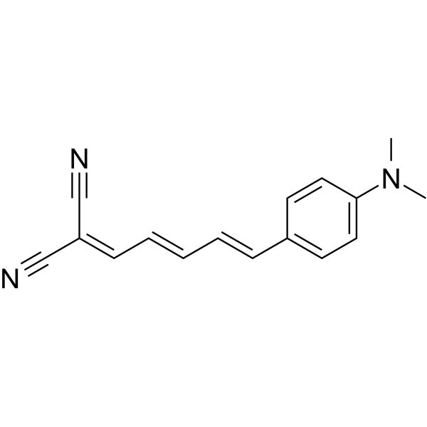
DCDAPH (Compound 2c) is a novel smart NIRF probe for detection of β-amyloid (Aβ) plaques (λex/λem=597/665 nm in PBS). DCDAPH shows high affinity for Aβ aggregates (Ki=37 nM, Kd=27 nM). DCDAPH shows good blood brain barrier permeation and can meet most of the requirements for the detection of Aβ plaques both in vitro and in vivo.
For research use only. We do not sell to patients.
- CAS No.: 125113-96-4
- Formula: C16H15N3
- Molecular Weight:249.31
-
Storage:
Please store the product under the recommended conditions in the Certificate of Analysis.

To place orders, for customer services and technical support, please contact: MedChemExpress USA
Tel: 609-228-6898 E-mail: [email protected] [email protected]


-
 Biological Activity
-
Chemical Information
- Protocols
- Purity & Documentation
- References
-
 Help & FAQs




Biological Activity
Description
In Vitro
DCDAPH (0-10 μM; 24 h) treatment shows no toxicity to human neuronal cells[1].
MedChemExpress (MCE) has not independently confirmed the accuracy of these methods. They are for reference only.
-
Cell Line:SH-SY5Y cells
-
Concentration:0, 0.1, 1, and 10 μM
-
Incubation Time:24 hours
-
Result:Showed no marked toxicity to this human neuronal cell line at 10 μM.
In Vivo
Fluorescent Staining of DCDAPH to Aβ Plaques in Mouse Brain[1].
1. Mouse should be i.v. injected with DCDAPH (0.4 mg/kg, 20% DMSO (HY-Y0320), 80% propylene glycol (HY-Y0921), 50 μL), and sacrificed at 30 min after injection.
2. The brains should be excised, embedded in optimum cutting temperature compound (OCT), and frozen in powdered dry ice immediately.
3. Frozen sections of 20 μm should be cut.
4. Fluorescent observation (Cy5 filter set).
MedChemExpress (MCE) has not independently confirmed the accuracy of these methods. They are for reference only.
Emission (Em)
665
Excitation (Ex)
597


Chemical Information
-
CAS No. 125113-96-4
-
Molecular Weight 249.31
-
Formula C16H15N3
-
SMILES
CN(C1=CC=C(/C=C/C=C/C=C(C#N)/C#N)C=C1)C
-
Shipping
Room temperature in continental US; may vary elsewhere.
-
Storage
Please store the product under the recommended conditions in the Certificate of Analysis.


Purity & Documentation
References

Calculators
Concentration (start) × Volume (start) = Concentration (final) × Volume (final)


DCDAPH Related Classifications
HY-D1684 Related Classifications
 Powered by Bioz