



ID-8
Based on 5 publication(s) in Google Scholar
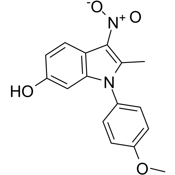
ID-8 is an inhibitor of dual-specificity tyrosine phosphorylation-regulated kinase (DYRK). ID-8 sustains embryonic stem cell (ESC) self-renewal and pluripotency. ID-8 enhances Wnt-mediated hESC survival and proliferation via inhibition of DYRKs.
For research use only. We do not sell to patients.
- Purity : 99.21%
- CAS No.: 147591-46-6
- Formula: C16H14N2O4
- Molecular Weight:298.29
-
Storage:Powder -20°C, 3 years , 4°C, 2 years ; In solvent -80°C, 2 years , -20°C, 1 year

To place orders, for customer services and technical support, please contact: MedChemExpress USA
Tel: 609-228-6898 E-mail: [email protected] [email protected]


-
 Biological Activity
-
Chemical Information
-
Solvent & Solubility
- Protocols
- Purity & Documentation
- References
-
 Help & FAQs


-
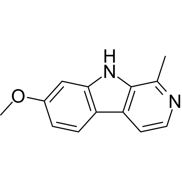
HY-N0737A
HarmineDYRK Inhibitor -
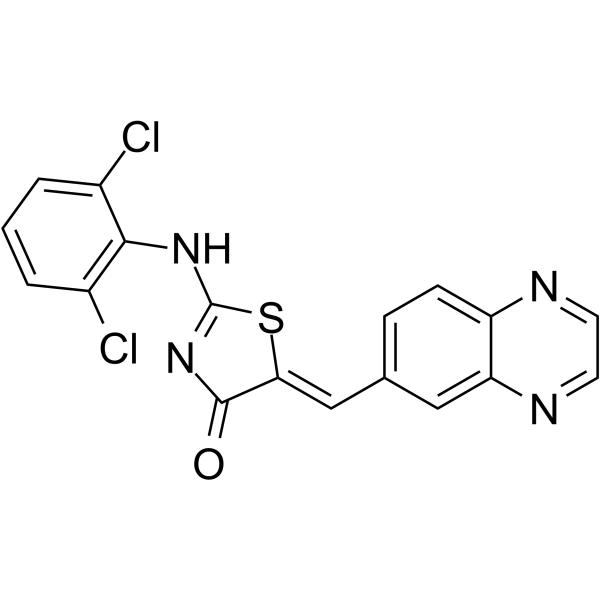
HY-105309
GSK-626616DYRK3 Inhibitor -
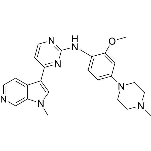
HY-12277
AZ191DYRK1B Inhibitor -
HY-108476
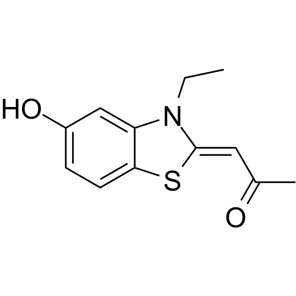
INDYDyrk1A And Dyrk1B Inhibitor -
HY-13756
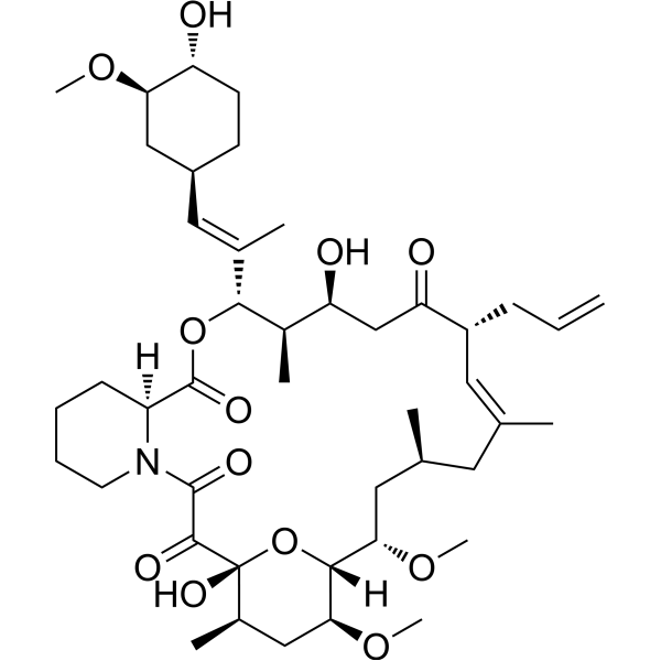
TacrolimusCalcineurin Inhibitor -
HY-59090
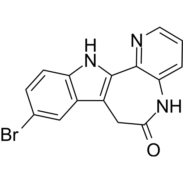
1-AzakenpaulloneGSK-3 Inhibitor -
HY-142295
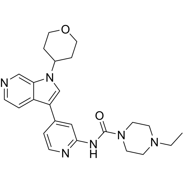
GNF2133DYRK1A Inhibitor -
HY-103193
NKH477Adenylate Cyclase Activator -
HY-13306
Pyrintegrinβ1-integrin Agonist -
HY-13455A
LDN-192960 hydrochlorideHaspin Inhibitor -

HY-N0737AR
Harmine (Standard)DYRK Inhibitor -

HY-N0737AG
Harmine (GMP)DYRK Inhibitor
Publications Citing Use of MedChemExpress (MCE) ID-8
More Customer Validation & Images

-
WB


Biological Activity
Description
IC50 & Target
DYRK[1]
Cellular Effect
|
Cell Line

|
Type | Value | Description | References |
|---|---|---|---|---|
| HT-22 | IC50 |
>100 μM
Compound: 44; ID-8
|
Cytotoxicity against mouse HT-22 cells assessed as reduction in cell viability incubated for 48 hrs by MTS assay
Cytotoxicity against mouse HT-22 cells assessed as reduction in cell viability incubated for 48 hrs by MTS assay
|
[PMID: 36876904] |
In Vitro


Chemical Information
-
CAS No. 147591-46-6
-
Appearance Solid
-
Molecular Weight 298.29
-
Formula C16H14N2O4
-
Color Light yellow to yellow
-
SMILES
OC1=CC2=C(C=C1)C([N+]([O-])=O)=C(C)N2C3=CC=C(OC)C=C3
-
Shipping
Room temperature in continental US; may vary elsewhere.
-
Storage
Powder -20°C 3 years 4°C 2 years In solvent -80°C 2 years -20°C 1 year


Publications (5)
-
Journal Impact Factor
-
Most Recent
-
Cell Chem Biol
In vivo drug discovery for increasing incretin-expressing cells identifies DYRK inhibitors that reinforce the enteroendocrine system. [Abstract]2022 Sep 15;29(9):1368-1380.e5. PMID: 35998625 -
Stem Cell Rev Rep
Dual-specificity Tyrosine Phosphorylation-regulated Kinase Inhibitor ID-8 Promotes Human Somatic Cell Reprogramming by Activating PDK4 Expression. [Abstract]2022 Aug;18(6):2074-2087. PMID: 35080746 -
Biochem Biophys Res Commun
β-catenin stimulates Tcf7l1 degradation through recruitment of casein kinase 2 in mouse embryonic stem cells. [Abstract]2020 Apr 2;524(2):280-287. PMID: 31987502
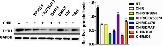
ID-8 purchased from MedChemExpress. Usage Cited in: Biochem Biophys Res Commun. 2020 Apr 2;524(2):280-287. [Abstract]
Western blot analysis of Tcf7l1 protein levels in mESCs pre-treated with the indicative different small molecules for 1 h and then treated with CHIR for 24 h.
-
-


Solvent & Solubility
In Vitro:
DMSO : ≥ 47 mg/mL (157.56 mM; Hygroscopic DMSO has a significant impact on the solubility of product, please use newly opened DMSO)
* "≥" means soluble, but saturation unknown.
Please refer to the solubility information to select the appropriate solvent. Once prepared, please aliquot and store the solution to prevent product inactivation from repeated freeze-thaw cycles.
Storage method and period of stock solution: -80°C, 2 years; -20°C, 1 year. When stored at -80°C, please use it within 2 years. When stored at -20°C, please use it within 1 year.
Please refer to the solubility information to select the appropriate solvent. Once prepared, please aliquot and store the solution to prevent product inactivation from repeated freeze-thaw cycles.
Storage method and period of stock solution: -80°C, 2 years; -20°C, 1 year. When stored at -80°C, please use it within 2 years. When stored at -20°C, please use it within 1 year.
Concentration (start) × Volume (start) = Concentration (final) × Volume (final)
In Vivo:
Select the appropriate dissolution method based on your experimental animal and administration route.
- For the following dissolution methods, please ensure to first prepare a clear stock solution using an In Vitro approach and then sequentially add co-solvents:
- To ensure reliable experimental results, the clarified stock solution can be appropriately stored based on storage conditions. As for the working solution for In Vivo experiments, it is recommended to prepare freshly and use it on the same day.
- The percentages shown for the solvents indicate their volumetric ratio in the final prepared solution. If precipitation or phase separation occurs during preparation, heat and/or sonication can be used to aid dissolution.
Add each solvent one by one: 10% DMSO 40% PEG300 5% Tween-80 45% Saline
Solubility: 2.5 mg/mL (8.38 mM); Suspended solution; Need ultrasonic
This protocol yields a suspended solution of 2.5 mg/mL. Suspended solution can be used for oral and intraperitoneal injection.
Taking 1 mL working solution as an example, add 100 μL DMSO stock solution (25.0 mg/mL) to 400 μL PEG300, and mix evenly; then add 50 μL Tween-80 and mix evenly; then add 450 μL Saline to adjust the volume to 1 mL.
Preparation of Saline: Dissolve 0.9 g sodium chloride in ddH₂O and dilute to 100 mL to obtain a clear Saline solution.
In Vivo Dissolution Calculator
Please enter the basic information of animal experiments:
-
-
-
-
Recommended: Prepare an additional quantity of animals to account for potential losses during experiments.
Please enter your animal formula composition:
-
%DMSO +
Recommended: Keep the proportion of DMSO in working solution below 2% if your animal is weak.
-
%+
-
+%Tween-80 + +
-
%Saline +
The co-solvents required include: DMSO, . All of co-solvents are available by MedChemExpress (MCE). , Tween 80. All of co-solvents are available by MedChemExpress (MCE).
Working solution concentration: 0.22 mg/mL
Method for preparing stock solution: mg drug dissolved in μL DMSO. Stock solution concentration: mg/mL.
1. Take μL DMSO stock solution;
2. Add μL .
μL , mix evenly;
3. Then add μL Tween 80, mix evenly;
4. Then add μL
Please ensure that the stock solution in the first step is dissolved to a clear state, and add co-solvents in sequence. You can use ultrasonic heating (ultrasonic cleaner, recommended frequency 20-40 kHz), vortexing, etc. to assist dissolution.


Purity & Documentation
-
Data Sheet (279 KB)
-
SDS (396 KB)
- English - EN (396 KB)
- Français - FR (396 KB)
- Deutsch - DE (396 KB)
- Norwegian - NO (396 KB)
- Español - ES (396 KB)
- Swedish - SV (396 KB)
- Italian - IT (396 KB)
- Korean - KR (396 KB)
- Portuguese - PT (396 KB)
-
Handling Instructions (2659 KB)
References
[1]. Hasegawa, K., et al., Wnt signaling orchestration with a small molecule DYRK inhibitor provides long-term xeno-free human pluripotent cell expansion. Stem Cells Transl Med, 2012. 1(1): p. 18-28. [Content Brief]
[2]. Miyabayashi, T., et al., Indole derivatives sustain embryonic stem cell self-renewal in long-term culture. Biosci Biotechnol Biochem, 2008. 72(5): p. 1242-8. [Content Brief]


Complete Stock Solution Preparation Table
Please refer to the solubility information to select the appropriate solvent. Once prepared, please aliquot and store the solution to prevent product inactivation from repeated freeze-thaw cycles.
Storage method and period of stock solution: -80°C, 2 years; -20°C, 1 year. When stored at -80°C, please use it within 2 years. When stored at -20°C, please use it within 1 year.
| Optional Solvent | Concentration Solvent Mass | 1 mg | 5 mg | 10 mg | 25 mg |
|---|---|---|---|---|---|
| DMSO | 1 mM | 3.3524 mL | 16.7622 mL | 33.5244 mL | 83.8111 mL |
| 5 mM | 0.6705 mL | 3.3524 mL | 6.7049 mL | 16.7622 mL | |
| 10 mM | 0.3352 mL | 1.6762 mL | 3.3524 mL | 8.3811 mL | |
| 15 mM | 0.2235 mL | 1.1175 mL | 2.2350 mL | 5.5874 mL | |
| 20 mM | 0.1676 mL | 0.8381 mL | 1.6762 mL | 4.1906 mL | |
| 25 mM | 0.1341 mL | 0.6705 mL | 1.3410 mL | 3.3524 mL | |
| 30 mM | 0.1117 mL | 0.5587 mL | 1.1175 mL | 2.7937 mL | |
| 40 mM | 0.0838 mL | 0.4191 mL | 0.8381 mL | 2.0953 mL | |
| 50 mM | 0.0670 mL | 0.3352 mL | 0.6705 mL | 1.6762 mL | |
| 60 mM | 0.0559 mL | 0.2794 mL | 0.5587 mL | 1.3969 mL | |
| 80 mM | 0.0419 mL | 0.2095 mL | 0.4191 mL | 1.0476 mL | |
| 100 mM | 0.0335 mL | 0.1676 mL | 0.3352 mL | 0.8381 mL |


ID-8 Related Classifications
HY-15838 Related Classifications
 Powered by Bioz