



Acifran
Based on 1 publication(s) in Google Scholar
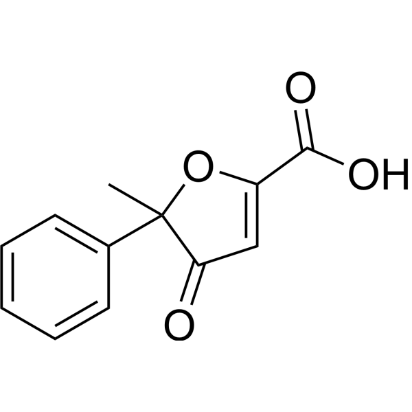
Acifran (AY 25712) is an orally active, full GPR109A and GPR109B agonist and hypolipidemic agent. Acifran reduces Forskolin (HY-15371)-induced cAMP elevation, triggers ERK1/ERK2 phosphorylation, and inhibits lipolysis in adipose tissue through Gi-coupled GPCR activation. Acifran is used for research on atherosclerosis, hyperlipidemia, and type 2 diabetes.
For research use only. We do not sell to patients.
- Purity : 99.03%
- CAS No.: 72420-38-3
- Formula: C12H10O4
- Molecular Weight:218.21
-
Storage:Powder -20°C, 3 years ; In solvent -80°C, 6 months , -20°C, 1 month

To place orders, for customer services and technical support, please contact: MedChemExpress USA
Tel: 609-228-6898 E-mail: [email protected] [email protected]


-
 Biological Activity
-
Chemical Information
-
Solvent & Solubility
- Protocols
- Purity & Documentation
- References
-
 Help & FAQs


-
HY-107581
MK-1903GPR109a/HCAR2 Agonist -

HY-107579R
Acifran (Standard)Reference Standard -
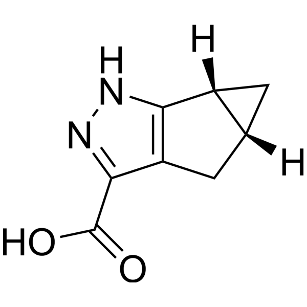
HY-107580
GPR109 receptor agonist-1GPR109b Receptor Agonist -
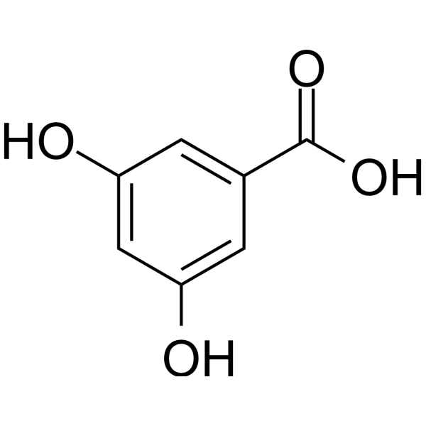
HY-W015560
3,5-Dihydroxybenzoic acidGPR81 Agonist -
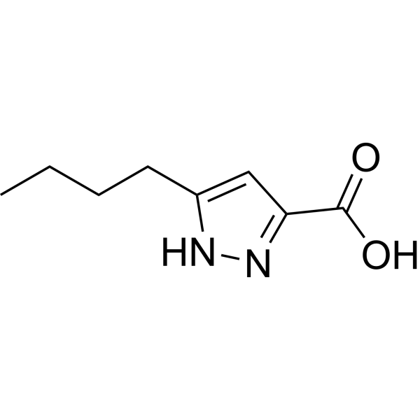
HY-100976
LUF6283HCA(2) Agonist -
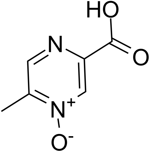
HY-B0283
AcipimoxAntilipolytic Agent -

HY-W015560R
3,5-Dihydroxybenzoic acid (Standard)GPR81 Agonist -
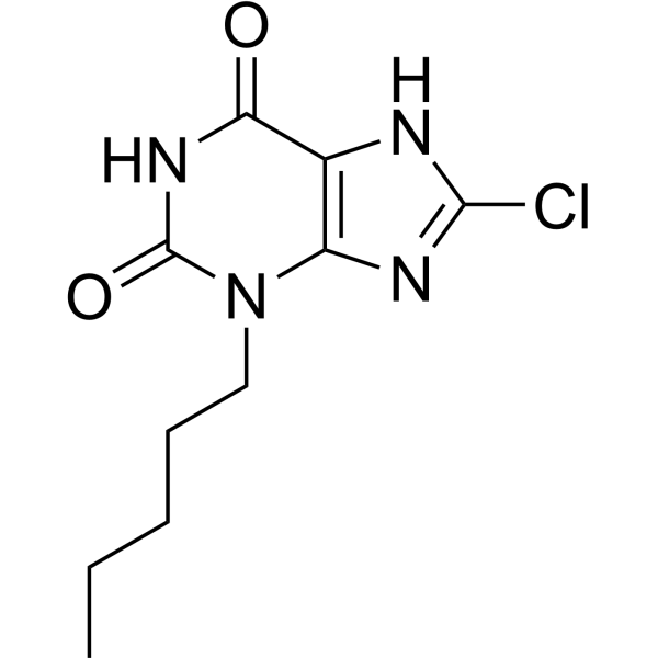
HY-119222
GSK256073GPR109A Agonist -

HY-107581R
MK-1903 (Standard)Reference Standard -

HY-B0283R
Acipimox (Standard)Antilipolytic Agent
Publications Citing Use of MedChemExpress (MCE) Acifran
More

Biological Activity
Description
IC50 & Target
[3]|
ERK1 |
ERK2 |
Cellular Effect
|
Cell Line

|
Type | Value | Description | References |
|---|---|---|---|---|
| CHO-K1 | EC50 |
160 nM
|
Phosphorylation of ERK1/ERK2 in CHO-K1 cells stably expressing HM74A incubated for 5 mins followed by ELISA.
Phosphorylation of ERK1/ERK2 in CHO-K1 cells stably expressing HM74A incubated for 5 mins followed by ELISA.
|
16389067 |
| CHO-K1 | EC50 |
316 nM
|
Phosphorylation of ERK1/ERK2 in CHO-K1 cells stably expressing HM74 incubated for 5 mins followed by ELISA.
Phosphorylation of ERK1/ERK2 in CHO-K1 cells stably expressing HM74 incubated for 5 mins followed by ELISA.
|
16389067 |
In Vitro
Acifran is a full agonist of GPR109a, with an EC50 of 1.3 μM in the cAMP whole-cell assay and an EC50 of 4.2 μM for GPR109b[1].
Acifran (10-50 μg/mL) shows weak binding to rat and dog serum proteins, and significantly displaces protein-bound Warfarin (HY-B0687) only in dog serum[2].
Acifran (5 min) activates ERK1/ERK2 phosphorylation with an EC50 of 160 nM in CHO-K1 cells expressing HM74A and an EC50 of 316 nM in CHO-K1 cells expressing HM74 [3].
Acifran (1 μM) induces strong phosphorylation of p-ERK1/ERK2 in CHO-K1 cells expressing HM74A and HM74, and this effect is completely inhibited by Pertussis toxin[3].
Acifran (20-200 μM; 5 min) induces phosphorylation of p-ERK1/ERK2 in CHO-K1 cells stably expressing HM74A[3].
Acifran (20-200 μM; 5 min) activates ERK1/ERK2 phosphorylation in CHO-K1 cells stably expressing HM74[3].
MedChemExpress (MCE) has not independently confirmed the accuracy of these methods. They are for reference only.
-
Cell Line:CHO-K1 cells stably expressing HM74A or HM74
-
Concentration:9-point log10 scale
-
Incubation Time:5 min
-
Result:Yielded an EC50 of 160 nM for HM74A.
Yielded an EC50 of 316 nM for HM74.
Showed a modest effect on phosphorylation of ERK1/ERK2 in parental CHO-K1 cells only at 100 μM.
-
Cell Line:CHO-K1 cells stably expressing HM74A
-
Concentration:20-200 μM
-
Incubation Time:5 min
-
Result:Elicited p-ERK1/ERK2 phosphorylation at both 20 μM and 200 μM, with a robust signal appearing at both concentrations.
-
Cell Line:CHO-K1 cells stably expressing HM74
-
Concentration:20-200 μM
-
Incubation Time:5 min
-
Result:Elicited phosphorylation of ERK1/ERK2 at both 20 μM and 200 μM, with robust activation observed even at 20 μM.
In Vivo
MedChemExpress (MCE) has not independently confirmed the accuracy of these methods. They are for reference only.
-
Animal Model:Sprague-Dawley (male, 175-200 g, fructose-fed)[3]
-
Dosage:1, 2.5, 5, 10, 15 mg/kg
-
Administration:p.o.; single dose
-
Result:Decreased serum triglycerides dose-dependently by 39% at 1 mg/kg, 44% at 2.5 mg/kg, 49% at 5 mg/kg, 57% at 10 mg/kg, and 61% at 15 mg/kg versus untreated control.


Chemical Information
-
CAS No. 72420-38-3
-
Appearance Solid
-
Molecular Weight 218.21
-
Formula C12H10O4
-
Color Off-white to light yellow
-
SMILES
O=C(C(OC1(C)C2=CC=CC=C2)=CC1=O)O
-
Synonyms
AY 25712
-
Shipping
Room temperature in continental US; may vary elsewhere.
-
Storage
Powder -20°C 3 years In solvent -80°C 6 months -20°C 1 month


Publications (1)
-
Journal Impact Factor
-
Most Recent
-
iScience
Comparative analysis reveals molecular adaptation of mammalian HCA2 to microbial metabolites. [Abstract]2026 May 22;29(6):116030. PMID: 42221821


Solvent & Solubility
In Vitro:
DMSO : 125 mg/mL (572.84 mM; Need ultrasonic; Hygroscopic DMSO has a significant impact on the solubility of product, please use newly opened DMSO)
Please refer to the solubility information to select the appropriate solvent. Once prepared, please aliquot and store the solution to prevent product inactivation from repeated freeze-thaw cycles.
Storage method and period of stock solution: -80°C, 6 months; -20°C, 1 month. When stored at -80°C, please use it within 6 months. When stored at -20°C, please use it within 1 month.
Please refer to the solubility information to select the appropriate solvent. Once prepared, please aliquot and store the solution to prevent product inactivation from repeated freeze-thaw cycles.
Storage method and period of stock solution: -80°C, 6 months; -20°C, 1 month. When stored at -80°C, please use it within 6 months. When stored at -20°C, please use it within 1 month.
Concentration (start) × Volume (start) = Concentration (final) × Volume (final)


Purity & Documentation
-
Data Sheet (292 KB)
-
SDS (254 KB)
- English - EN (254 KB)
- Français - FR (254 KB)
- Deutsch - DE (254 KB)
- Norwegian - NO (254 KB)
- Español - ES (254 KB)
- Swedish - SV (254 KB)
- Italian - IT (254 KB)
- Korean - KR (254 KB)
- Portuguese - PT (254 KB)
-
Handling Instructions (2659 KB)
References
[1]. Jung JK, et al. Analogues of acifran: agonists of the high and low affinity niacin receptors, GPR109a and GPR109b. Journal of medicinal chemistry. 2007 Apr 05;50(7):1445-8. [Content Brief]
[2]. Cayen MN, et al. The metabolic disposition of acifran, a new antihyperlipidemic agent, in rats and dogs. Xenobiotica; the fate of foreign compounds in biological systems. 1986 Mar;16(3):251-63. [Content Brief]


Complete Stock Solution Preparation Table
Please refer to the solubility information to select the appropriate solvent. Once prepared, please aliquot and store the solution to prevent product inactivation from repeated freeze-thaw cycles.
Storage method and period of stock solution: -80°C, 6 months; -20°C, 1 month. When stored at -80°C, please use it within 6 months. When stored at -20°C, please use it within 1 month.
| Optional Solvent | Concentration Solvent Mass | 1 mg | 5 mg | 10 mg | 25 mg |
|---|---|---|---|---|---|
| DMSO | 1 mM | 4.5827 mL | 22.9137 mL | 45.8274 mL | 114.5685 mL |
| 5 mM | 0.9165 mL | 4.5827 mL | 9.1655 mL | 22.9137 mL | |
| 10 mM | 0.4583 mL | 2.2914 mL | 4.5827 mL | 11.4569 mL | |
| 15 mM | 0.3055 mL | 1.5276 mL | 3.0552 mL | 7.6379 mL | |
| 20 mM | 0.2291 mL | 1.1457 mL | 2.2914 mL | 5.7284 mL | |
| 25 mM | 0.1833 mL | 0.9165 mL | 1.8331 mL | 4.5827 mL | |
| 30 mM | 0.1528 mL | 0.7638 mL | 1.5276 mL | 3.8190 mL | |
| 40 mM | 0.1146 mL | 0.5728 mL | 1.1457 mL | 2.8642 mL | |
| 50 mM | 0.0917 mL | 0.4583 mL | 0.9165 mL | 2.2914 mL | |
| 60 mM | 0.0764 mL | 0.3819 mL | 0.7638 mL | 1.9095 mL | |
| 80 mM | 0.0573 mL | 0.2864 mL | 0.5728 mL | 1.4321 mL | |
| 100 mM | 0.0458 mL | 0.2291 mL | 0.4583 mL | 1.1457 mL |


Acifran Related Classifications
HY-107579 Related Classifications
 Powered by Bioz